
Rabbit Anti-CK7 antibody
CK 7; CK-7; Cytokeratin 7; Cytokeratin-7; Cytokeratin7; D15Wsu77e; K2C7; K2C7_HUMAN; K7; Keratin 55k type ii cytoskeletal; Keratin 7; Keratin simple epithelial type 1 k7; Keratin type II cytoskeletal 7; Keratin type ii cytoskletal 7; Keratin, 55K type II
View History [Clear]
Details
Product Name CK7 Chinese Name 细胞角蛋白7抗体 Alias CK 7; CK-7; Cytokeratin 7; Cytokeratin-7; Cytokeratin7; D15Wsu77e; K2C7; K2C7_HUMAN; K7; Keratin 55k type ii cytoskeletal; Keratin 7; Keratin simple epithelial type 1 k7; Keratin type II cytoskeletal 7; Keratin type ii cytoskletal 7; Keratin, 55K type II cytoskeletal; Keratin, simple epithelial; Keratin, simple epithelial type I, K7; Keratin, type II cytoskeletal 7; Keratin-7; Keratin7; KRT 7; Krt2-7; KRT7; MGC11625; MGC129731; mgc3625; Sarcolectin; SCL; Type ii mesothelial keratin k7; Type-II keratin Kb7 literatures Research Area Tumour Cell biology Signal transduction Immunogen Species Rabbit Clonality Polyclonal React Species Human, Mouse, Rat, Applications WB=1:500-2000 ELISA=1:5000-10000 IHC-P=1:100-500 IHC-F=1:100-500 Flow-Cyt=1ug/Test ICC=1:100 IF=1:100-500 (Paraffin sections need antigen repair)
not yet tested in other applications.
optimal dilutions/concentrations should be determined by the end user.Theoretical molecular weight 54kDa Cellular localization cytoplasmic Form Liquid Concentration 1mg/ml immunogen KLH conjugated synthetic peptide derived from the middle of mouse CK7: 251-350/469 Lsotype IgG Purification affinity purified by Protein A Buffer Solution 0.01M TBS(pH7.4) with 1% BSA, 0.03% Proclin300 and 50% Glycerol. Storage Shipped at 4℃. Store at -20 °C for one year. Avoid repeated freeze/thaw cycles. Attention This product as supplied is intended for research use only, not for use in human, therapeutic or diagnostic applications. PubMed PubMed Product Detail The protein encoded by this gene is a member of the keratin gene family. The type II cytokeratins consist of basic or neutral proteins which are arranged in pairs of heterotypic keratin chains coexpressed during differentiation of simple and stratified epithelial tissues. This type II cytokeratin is specifically expressed in the simple epithelia lining the cavities of the internal organs and in the gland ducts and blood vessels. The genes encoding the type II cytokeratins are clustered in a region of chromosome 12q12-q13. Alternative splicing may result in several transcript variants; however, not all variants have been fully described. [provided by RefSeq, Jul 2008]
Function:
Blocks interferon-dependent interphase and stimulates DNA synthesis in cells. Involved in the translational regulation of the human papillomavirus type 16 E7 mRNA (HPV16 E7).
Subunit:
Heterotetramer of two type I and two type II keratins. Interacts with eukaryotic translation initiator factor 3 (eIF3) subunit EIF3S10 and with HPV16 E7.
Subcellular Location:
Cytoplasm.
Tissue Specificity:
Expressed in cultured epidermal, bronchial and mesothelial cells but absent in colon, ectocervix and liver. Observed throughout the glandular cells in the junction between stomach and esophagus but is absent in the esophagus.
Post-translational modifications:
Arg-20 is dimethylated, probably to asymmetric dimethylarginine.
Similarity:
Belongs to the intermediate filament family.
SWISS:
Q9DCV7
Gene ID:
110310
Database links:Entrez Gene: 3855 Human
Entrez Gene: 110310 Mouse
Omim: 148059 Human
SwissProt: P08729 Human
SwissProt: Q9DCV7 Mouse
Unigene: 411501 Human
Unigene: 670221 Human
Unigene: 289377 Mouse
Unigene: 7913 Rat
结构蛋白(Structural Proteins)
CK-7是一种 54KDa 的中间丝蛋白,存在于大多数正常组织的腺上皮和移行epithelial cells中。 该抗体与多种良/恶性上皮性Tumour反应。腺癌中的卵巢、乳腺、肺的腺癌呈阳性反应,而胃肠道的腺癌阴性。移行细胞Tumour、前列腺癌也呈阳性反应。通常认为 CK7是腺癌和移行epithelial cells癌的比较特异性的标志。Product Picture 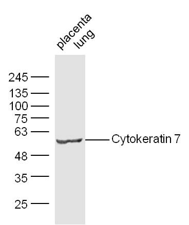 Sample:
Placenta (Mouse) Lysate at 30 ug
Lung (Mouse) Lysate at 30 ug
Primary: Anti-Cytokeratin 7 (SL1744R) at 1/300 dilution
Secondary: IRDye800CW Goat Anti-Mouse IgG at 1/10000 dilution
Predicted band size: 54 kD
Observed band size: 54 kD
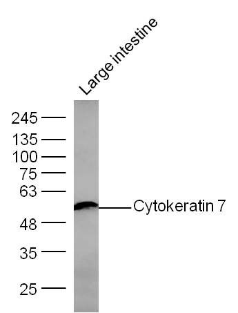 Sample: Large intestine (Mouse) Lysate at 30 ug
Primary: Anti- Cytokeratin 7 (SL1744R) at 1/300 dilution
Secondary: IRDye800CW Goat Anti-Mouse IgG at 1/10000 dilution
Predicted band size: 54 kD
Observed band size: 54 kD
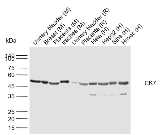 Sample:
Lane 1: Mouse Urinary bladder tissue lysates
Lane 2: Mouse Breast tissue lysates
Lane 3: Mouse Placenta tissue lysates
Lane 4: Mouse trachea tissue lysates
Lane 5: Rat Urinary bladder tissue lysates
Lane 6: Rat Placenta tissue lysates
Lane 7: Human Hela cell lysates
Lane 8: Human HepG2 cell lysates
Lane 9: Human Siha cell lysates
Lane 10: Human Huvec cell lysates
Primary: Anti-CK7 (SL1744R) at 1/1000 dilution
Secondary: IRDye800CW Goat Anti-Rabbit IgG at 1/20000 dilution
Predicted band size: 54 kDa
Observed band size: 52 kDa
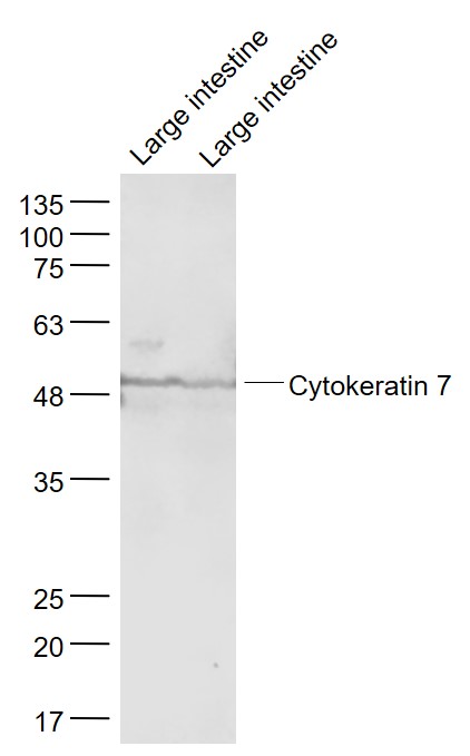 Sample:
Large intestine (Mouse) Lysate at 40 ug
Large intestine(Rat) Lysate at 40 ug
Primary: Anti- Cytokeratin 7 (SL1744R) at 1/1000 dilution
Secondary: IRDye800CW Goat Anti-Rabbit IgG at 1/20000 dilution
Predicted band size: 54 kD
Observed band size: 54 kD
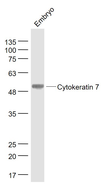 Sample:
Embryo (Mouse) Lysate at 40 ug
Primary: Anti- Cytokeratin 7 (SL1744R) at 1/1000 dilution
Secondary: IRDye800CW Goat Anti-Rabbit IgG at 1/20000 dilution
Predicted band size: 54 kD
Observed band size: 54 kD
 Tissue/cell: human lung carcinoma; 4% Paraformaldehyde-fixed and paraffin-embedded;
Antigen retrieval: citrate buffer ( 0.01M, pH 6.0 ), Boiling bathing for 15min; Block endogenous peroxidase by 3% Hydrogen peroxide for 30min; Blocking buffer (normal goat serum,C-0005) at 37℃ for 20 min;
Incubation: Anti-Cytokeratin 7 Polyclonal Antibody, Unconjugated(SL1744R) 1:200, overnight at 4°C, followed by conjugation to the secondary antibody(SP-0023) and DAB(C-0010) staining
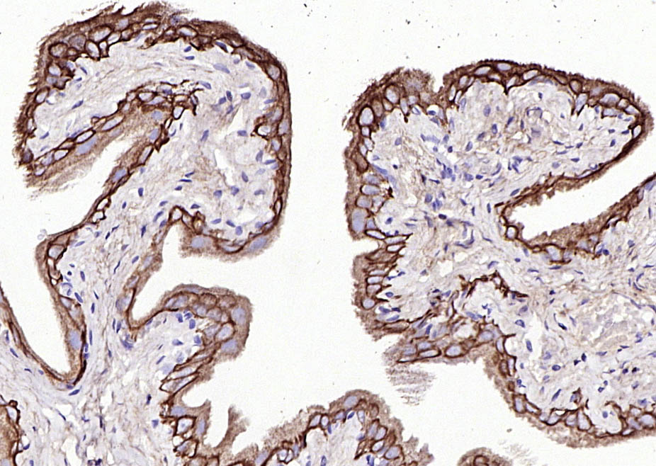 Paraformaldehyde-fixed, paraffin embedded (Rat bladder); Antigen retrieval by boiling in sodium citrate buffer (pH6.0) for 15min; Block endogenous peroxidase by 3% hydrogen peroxide for 20 minutes; Blocking buffer (normal goat serum) at 37°C for 30min; Antibody incubation with (CK7) Polyclonal Antibody, Unconjugated (SL1744R) at 1:200 overnight at 4°C, followed by operating according to SP Kit(Rabbit) (sp-0023) instructionsand DAB staining.
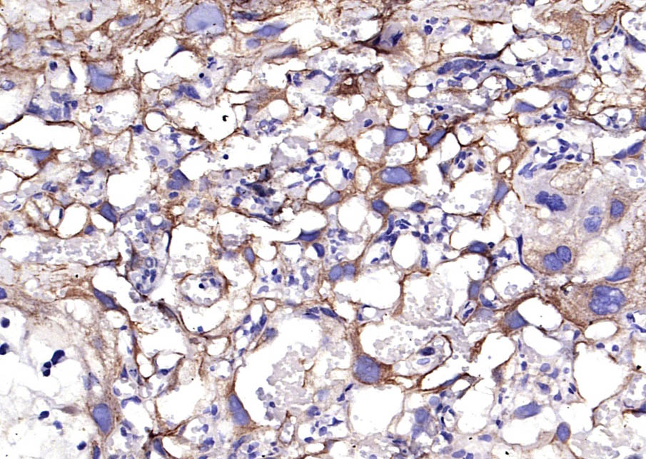 Paraformaldehyde-fixed, paraffin embedded (Rat placenta); Antigen retrieval by boiling in sodium citrate buffer (pH6.0) for 15min; Block endogenous peroxidase by 3% hydrogen peroxide for 20 minutes; Blocking buffer (normal goat serum) at 37°C for 30min; Antibody incubation with (CK7) Polyclonal Antibody, Unconjugated (SL1744R) at 1:200 overnight at 4°C, followed by operating according to SP Kit(Rabbit) (sp-0023) instructionsand DAB staining.
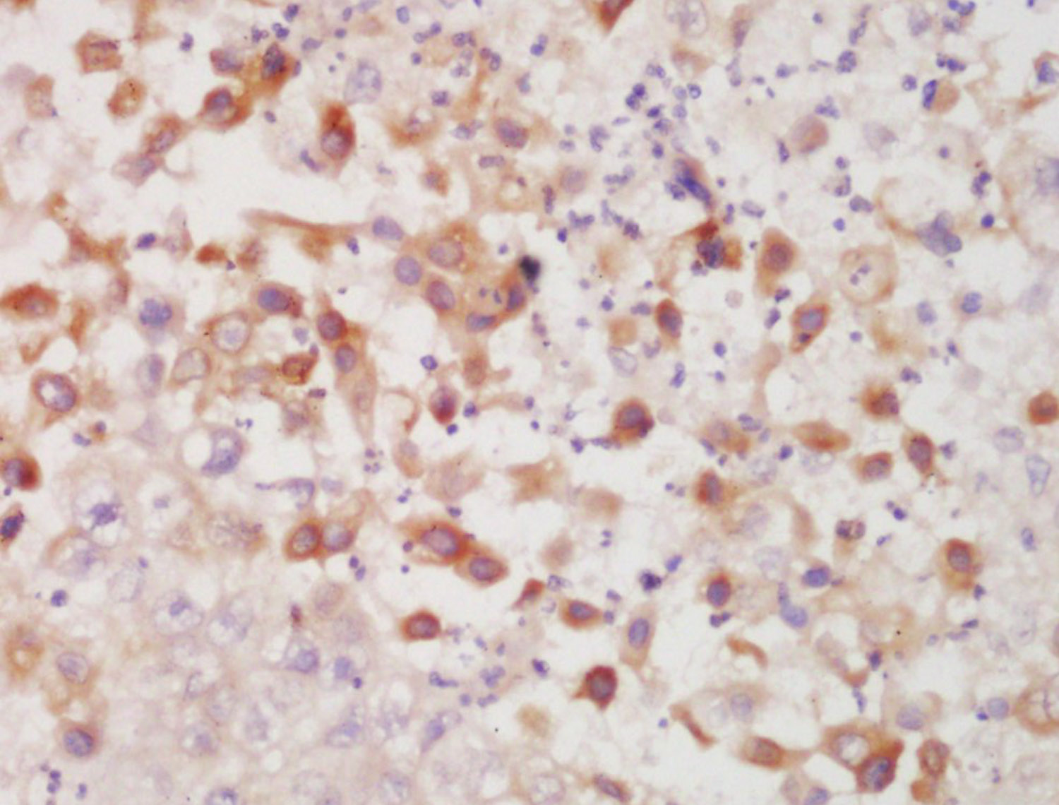
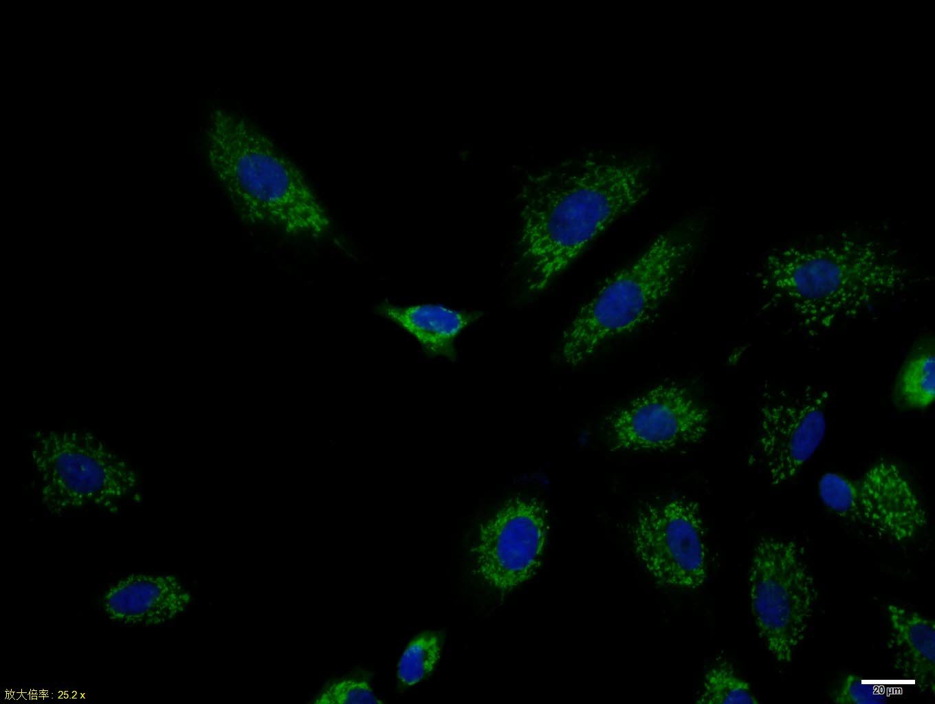 A549 cell; 4% Paraformaldehyde-fixed; Triton X-100 at room temperature for 20 min; Blocking buffer (normal goat serum, C-0005) at 37°C for 20 min; Antibody incubation with (Cytokeratin 7) polyclonal Antibody, Unconjugated (SL1744R) 1:100, 90 minutes at 37°C; followed by a conjugated Goat Anti-Rabbit IgG antibody at 37°C for 90 minutes, DAPI (blue, C02-04002) was used to stain the cell nuclei.
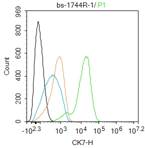 Blank control:Hela.
Primary Antibody (green line): Rabbit Anti-Cytokeratin 7 antibody (SL1744R)
Dilution: 1ug/Test;
Secondary Antibody : Goat anti-rabbit IgG-FITC
Dilution: 0.5ug/Test.
Protocol
The cells were fixed with 4% PFA (10min at room temperature)and then permeabilized with 90% ice-cold methanol for 20 min at -20℃.The cells were then incubated in 5%BSA to block non-specific protein-protein interactions for 30 min at room temperature .Cells stained with Primary Antibody for 30 min at room temperature. The secondary antibody used for 40 min at room temperature. Acquisition of 20,000 events was performed.
Cartpieces
Totalgoods,subtotals:¥Checkout
Bought notes(bought amounts latest0)
No one bought this product
User Comment(Total0User Comment Num)
- No comment


 +86 571 56623320
 +86 18668110335

