
Rabbit Anti-alpha Actinin 4 antibody
actinin 4; Actinin alpha 4; Actinin alpha-4; Alpha-actinin-4; α-Actinin-4; αActinin-4; actinin4; ACTN 4; ACTN4; ACTN4_HUMAN; DKFZp686K23158; F actin cross linking protein; F-actin cross-linking protein; Focal segmental glomerulosclerosis 1; FSGS 1; FSGS;
View History [Clear]
Details
Product Name alpha Actinin 4 Chinese Name α-辅肌动蛋白4抗体 Alias actinin 4; Actinin alpha 4; Actinin alpha-4; Alpha-actinin-4; α-Actinin-4; αActinin-4; actinin4; ACTN 4; ACTN4; ACTN4_HUMAN; DKFZp686K23158; F actin cross linking protein; F-actin cross-linking protein; Focal segmental glomerulosclerosis 1; FSGS 1; FSGS; FSGS1; Non muscle alpha actinin 4; Non-muscle alpha-actinin 4. literatures Research Area immunology Developmental biology Signal transduction Stem cells Binding protein Cytoskeleton Immunogen Species Rabbit Clonality Polyclonal React Species Human, Mouse, Rat, (predicted: Chicken, Pig, Cow, Rabbit, ) Applications WB=1:500-2000 ELISA=1:5000-10000 IHC-P=1:100-500 IHC-F=1:100-500 Flow-Cyt=2ug/Test IF=1:100-500 (Paraffin sections need antigen repair)
not yet tested in other applications.
optimal dilutions/concentrations should be determined by the end user.Theoretical molecular weight 105kDa Cellular localization The nucleus cytoplasmic Form Liquid Concentration 1mg/ml immunogen KLH conjugated synthetic peptide derived from human Actinin alpha 4: 811-911/911 Lsotype IgG Purification affinity purified by Protein A Buffer Solution 0.01M TBS(pH7.4) with 1% BSA, 0.03% Proclin300 and 50% Glycerol. Storage Shipped at 4℃. Store at -20 °C for one year. Avoid repeated freeze/thaw cycles. Attention This product as supplied is intended for research use only, not for use in human, therapeutic or diagnostic applications. PubMed PubMed Product Detail Alpha actinins belong to the spectrin gene superfamily which represents a diverse group of cytoskeletal proteins, including the alpha and beta spectrins and dystrophins. Alpha actinin is an actin-binding protein with multiple roles in different cell types. In nonmuscle cells, the cytoskeletal isoform is found along microfilament bundles and adherens-type junctions, where it is involved in binding actin to the membrane. In contrast, skeletal, cardiac, and smooth muscle isoforms are localized to the Z-disc and analogous dense bodies, where they help anchor the myofibrillar actin filaments. This gene encodes a nonmuscle, alpha actinin isoform which is concentrated in the cytoplasm, and thought to be involved in metastatic processes. Mutations in this gene have been associated with focal and segmental glomerulosclerosis. [provided by RefSeq, Jul 2008].
Function:
F-actin cross-linking protein which is thought to anchor actin to a variety of intracellular structures. This is a bundling protein. Probably involved in vesicular trafficking via its association with the CART complex. The CART complex is necessary for efficient transferrin receptor recycling but not for EGFR degradation.
Subunit:
Homodimer; antiparallel. Binds TRIM3 at the N-terminus. Identified in a complex with CASK, IQGAP1, MAGI2, NPHS1, SPTAN1 and SPTBN1. Identified in a mRNP granule complex, at least composed of ACTB, ACTN4, DHX9, ERG, HNRNPA1, HNRNPA2B1, HNRNPAB, HNRNPD, HNRNPL, HNRNPR, HNRNPU, HSPA1, HSPA8, IGF2BP1, ILF2, ILF3, NCBP1, NCL, PABPC1, PABPC4, PABPN1, RPLP0, RPS3, RPS3A, RPS4X, RPS8, RPS9, SYNCRIP, TROVE2, YBX1 and untranslated mRNAs. Component of the CART complex, at least composed of ACTN4, HGS/HRS, MYO5B and TRIM3. Interacts with BAIAP1 and PDLIM2.
Subcellular Location:
Nucleus. Cytoplasm. Note=Localized in cytoplasmic mRNP granules containing untranslated mRNAs. Colocalizes with actin stress fibers. Nuclear translocation can be induced by the PI3 kinase inhibitor wortmannin or by cytochalasin D. Exclusively localized in the nucleus in a limited number of cell lines (breast cancer cell line MCF-7, oral floor cancer IMC-2, and bladder cancer KU-7).
Tissue Specificity:
Widely expressed.
DISEASE:
Defects in ACTN4 are the cause of focal segmental glomerulosclerosis type 1 (FSGS1) [MIM:603278]. A renal pathology defined by the presence of segmental sclerosis in glomeruli and resulting in proteinuria, reduced glomerular filtration rate and edema. Renal insufficiency often progresses to end-stage renal disease, a highly morbid state requiring either dialysis therapy or kidney transplantation.
Similarity:
Belongs to the alpha-actinin family.
Contains 1 actin-binding domain.
Contains 2 CH (calponin-homology) domains.
Contains 2 EF-hand domains.
Contains 4 spectrin repeats.
SWISS:
O43707
Gene ID:
81
Database links:Entrez Gene: 81 Human
Entrez Gene: 60595 Mouse
Omim: 604638 Human
SwissProt: O43707 Human
SwissProt: P57780 Mouse
Unigene: 270291 Human
Unigene: 81144 Mouse
Unigene: 15777 Rat
Product Picture 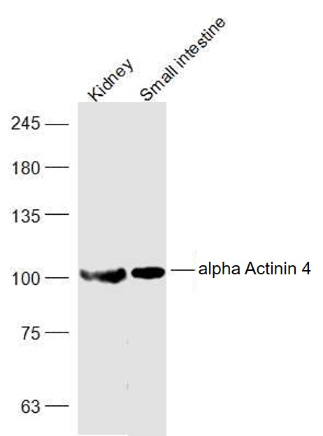 Sample:
Kidney (Rat) Lysate at 40 ug
Small intestine (Mouse) Lysate at 40 ug
Primary: Anti-alpha Actinin 4-Loading Control (SL1741R) at 1/1000 dilution
Secondary: IRDye800CW Goat Anti-Rabbit IgG at 1/20000 dilution
Predicted band size: 105 kD
Observed band size: 105 kD
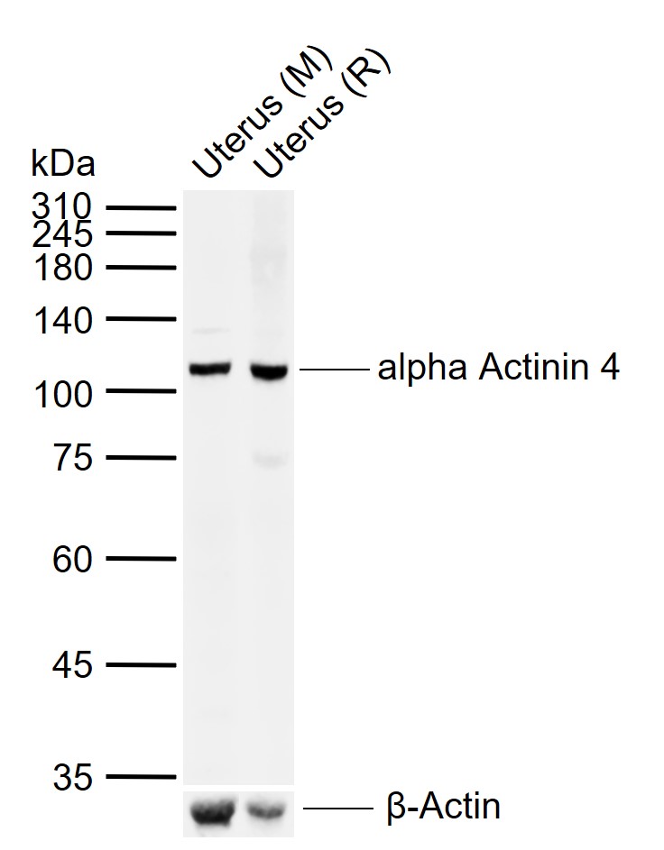 Sample:
Lane 1: Mouse Uterus tissue lysates
Lane 2: Rat Uterus tissue lysates
Primary: Anti-alpha Actinin 4 (SL1741R) at 1/1000 dilution
Secondary: IRDye800CW Goat Anti-Rabbit IgG at 1/20000 dilution
Predicted band size: 105 kDa
Observed band size: 105 kDa
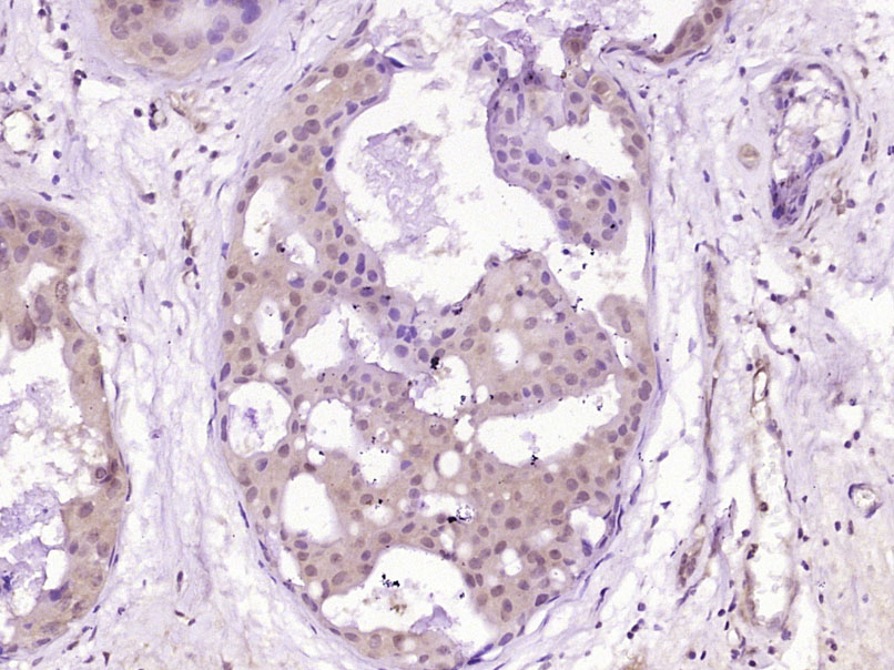 Paraformaldehyde-fixed, paraffin embedded (Human breast carcinoma); Antigen retrieval by boiling in sodium citrate buffer (pH6.0) for 15min; Block endogenous peroxidase by 3% hydrogen peroxide for 20 minutes; Blocking buffer (normal goat serum) at 37°C for 30min; Antibody incubation with (alpha Actinin 4-Loading Control) Polyclonal Antibody, Unconjugated (SL1741R) at 1:400 overnight at 4°C, followed by operating according to SP Kit(Rabbit) (sp-0023) instructionsand DAB staining.
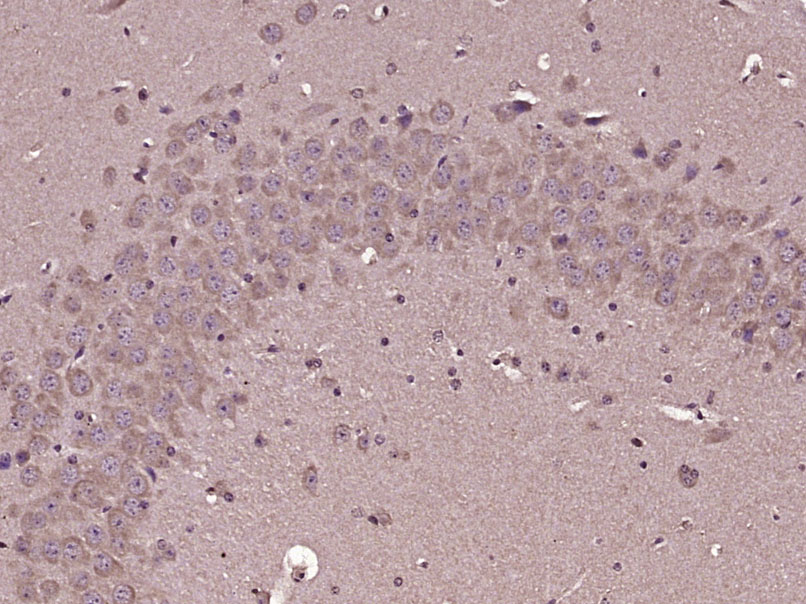
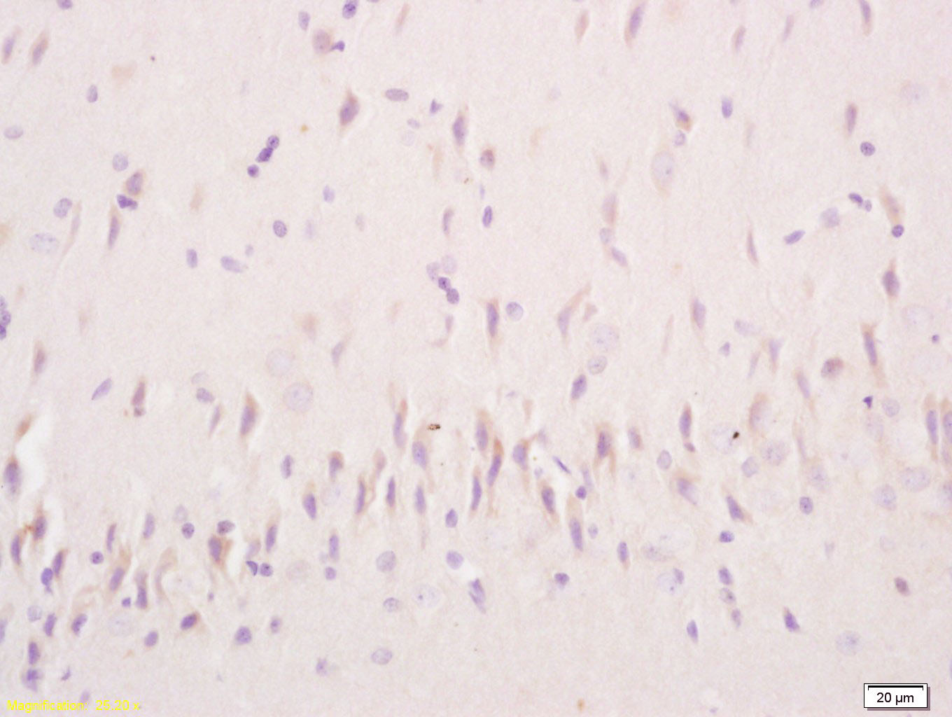 Tissue/cell: rat brain tissue; 4% Paraformaldehyde-fixed and paraffin-embedded;
Antigen retrieval: citrate buffer ( 0.01M, pH 6.0 ), Boiling bathing for 15min; Block endogenous peroxidase by 3% Hydrogen peroxide for 30min; Blocking buffer (normal goat serum,C-0005) at 37℃ for 20 min;
Incubation: Anti-alpha-Actinin-4 Polyclonal Antibody, Unconjugated(SL1741R) 1:200, overnight at 4°C, followed by conjugation to the secondary antibody(SP-0023) and DAB(C-0010) staining
 Paraformaldehyde-fixed, paraffin embedded (Mouse brain); Antigen retrieval by boiling in sodium citrate buffer (pH6.0) for 15min; Block endogenous peroxidase by 3% hydrogen peroxide for 20 minutes; Blocking buffer (normal goat serum) at 37°C for 30min; Antibody incubation with (alpha Actinin 4-Loading Control) Polyclonal Antibody, Unconjugated (SL1741R) at 1:400 overnight at 4°C, followed by operating according to SP Kit(Rabbit) (sp-0023) instructionsand DAB staining.
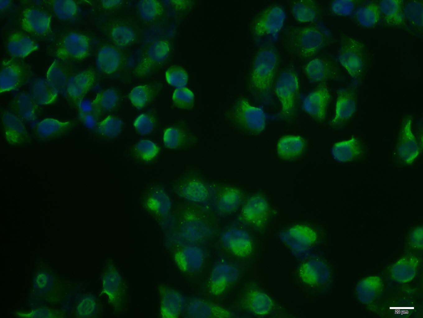 Hela cell; 4% Paraformaldehyde-fixed; Triton X-100 at room temperature for 20 min; Blocking buffer (normal goat serum, C-0005) at 37°C for 20 min; Antibody incubation with (alpha Actinin 4) polyclonal Antibody, Unconjugated (SL1741R) 1:100, 90 minutes at 37°C; followed by a conjugated Goat Anti-Rabbit IgG antibody at 37°C for 90 minutes, DAPI (blue, C02-04002) was used to stain the cell nuclei.
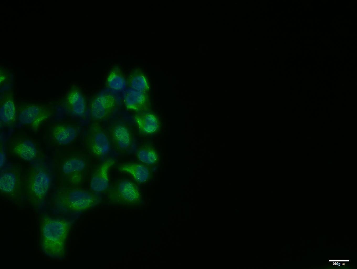 Hela cell; 4% Paraformaldehyde-fixed; Triton X-100 at room temperature for 20 min; Blocking buffer (normal goat serum, C-0005) at 37°C for 20 min; Antibody incubation with (alpha Actinin 4) polyclonal Antibody, Unconjugated (SL1741R) 1:100, 90 minutes at 37°C; followed by a conjugated Goat Anti-Rabbit IgG antibody at 37°C for 90 minutes, DAPI (blue, C02-04002) was used to stain the cell nuclei.
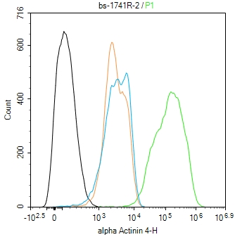 Blank control(black line):U251.
Primary Antibody (green line): Rabbit Anti-alpha Actinin 4 antibody (SL1741R)
Dilution:2ug/Test;
Secondary Antibody(white blue line): Goat anti-rabbit IgG-AF488
Dilution: 0.5ug/Test.
Isotype control(orange line): Normal Rabbit IgG
Protocol
The cells were fixed with 4% PFA (10min at room temperature)and then permeabilized with 90% ice-cold methanol for 20 min at -20℃, The cells were then incubated in 5%BSA to block non-specific protein-protein interactions for 30 min at room temperature .Cells stained with Primary Antibody for 30 min at room temperature. The secondary antibody used for 40 min at room temperature. Acquisition of 20,000 events was performed.
Cartpieces
Totalgoods,subtotals:¥Checkout
References (0)
No References
Bought notes(bought amounts latest0)
No one bought this product
User Comment(Total0User Comment Num)
- No comment


 +86 571 56623320
 +86 18668110335

