
Rabbit Anti-phospho-STAT3 (Tyr705)antibody
STAT3 (phospho Y705); p-STAT3 (phospho Y705); Phospho-Stat3(pTyr705); STAT3(Phospho-Tyr705); p-STAT3(Tyr705); Phosphorylated Stat3(pTyr705); p-Stat3; Acute Phase Response Factor; APRF; DNA binding protein APRF; FLJ20882; MGC16063; Signal Transductor and
View History [Clear]
Details
Product Name phospho-STAT3 (Tyr705) Chinese Name 磷酸化Signal transduction和转录激活因子3抗体 Alias STAT3 (phospho Y705); p-STAT3 (phospho Y705); Phospho-Stat3(pTyr705); STAT3(Phospho-Tyr705); p-STAT3(Tyr705); Phosphorylated Stat3(pTyr705); p-Stat3; Acute Phase Response Factor; APRF; DNA binding protein APRF; FLJ20882; MGC16063; Signal Transductor and Activator of Transcription 3; STAT 3; STAT3_HUMAN. literatures Product Type Phosphorylated anti Research Area Tumour Cardiovascular Cell biology immunology Signal transduction Stem cells Apoptosis transcriptional regulatory factor Epigenetics Immunogen Species Rabbit Clonality Polyclonal React Species Human, Mouse, Rat, Chicken, (predicted: Dog, Pig, Cow, Rabbit, Sheep, Guinea Pig, ) Applications WB=1:500-2000 ELISA=1:5000-10000 IHC-P=1:100-500 IHC-F=1:100-500 Flow-Cyt=1μg /test IF=1:100-500 (Paraffin sections need antigen repair)
not yet tested in other applications.
optimal dilutions/concentrations should be determined by the end user.Theoretical molecular weight 85kDa Cellular localization The nucleus cytoplasmic Form Liquid Concentration 1mg/ml immunogen KLH conjugated Synthesised phosphopeptide derived from human STAT3 around the phosphorylation site of Tyr705: AP(p-Y)LK Lsotype IgG Purification affinity purified by Protein A Buffer Solution 0.01M TBS(pH7.4) with 1% BSA, 0.03% Proclin300 and 50% Glycerol. Storage Shipped at 4℃. Store at -20 °C for one year. Avoid repeated freeze/thaw cycles. Attention This product as supplied is intended for research use only, not for use in human, therapeutic or diagnostic applications. PubMed PubMed Product Detail The protein encoded by this gene is a member of the STAT protein family. In response to cytokines and growth factors, STAT family members are phosphorylated by the receptor associated kinases, and then form homo- or heterodimers that translocate to the cell nucleus where they act as transcription activators. This protein is activated through phosphorylation in response to various cytokines and growth factors including IFNs, EGF, IL5, IL6, HGF, LIF and BMP2. This protein mediates the expression of a variety of genes in response to cell stimuli, and thus plays a key role in many cellular processes such as cell growth and apoptosis. The small GTPase Rac1 has been shown to bind and regulate the activity of this protein. PIAS3 protein is a specific inhibitor of this protein. Mutations in this gene are associated with infantile-onset multisystem autoimmune disease and hyper-immunoglobulin E syndrome. Alternative splicing results in multiple transcript variants encoding distinct isoforms. [provided by RefSeq, Sep 2015]
Function:
Transcription factor that binds to the interleukin-6 (IL-6)-responsive elements identified in the promoters of various acute-phase protein genes. Activated by IL31 through IL31RA.
Subcellular Location:
Cytoplasm. Nucleus. Shuttles between the nucleus and the cytoplasm. Constitutive nuclear presence is independent of tyrosine phosphorylation. Predominantly present in the cytoplasm without stimuli. Upon leukemia inhibitory factor (LIF) stimulation, accumulates in the nucleus. The complex composed of BART and ARL2 plays an important role in the nuclear translocation and retention of STAT3.
Tissue Specificity:
Heart, brain, placenta, lung, liver, skeletal muscle, kidney and pancreas.
Post-translational modifications:
Tyrosine phosphorylated upon stimulation with EGF (By similarity). Tyrosine phosphorylated in response to IL-6, IL-11, CNTF, LIF, CSF-1, EGF, PDGF, IFN-alpha and OSM. Phosphorylated on serine upon DNA damage, probably by ATM or ATR. Serine phosphorylation is important for the formation of stable DNA-binding STAT3 homodimers and maximal transcriptional activity. ARL2BP may participate in keeping the phosphorylated state of STAT3 within the nucleus.
DISEASE:
Defects in STAT3 are the cause of hyperimmunoglobulin E recurrent infection syndrome autosomal dominant (AD-HIES) [MIM:147060]; also known as hyper-IgE syndrome or Job syndrome. AD-HIES is a rare disorder of immunity and connective tissue characterized by immunodeficiency, chronic eczema, recurrent Staphylococcal infections, increased serum IgE, eosinophilia, distinctive coarse facial appearance, abnormal dentition, hyperextensibility of the joints, and bone fractures.
SWISS:
P40763
Gene ID:
6774
Database links:Entrez Gene: 6774 Human
Entrez Gene: 20848 Mouse
Omim: 102582 Human
SwissProt: P40763 Human
SwissProt: P42227 Mouse
Unigene: 463059 Human
transcriptional regulatory factor(Transcriptin Regulators)Product Picture 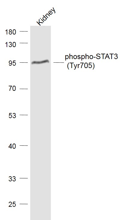 Sample:
Kidney (Mouse) Lysate at 40 ug
Primary: Anti-phospho-STAT3 (Tyr705) (SL1658R) at 1/1000 dilution
Secondary: IRDye800CW Goat Anti-Rabbit IgG at 1/20000 dilution
Predicted band size: 85 kD
Observed band size: 85 kD
 Sample:
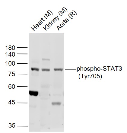
Lane 1: Heart (Mouse) Lysate at 40 ug
Lane 2: Kidney (Mouse) Lysate at 40 ug
Lane 3: Aorta (Rat) Lysate at 40 ug
Primary:
Anti-phospho-STAT3 (Tyr705) (SL1658R) at 1/1000 dilution
Secondary: IRDye800CW Goat Anti-Rabbit IgG at 1/20000 dilution
Predicted band size: 92/84 kD
Observed band size: 84 kD
 Sample:
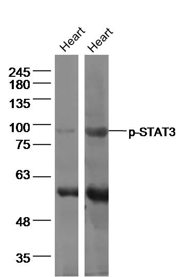
Heart (Mouse) Lysate at 40 ug
Heart (Rat) Lysate at 40 ug
Primary: Anti-p-STAT3 (SL1658R) at 1/500 dilution
Secondary: IRDye800CW Goat Anti-Rabbit IgG at 1/20000 dilution
Predicted band size: 85 kD
Observed band size: 85 kD
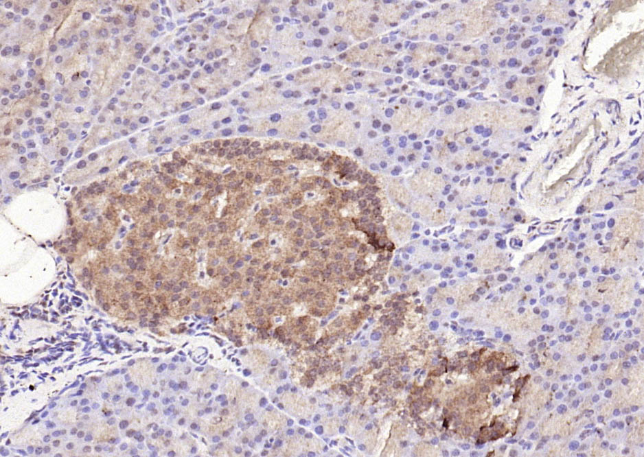 Paraformaldehyde-fixed, paraffin embedded (rat pancreas); Antigen retrieval by boiling in sodium citrate buffer (pH6.0) for 15min; Block endogenous peroxidase by 3% hydrogen peroxide for 20 minutes; Blocking buffer (normal goat serum) at 37°C for 30min; Antibody incubation with (phospho-STAT3 (Tyr705)) Polyclonal Antibody, Unconjugated (SL1658R) at 1:200 overnight at 4°C, followed by operating according to SP Kit(Rabbit) (sp-0023) instructionsand DAB staining.
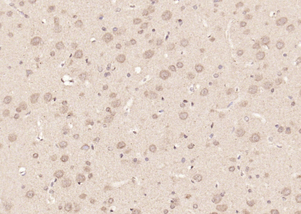 Paraformaldehyde-fixed, paraffin embedded (rat brain); Antigen retrieval by boiling in sodium citrate buffer (pH6.0) for 15min; Block endogenous peroxidase by 3% hydrogen peroxide for 20 minutes; Blocking buffer (normal goat serum) at 37°C for 30min; Antibody incubation with (phospho-STAT3 (Tyr705)) Polyclonal Antibody, Unconjugated (SL1658R) at 1:200 overnight at 4°C, followed by operating according to SP Kit(Rabbit) (sp-0023) instructionsand DAB staining.
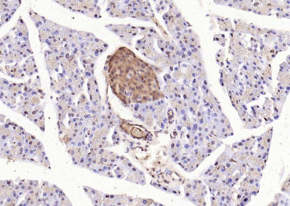 Paraformaldehyde-fixed, paraffin embedded (mouse pancreas); Antigen retrieval by boiling in sodium citrate buffer (pH6.0) for 15min; Block endogenous peroxidase by 3% hydrogen peroxide for 20 minutes; Blocking buffer (normal goat serum) at 37°C for 30min; Antibody incubation with (phospho-STAT3 (Tyr705)) Polyclonal Antibody, Unconjugated (SL1658R) at 1:200 overnight at 4°C, followed by operating according to SP Kit(Rabbit) (sp-0023) instructionsand DAB staining.
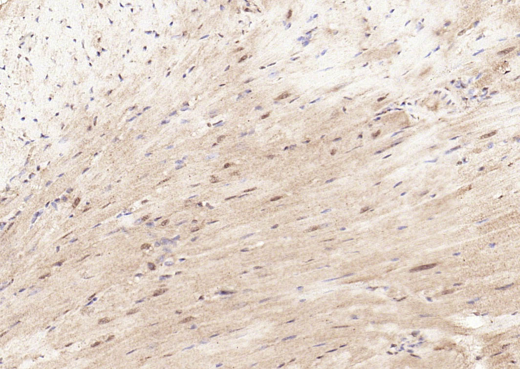 Paraformaldehyde-fixed, paraffin embedded (mouse heart); Antigen retrieval by boiling in sodium citrate buffer (pH6.0) for 15min; Block endogenous peroxidase by 3% hydrogen peroxide for 20 minutes; Blocking buffer (normal goat serum) at 37°C for 30min; Antibody incubation with (phospho-STAT3 (Tyr705)) Polyclonal Antibody, Unconjugated (SL1658R) at 1:200 overnight at 4°C, followed by operating according to SP Kit(Rabbit) (sp-0023) instructionsand DAB staining.
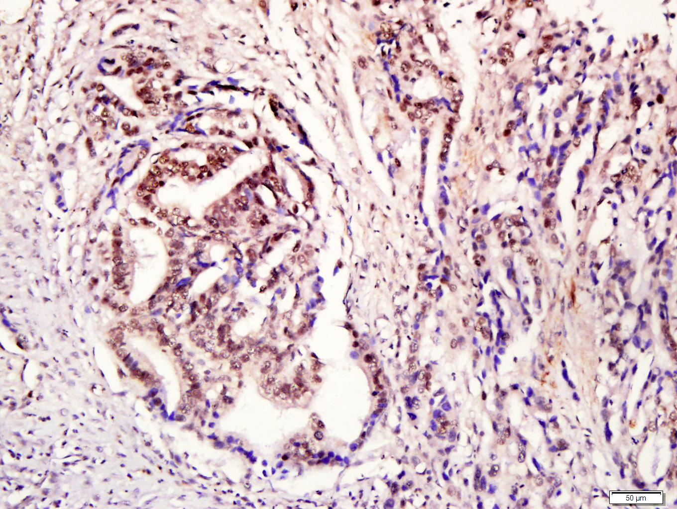 Tissue/cell: human gastric cancer; 4% Paraformaldehyde-fixed and paraffin-embedded;
Antigen retrieval: citrate buffer ( 0.01M, pH 6.0 ), Boiling bathing for 15min; Block endogenous peroxidase by 3% Hydrogen peroxide for 30min; Blocking buffer (normal goat serum,C-0005) at 37℃ for 20 min;
Incubation: Anti- phospho-STAT3 Polyclonal Antibody, Unconjugated(SL1658R) 1:200, overnight at 4°C, followed by conjugation to the secondary antibody(SP-0023) and DAB(C-0010) staining
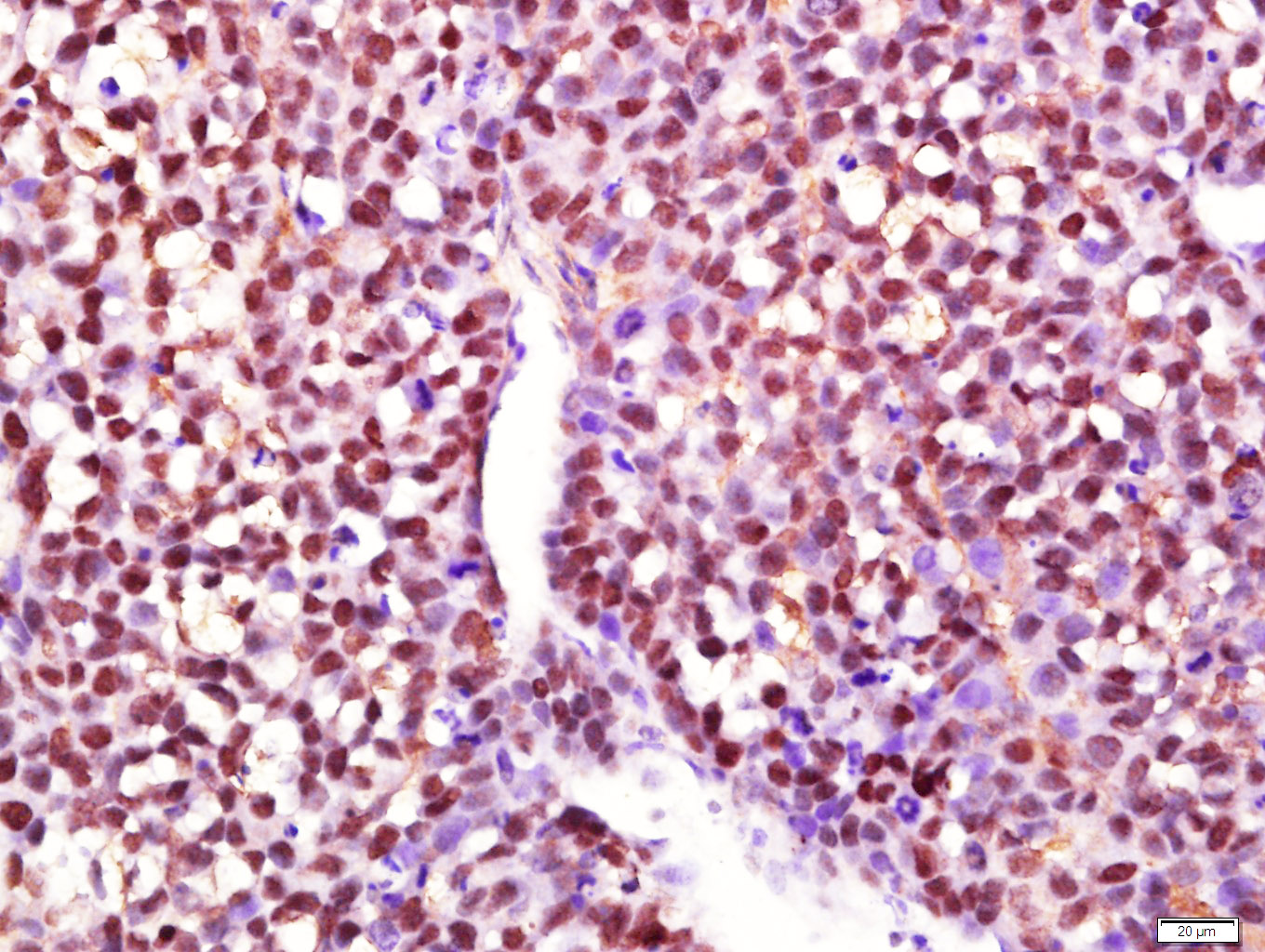 Paraformaldehyde-fixed, paraffin embedded (Transplantation tumor); Antigen retrieval by boiling in sodium citrate buffer (pH6.0) for 15min; Block endogenous peroxidase by 3% hydrogen peroxide for 20 minutes; Blocking buffer (normal goat serum) at 37°C for 30min; Antibody incubation with (Phosphorylated signal Transductor and Activator of Transcription 3; p-STAT3) Polyclonal Antibody, Unconjugated (SL1658R) at 1:400 overnight at 4°C, followed by a conjugated secondary antibody (sp-0023) for 20 minutes and DAB staining.
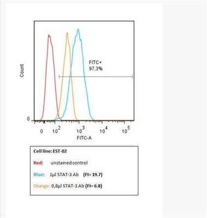 This image has been kindly submitted by Tübingen Ageing and Tumour Immunology Group (TATI), Centre for Medical Research (ZMF), University of Tübingen Medical School www.tati-group.de via our distributor Biozol.
Human Melanoma cell line, EST02, was stained with Rabbit Anti-phospho-STAT3 (Tyr705) Polyclonal Antibody, FITC Conjugated (SL1658R-FITC)for 30 minutes, on ice.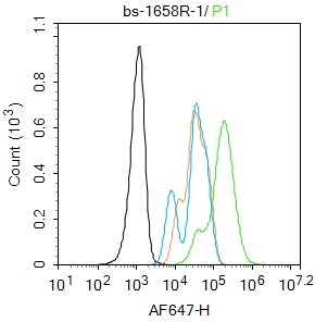 Blank control: A431.
Primary Antibody (green line): Rabbit Anti-phospho-STAT3 (Tyr705) antibody (SL1658R)
Dilution: 1μg /10^6 cells;
Isotype Control Antibody (orange line): Rabbit IgG .
Secondary Antibody : Goat anti-rabbit IgG-AF647
Dilution: 1μg /test.
Protocol
The cells were fixed with 4% PFA (10min at room temperature)and then permeabilized with 90% ice-cold methanol for 20 min at-20℃. The cells were then incubated in 5%BSA to block non-specific protein-protein interactions for 30 min at room temperature .Cells stained with Primary Antibody for 30 min at room temperature. The secondary antibody used for 40 min at room temperature. Acquisition of 20,000 events was performed.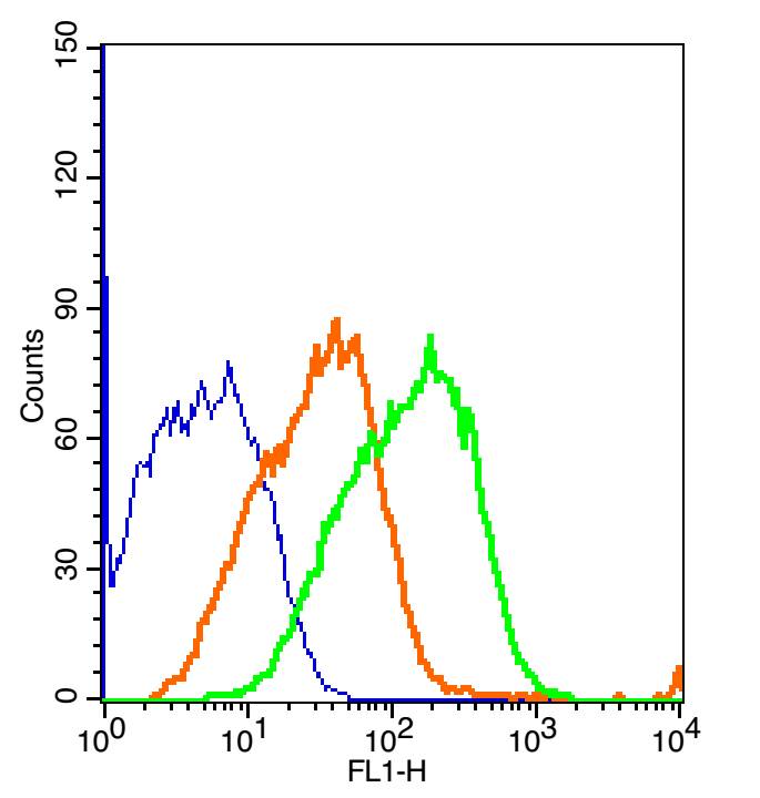 The blue histogram is unstained cells (mouse kidney).
The Orange histogram is cells stained with Rabbit IgG/FITC (SL0295P-FITC)isotype control antibody.
The green histogram is cells stained with Rabbit Anti-phospho-STAT3 (Tyr705)/FITC Conjugated antibody (SL1658R-FITC).
Concentration: 2μg/10^6 cells or 5μg/10^6 cells.
Cartpieces
Totalgoods,subtotals:¥Checkout
Bought notes(bought amounts latest0)
No one bought this product
User Comment(Total0User Comment Num)
- No comment


 +86 571 56623320
 +86 18668110335

