
Rabbit Anti-phospho-STAT1 (Tyr701)antibody
STAT1 (phospho Y701); p-STAT1 (phospho Y701); Phospho-Stat1(pTyr701); STAT1(Phospho-Tyr701); Signal transducer and activator of transcription 1-alpha/beta; Transcription factor ISGF-3 components p91/p84; STAT1_HUMAN;
View History [Clear]
Details
Product Name phospho-STAT1 (Tyr701) Chinese Name 磷酸化Signal transduction与转录激活因子1抗体 Alias STAT1 (phospho Y701); p-STAT1 (phospho Y701); Phospho-Stat1(pTyr701); STAT1(Phospho-Tyr701); Signal transducer and activator of transcription 1-alpha/beta; Transcription factor ISGF-3 components p91/p84; STAT1_HUMAN; literatures Product Type Phosphorylated anti Research Area Tumour Cell biology immunology Signal transduction Apoptosis transcriptional regulatory factor Epigenetics Immunogen Species Rabbit Clonality Polyclonal React Species Human, Mouse, (predicted: Rat, Dog, Pig, Cow, Rabbit, Sheep, ) Applications ELISA=1:5000-10000 IHC-P=1:100-500 (Paraffin sections need antigen repair)
not yet tested in other applications.
optimal dilutions/concentrations should be determined by the end user.Theoretical molecular weight 82kDa Cellular localization The nucleus cytoplasmic Form Liquid Concentration 1mg/ml immunogen KLH conjugated Synthesised phosphopeptide derived from human STAT1 around the phosphorylation site of Tyr701: TG(p-Y)IK Lsotype IgG Purification affinity purified by Protein A Buffer Solution 0.01M TBS(pH7.4) with 1% BSA, 0.03% Proclin300 and 50% Glycerol. Storage Shipped at 4℃. Store at -20 °C for one year. Avoid repeated freeze/thaw cycles. Attention This product as supplied is intended for research use only, not for use in human, therapeutic or diagnostic applications. PubMed PubMed Product Detail The protein encoded by this gene is a member of the STAT protein family. In response to cytokines and growth factors, STAT family members are phosphorylated by the receptor associated kinases, and then form homo- or heterodimers that translocate to the cell nucleus where they act as transcription activators. The protein encoded by this gene can be activated by various ligands including interferon-alpha, interferon-gamma, EGF, PDGF and IL6. This protein mediates the expression of a variety of genes, which is thought to be important for cell viability in response to different cell stimuli and pathogens. The protein plays an important role in immune responses to viral, fungal and mycobacterial pathogens. Mutations in this gene are associated with Immunodeficiency 31B, 31A, and 31C. [provided by RefSeq, Jun 2020]
Function:
Signal transducer and activator of transcription that mediates signaling by interferons (IFNs). Following type I IFN (IFN-alpha and IFN-beta) binding to cell surface receptors, Jak kinases (TYK2 and JAK1) are activated, leading to tyrosine phosphorylation of STAT1 and STAT2. The phosphorylated STATs dimerize, associate with ISGF3G/IRF-9 to form a complex termed ISGF3 transcription factor, that enters the nucleus. ISGF3 binds to the IFN stimulated response element (ISRE) to activate the transcription of interferon stimulated genes, which drive the cell in an antiviral state. In response to type II IFN (IFN-gamma), STAT1 is tyrosine- and serine-phosphorylated. It then forms a homodimer termed IFN-gamma-activated factor (GAF), migrates into the nucleus and binds to the IFN gamma activated sequence (GAS) to drive the expression of the target genes, inducing a cellular antiviral state.
Subcellular Location:
Cytoplasm. Nucleus. Translocated into the nucleus in response to IFN-gamma-induced tyrosine phosphorylation and dimerization.
Post-translational modifications:
Post-translational modificationsPhosphorylated on tyrosine and serine residues in response to IFN-alpha, IFN-gamma, PDGF and EGF. Phosphorylation on Tyr-701 (lacking in beta form) by JAK promotes dimerization and subsequent translocation to the nucleus. Phosphorylation on Ser-727 by several kinases including MAPK14, ERK1/2 and CAMKII on IFN-gamma stimulation, regulates STAT1 transcriptional activity. Phosphorylation on Ser-727 promotes sumoylation though increasing interaction with PIAS. Phosphorylation on Ser-727 by PKCdelta induces apoptosis in response to DNA-damaging agents. Sumoylated by SUMO1, SUMO2 and SUMO3. Sumoylation is enhanced by IFN-gamma-induced phosphorylation on Ser-727, and by interaction with PIAS proteins. Enhances the transactivation activity. ISGylated.
DISEASE:
Note=STAT1 deficiency results in impaired immune response leading to severe mycobacterial and viral diseases. In the case of complete deficiency, patients can die of viral disease.
Defects in STAT1 are a cause of mendelian susceptibility to mycobacterial disease (MSMD) [MIM:209950]; also known as familial disseminated atypical mycobacterial infection. This rare condition confers predisposition to illness caused by moderately virulent mycobacterial species, such as Bacillus Calmette-Guerin (BCG) vaccine and environmental non-tuberculous mycobacteria, and by the more virulent Mycobacterium tuberculosis. Other microorganisms rarely cause severe clinical disease in individuals with susceptibility to mycobacterial infections, with the exception of Salmonella which infects less than 50% of these individuals. The pathogenic mechanism underlying MSMD is the impairment of interferon-gamma mediated immunity whose severity determines the clinical outcome. Some patients die of overwhelming mycobacterial disease with lepromatous-like lesions in early childhood, whereas others develop, later in life, disseminated but curable infections with tuberculoid granulomas. MSMD is a genetically heterogeneous disease with autosomal recessive, autosomal dominant or X-linked inheritance.
Similarity:
Belongs to the transcription factor STAT family.
Contains 1 SH2 domain.
SWISS:
P42224
Gene ID:
6772
Database links:Entrez Gene: 6772 Human
Entrez Gene: 20846 Mouse
SwissProt: P42224 Human
SwissProt: P42225 Mouse
Product Picture 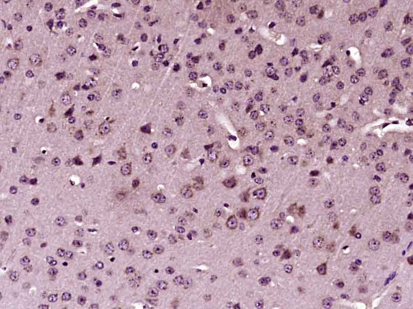 Paraformaldehyde-fixed, paraffin embedded (Mouse brain); Antigen retrieval by boiling in sodium citrate buffer (pH6.0) for 15min; Block endogenous peroxidase by 3% hydrogen peroxide for 20 minutes; Blocking buffer (normal goat serum) at 37°C for 30min; Antibody incubation with (phospho-STAT1 (Tyr701)) Polyclonal Antibody, Unconjugated (SL1657R) at 1:400 overnight at 4°C, followed by operating according to SP Kit(Rabbit) (sp-0023) instructionsand DAB staining.
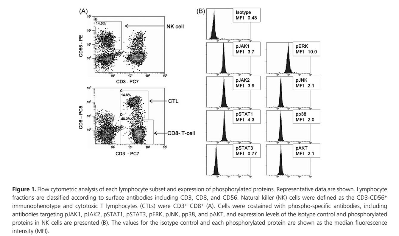 From 《Cancer Medicine》(2016.6): PublitionDirect effect of dasatinib on signal transduction pathways associated with a rapid mobilization of cytotoxic lymphocytes , IF:2.5
Author: Noriyoshi Iriyama, Yoshihiro Hatta & Masami Takei
Division of Hematology and Rheumatology, Department of Medicine, Nihon University School of Medicine, Tokyo, Japan
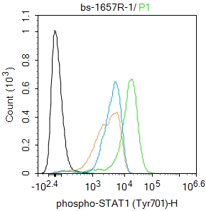 Blank control:THP-1.
Primary Antibody (green line): Rabbit Anti-phospho-STAT1 (Tyr701) antibody (SL1657R)
Dilution: 1μg /10^6 cells;
Isotype Control Antibody (orange line): Rabbit IgG .
Secondary Antibody : Goat anti-rabbit IgG-FITC
Dilution: 0.5μg /test.
Protocol
The cells were fixed with 4% PFA (10min at room temperature)and then permeabilized with 90% ice-cold methanol for 20 min at-20℃. The cells were then incubated in 5%BSA to block non-specific protein-protein interactions for 30 min at room temperature .Cells stained with Primary Antibody for 30 min at room temperature. The secondary antibody used for 40 min at room temperature. Acquisition of 20,000 events was performed.
Cartpieces
Totalgoods,subtotals:¥Checkout
Bought notes(bought amounts latest0)
No one bought this product
User Comment(Total0User Comment Num)
- No comment


 +86 571 56623320
 +86 18668110335

