
Rabbit Anti-phospho-Erk1 (Thr202 + Tyr204) antibody
Erk1 (pT202/pY204); ERK/MAPK(phospho T202/Y204); ERK1 (phospho T202); p-ERK1 (phospho T202); p44/42 MAP Kinase(Phospho-Thr202); ERK; ERK-1; ERT 2; ERT2; Extracellular Signal Regulated Kinase 1; Extracellular signal related kinase 1; Extracellular signal-
View History [Clear]
Details
Product Name phospho-Erk1 (Thr202 + Tyr204) Chinese Name 磷酸化丝裂原活化蛋白激酶1抗体 Alias Erk1 (pT202/pY204); ERK/MAPK(phospho T202/Y204); ERK1 (phospho T202); p-ERK1 (phospho T202); p44/42 MAP Kinase(Phospho-Thr202); ERK; ERK-1; ERT 2; ERT2; Extracellular Signal Regulated Kinase 1; Extracellular signal related kinase 1; Extracellular signal-regulated kinase 1; HGNC6877; HS44KDAP; HUMKER1A; Insulin Stimulated MAP2 Kinase; Insulin-stimulated MAP2 kinase; MAP kinase 1; MAP kinase 3; MAP Kinase; MAP kinase isoform p44; MAPK 1; MAPK 3; MAPK; MAPK1; Mapk3; MGC20180; Microtubule Associated Protein 2 Kinase; Microtubule-associated protein 2 kinase; Mitogen Activated Protein Kinase 3; Mitogen-activated protein kinase 1; Mitogen-activated protein kinase 3; MK03_HUMAN; OTTHUMP00000174538; OTTHUMP00000174541; p44 ERK1; p44 MAPK; p44-ERK1; p44-MAPK; P44ERK1; P44MAPK; PRKM 3; PRKM3; Protein Kinase Mitogen Activated 3. literatures Product Type Phosphorylated anti Research Area immunology Neurobiology Signal transduction Stem cells Kinases and Phosphatases Immunogen Species Rabbit Clonality Polyclonal React Species Human, Mouse, Rat, (predicted: Chicken, Dog, Cow, Horse, Rabbit, Guinea Pig, ) Applications ELISA=1:5000-10000 IHC-P=1:100-500 IHC-F=1:100-500 Flow-Cyt=2ug/Test IF=1:100-500 (Paraffin sections need antigen repair)
not yet tested in other applications.
optimal dilutions/concentrations should be determined by the end user.Theoretical molecular weight 43kDa Cellular localization cytoplasmic Form Liquid Concentration 1mg/ml immunogen KLH conjugated Synthesised phosphopeptide derived from rat ERK1 around the phosphorylation site of Thr201/204: FL(p-T)E(p-Y)VA Lsotype IgG Purification affinity purified by Protein A Buffer Solution 0.01M TBS(pH7.4) with 1% BSA, 0.03% Proclin300 and 50% Glycerol. Storage Shipped at 4℃. Store at -20 °C for one year. Avoid repeated freeze/thaw cycles. Attention This product as supplied is intended for research use only, not for use in human, therapeutic or diagnostic applications. PubMed PubMed Product Detail The protein encoded by this gene is a member of the MAP kinase family. MAP kinases, also known as extracellular signal-regulated kinases (ERKs), act in a signaling cascade that regulates various cellular processes such as proliferation, differentiation, and cell cycle progression in response to a variety of extracellular signals. This kinase is activated by upstream kinases, resulting in its translocation to the nucleus where it phosphorylates nuclear targets. Alternatively spliced transcript variants encoding different protein isoforms have been described.
Function:
Serine/threonine kinase which acts as an essentialcomponent of the MAP kinase signal transduction pathway. MAPK1/ERK2and MAPK3/ERK1 are the 2 MAPKs which play an important role in theMAPK/ERK cascade. They participate also in a signaling cascadeinitiated by activated KIT and KITLG/SCF. Depending on the cellularcontext, the MAPK/ERK cascade mediates diverse biological functionssuch as cell growth, adhesion, survival and differentiation throughthe regulation of transcription, translation, cytoskeletalrearrangements. The MAPK/ERK cascade plays also a role ininitiation and regulation of meiosis, mitosis, and postmitoticfunctions in differentiated cells by phosphorylating a number oftranscription factors. About 160 substrates have already beendiscovered for ERKs. Many of these substrates are localized in thenucleus, and seem to participate in the regulation of transcriptionupon stimulation. However, other substrates are found in thecytosol as well as in other cellular organelles, and those areresponsible for processes such as translation, mitosis andapoptosis. Moreover, the MAPK/ERK cascade is also involved in theregulation of the endosomal dynamics, including lysosome processingand endosome cycling through the perinuclear recycling compartment(PNRC); as well as in the fragmentation of the Golgi apparatusduring mitosis. The substrates include transcription factors (suchas ATF2, BCL6, ELK1, ERF, FOS, HSF4 or SPZ1), cytoskeletal elements(such as CANX, CTTN, GJA1, MAP2, MAPT, PXN, SORBS3 or STMN1),regulators of apoptosis (such as BAD, BTG2, CASP9, DAPK1, IER3,MCL1 or PPARG), regulators of translation (such as EIF4EBP1) and avariety of other signaling-related molecules (like ARHGEF2, FRS2 orGRB10). Protein kinases (such as RAF1, RPS6KA1/RSK1, RPS6KA3/RSK2,RPS6KA2/RSK3, RPS6KA6/RSK4, SYK, MKNK1/MNK1, MKNK2/MNK2,RPS6KA5/MSK1, RPS6KA4/MSK2, MAPKAPK3 or MAPKAPK5) and phosphatases(such as DUSP1, DUSP4, DUSP6 or DUSP16) are other substrates whichenable the propagation the MAPK/ERK signal to additional cytosolicand nuclear targets, thereby extending the specificity of thecascade.
Subunit:
Binds both upstream activators and downstream substratesin multimolecular complexes. Found in a complex with at least BRAF,HRAS1, MAP2K1/MEK1, MAPK3 and RGS14. Interacts with ADAM15, ARRB2,CANX, DAPK1 (via death domain), HSF4, IER3, MAP2K1/MEK1, MORG1,NISCH, PEA15, SGK1 and MKNK2 (By similarity). MKNK2 isoform 1binding prevents from dephosphorylation and inactivation. Interactswith TPR (By similarity).
Subcellular Location:
Cytoplasm (By similarity). Nucleus.Note=Autophosphorylation at Thr-207 promotes nuclear localization(By similarity). PEA15-binding redirects the biological outcome ofMAPK3 kinase-signaling by sequestering MAPK3 into the cytoplasm (Bysimilarity).
Isoform 2: Nucleus.
Tissue Specificity:
Highest levels within the nervous system,expressed in different tissues, mostly in intestine, placenta andlung.
Post-translational modifications:
Phosphorylated upon FLT3 and KIT signaling. Ligand-activatedALK induces tyrosine phosphorylation (By similarity).Dephosphorylated by PTPRJ at Tyr-205 (By similarity). Duallyphosphorylated on Thr-203 and Tyr-205, which activates the enzyme.
Similarity:
Belongs to the protein kinase superfamily. CMGCSer/Thr protein kinase family. MAP kinase subfamily.
Contains 1 protein kinase domain.
SWISS:
P27361
Gene ID:
5595
Database links:Entrez Gene: 5594 Human
Entrez Gene: 5595 Human
SwissProt: P27361 Human
SwissProt: P28482 Human
Product Picture 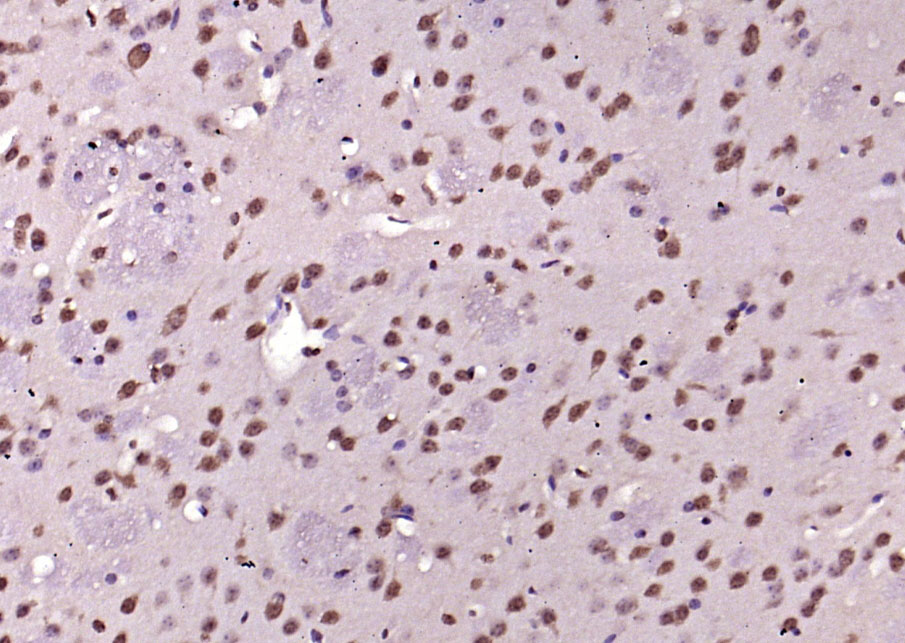 Paraformaldehyde-fixed, paraffin embedded (mouse brain); Antigen retrieval by boiling in sodium citrate buffer (pH6.0) for 15min; Block endogenous peroxidase by 3% hydrogen peroxide for 20 minutes; Blocking buffer (normal goat serum) at 37°C for 30min; Antibody incubation with (phospho-Erk1 (Thr202 + Tyr204)) Polyclonal Antibody, Unconjugated (SL1645R) at 1:200 overnight at 4°C, followed by operating according to SP Kit(Rabbit) (sp-0023) instructionsand DAB staining.
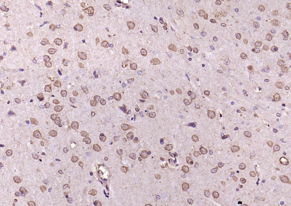 Paraformaldehyde-fixed, paraffin embedded (rat brain); Antigen retrieval by boiling in sodium citrate buffer (pH6.0) for 15min; Block endogenous peroxidase by 3% hydrogen peroxide for 20 minutes; Blocking buffer (normal goat serum) at 37°C for 30min; Antibody incubation with (phospho-Erk1 (Thr202 + Tyr204)) Polyclonal Antibody, Unconjugated (SL1645R) at 1:200 overnight at 4°C, followed by operating according to SP Kit(Rabbit) (sp-0023) instructionsand DAB staining.
 Tissue/cell: rat brain tissue; 4% Paraformaldehyde-fixed and paraffin-embedded;
Antigen retrieval: citrate buffer ( 0.01M, pH 6.0 ), Boiling bathing for 15min; Block endogenous peroxidase by 3% Hydrogen peroxide for 30min; Blocking buffer (normal goat serum,C-0005) at 37℃ for 20 min;
Incubation: Anti-phospho-Erk1(Thr202+Tyr204) Polyclonal Antibody, Unconjugated(SL1645R) 1:200, overnight at 4°C, followed by conjugation to the secondary antibody(SP-0023) and DAB(C-0010) staining
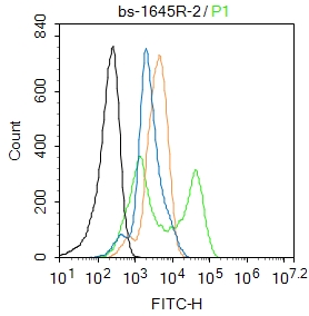 Blank control:MCF7.
Primary Antibody (green line): Rabbit Anti-phospho-Erk1 (Thr202 + Tyr204) antibody (SL 1645R)
Dilution: 2μg /10^6 cells;
Isotype Control Antibody (orange line): Rabbit IgG .
Secondary Antibody : Goat anti-rabbit IgG-FITC
Dilution: 1μg /test.
Protocol
The cells were fixed with 4% PFA (10min at room temperature)and then permeabilized with 0.1%PBST for 20 min at room temperature. The cells were then incubated in 5%BSA to block non-specific protein-protein interactions for 30 min at room temperature .Cells stained with Primary Antibody for 30 min at room temperature. The secondary antibody used for 40 min at room temperature. Acquisition of 20,000 events was performed.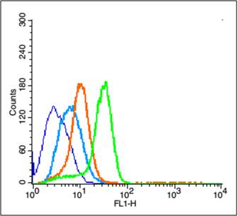 Blank control (blue line): U251 (blue).
Primary Antibody (green line): Rabbit Anti-phospho-Erk1 (Thr202 + Tyr204) antibody (SL1645R)
Dilution: 3μg /10^6 cells;
Isotype Control Antibody (orange line): Rabbit IgG .
Secondary Antibody (white blue line): Goat anti-rabbit IgG-PE
Dilution: 1μg /test.
Protocol
The cells were fixed with 2% paraformaldehyde (10 min)and then permeabilized with 0.1% PBS-Tween for 20 min at room temperature. Cells stained with Primary Antibody for 30 min at room temperature. The cells were then incubated in 1 X PBS/2%BSA/10% goat serum to block non-specific protein-protein interactions followed by the antibody for 15 min at room temperature. The secondary antibody used for 40 min at room temperature. Acquisition of 20,000 events was performed.
Cartpieces
Totalgoods,subtotals:¥Checkout
Bought notes(bought amounts latest0)
No one bought this product
User Comment(Total0User Comment Num)
- No comment


 +86 571 56623320
 +86 18668110335

