
Rabbit Anti-VEGF-C antibody
Vascuoar endothelial growth factor-C; AW228853; Flt4 ligand; Flt4-L; VEGF2; VEGFC; VRP; VEGFC_HUMAN.
View History [Clear]
Details
Product Name VEGF-C Chinese Name 血管内皮生长因子C型抗体 Alias Vascuoar endothelial growth factor-C; AW228853; Flt4 ligand; Flt4-L; VEGF2; VEGFC; VRP; VEGFC_HUMAN. literatures Research Area Cell biology vascular endothelial cell Immunogen Species Rabbit Clonality Polyclonal React Species Human, Mouse, Rat, Applications WB=1:500-2000 ELISA=1:5000-10000 IHC-P=1:100-500 IHC-F=1:100-500 Flow-Cyt=1ug/Test IF=1:100-500 (Paraffin sections need antigen repair)
not yet tested in other applications.
optimal dilutions/concentrations should be determined by the end user.Theoretical molecular weight 46kDa Cellular localization Secretory protein Form Liquid Concentration 1mg/ml immunogen KLH conjugated synthetic peptide derived from human VEGF-C: 321-415/415 Lsotype IgG Purification affinity purified by Protein A Buffer Solution 0.01M TBS(pH7.4) with 1% BSA, 0.03% Proclin300 and 50% Glycerol. Storage Shipped at 4℃. Store at -20 °C for one year. Avoid repeated freeze/thaw cycles. Attention This product as supplied is intended for research use only, not for use in human, therapeutic or diagnostic applications. PubMed PubMed Product Detail Vascular endothelial growth factors (VEGFs), also known as vasculotropins, are a family of closely related growth factors having a conserved pattern of eight cysteine residues and sharing common VEGF receptors. VEGFs stimulate the proliferation of endothelial cells, induce angiogenesis, promote cell migration, increase vascular permeability, and inhibit apoptosis. The mitogenic activity of VEGFs appears to be mediated by specific VEGF receptors. The target cell specificity of VEGF is restricted to vascular endothelial cells. Vascular Endothelial Growth Factor C (VEGFC) is a member of the VEGF subfamily of PDGF-related growth factors. It is the ligand for Flt4 (VEGFR3) and KDR (VEGFR2). VEGFC binds Flt4 and induces tyrosine autophosphorylation of VEGFR3 and VEGFR2. VEGFC also stimulates the migration of bovine capillary endothelial cells in collagen gel. It is a specific growth factor for the lymphatic vascular system and mediates lymphangiogenesis. VEGFC is abundantly expressed in heart and skeletal muscle. Other tissues such as lung and kidney also express VEGFC.
Subunit:
Homodimer; non-covalent and antiparallel.
Subcellular Location:
Secreted.
Tissue Specificity:
Spleen, lymph node, thymus, appendix, bone marrow, heart, placenta, ovary, skeletal muscle, prostate, testis, colon and small intestine and fetal liver, lung and kidney, but not in peripheral blood lymphocyte.
Similarity:
Belongs to the PDGF/VEGF growth factor family.
SWISS:
P97953
Gene ID:
7424
Database links:Entrez Gene: 7424 Human
Entrez Gene: 22341 Mouse
Omim: 601528 Human
SwissProt: P49767 Human
SwissProt: P97953 Mouse
Unigene: 435215 Human
Unigene: 1402 Mouse
Unigene: 6913 Rat
Product Picture 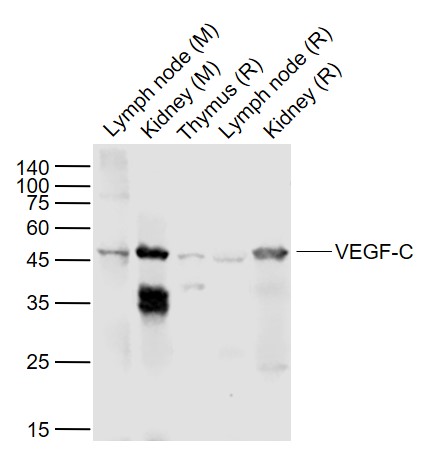 Sample:
Lane 1: Lymph node (Mouse) Lysate at 40 ug
Lane 2: Kidney (Mouse) Lysate at 40 ug
Lane 3: Thymus (Rat) Lysate at 40 ug
Lane 4: Lymph node (Rat) Lysate at 40 ug
Lane 5: Kidney (Rat) Lysate at 40 ug
Primary:
Anti-VEGF-C (SL1586R) at 1/1000 dilution
Secondary: IRDye800CW Goat Anti-Rabbit IgG at 1/20000 dilution
Predicted band size: 46 kD
Observed band size: 46 kD
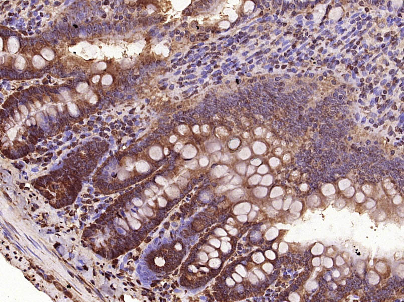 Paraformaldehyde-fixed, paraffin embedded (Rat small intestine); Antigen retrieval by microwave in sodium citrate buffer (pH6.0) ; Block endogenous peroxidase by 3% hydrogen peroxide for 30 minutes; Blocking buffer (3% BSA) at RT for 30min; Antibody incubation with (VEGF-C) Polyclonal Antibody, Unconjugated (SL1586R) at 1:400 overnight at 4℃, followed by conjugation to the secondary antibody (labeled with HRP)and DAB staining.
 Blank control: HepG2.
Primary Antibody (green line): Rabbit Anti-VEGF-C antibody (SL1586R)
Dilution: 1μg /10^6 cells;
Isotype Control Antibody (orange line): Rabbit IgG .
Secondary Antibody : Goat anti-rabbit IgG-AF647
Dilution: 1μg /test.
Protocol
The cells were fixed with 4% PFA (10min at room temperature)and then permeabilized with 0.1% PBST for 20 min at room temperature. The cells were then incubated in 5%BSA to block non-specific protein-protein interactions for 30 min at room temperature .Cells stained with Primary Antibody for 30 min at room temperature. The secondary antibody used for 40 min at room temperature. Acquisition of 20,000 events was performed.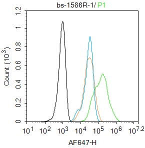 Blank control: HepG2.
Primary Antibody (green line): Rabbit Anti-VEGF-C antibody (SL1586R)
Dilution: 1μg /10^6 cells;
Isotype Control Antibody (orange line): Rabbit IgG .
Secondary Antibody : Goat anti-rabbit IgG-AF647
Dilution: 1μg /test.
Protocol
The cells were fixed with 4% PFA (10min at room temperature)and then permeabilized with 0.1% PBST for 20 min at room temperature. The cells were then incubated in 5%BSA to block non-specific protein-protein interactions for 30 min at room temperature .Cells stained with Primary Antibody for 30 min at room temperature. The secondary antibody used for 40 min at room temperature. Acquisition of 20,000 events was performed.Blank control: HepG2.
Primary Antibody (green line): Rabbit Anti-VEGF-C antibody (SL1586R)
Dilution: 1μg /10^6 cells;
Isotype Control Antibody (orange line): Rabbit IgG .
Secondary Antibody : Goat anti-rabbit IgG-AF647
Dilution: 1μg /test.
Protocol
The cells were fixed with 4% PFA (10min at room temperature)and then permeabilized with 0.1% PBST for 20 min at room temperature. The cells were then incubated in 5%BSA to block non-specific protein-protein interactions for 30 min at room temperature .Cells stained with Primary Antibody for 30 min at room temperature. The secondary antibody used for 40 min at room temperature. Acquisition of 20,000 events was performed.
Cartpieces
Totalgoods,subtotals:¥Checkout
Bought notes(bought amounts latest0)
No one bought this product
User Comment(Total0User Comment Num)
- No comment


 +86 571 56623320
 +86 18668110335

