
Rabbit Anti-ENPP3 antibody
ENPP3; CD203c; ectonucleotide pyrophosphatase/phosphodiesterase 3; RP5-988G15.3; B10; CD203c; NPP3; PD-IBETA; PDNP3; ENPP3_HUMAN.
View History [Clear]
Details
Product Name ENPP3 Chinese Name ENPP3蛋白抗体 Alias ENPP3; CD203c; ectonucleotide pyrophosphatase/phosphodiesterase 3; RP5-988G15.3; B10; CD203c; NPP3; PD-IBETA; PDNP3; ENPP3_HUMAN. Research Area immunology Channel protein Immunogen Species Rabbit Clonality Polyclonal React Species Human, Rat, (predicted: Mouse, Dog, Pig, Cow, ) Applications ELISA=1:5000-10000 IHC-P=1:100-500 IHC-F=1:100-500 Flow-Cyt=2μg /test IF=1:200-800 (Paraffin sections need antigen repair)
not yet tested in other applications.
optimal dilutions/concentrations should be determined by the end user.Theoretical molecular weight 100kDa Cellular localization The cell membrane Secretory protein Form Liquid Concentration 1mg/ml immunogen KLH conjugated synthetic peptide derived from human ENPP3: 40-140/875 <Extracellular> Lsotype IgG Purification affinity purified by Protein A Buffer Solution 0.01M TBS(pH7.4) with 1% BSA, 0.03% Proclin300 and 50% Glycerol. Storage Shipped at 4℃. Store at -20 °C for one year. Avoid repeated freeze/thaw cycles. Attention This product as supplied is intended for research use only, not for use in human, therapeutic or diagnostic applications. PubMed PubMed Product Detail The protein encoded by this gene belongs to a series of ectoenzymes that are involved in hydrolysis of extracellular nucleotides. These ectoenzymes possess ATPase and ATP pyrophosphatase activities and are type II transmembrane proteins. Expression of the related rat mRNA has been found in a subset of immature glial cells and in the alimentary tract. The corresponding rat protein has been detected in the pancreas, small intestine, colon, and liver. The human mRNA is expressed in glioma cells, prostate, and uterus. Expression of the human protein has been detected in uterus, basophils, and mast cells.
Function:
Cleaves a variety of phosphodiester and phosphosulfate bonds including deoxynucleotides, nucleotide sugars, and NAD.
Subcellular Location:
Membrane; Single-pass type II membrane protein (Potential). Secreted. Note=Located to the apical surface in intestinal and kidney epithelial cells. Located to the cell surface of basophils, and to the apical plasma membrane of bile duct cells. Secreted in serum, and in lumen of epithelial cells.
Tissue Specificity:
Expressed in bile ducts, prostate, uterus and colon. Exclusively expressed on basophils, mast cells and their progenitors.
Post-translational modifications:
N-glycosylation is necessary for correct trafficking to the apical surface, but is not the apical targeting signal.
It has been suggested that the active SMB domain may be permitted considerable disulfide bond heterogeneity or variability, thus two alternate disulfide patterns based on 3D structures are described with 1 disulfide bond conserved in both.
Similarity:
Belongs to the nucleotide pyrophosphatase/phosphodiesterase family.
Contains 2 SMB (somatomedin-B) domains.
SWISS:
O14638
Gene ID:
5169
Database links:Entrez Gene: 5169 Human
SwissProt: O14638 Human
CD203c蛋白是一个糖基化的2型Transmembrane protein分子,CD203c的表达主要在嗜碱细胞、肥大细胞和它们的前体细胞上。Product Picture 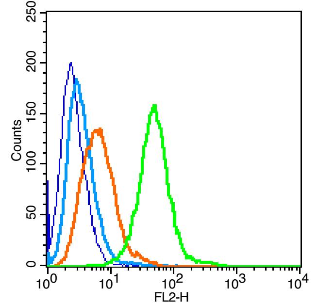 Blank control: Raji (blue).
Primary Antibody: Rabbit Anti-ENPP3 antibody(SL1568R), Dilution: 1μg in 100 μL 1X PBS containing 0.5% BSA;
Isotype Control Antibody: Rabbit IgG(orange),used under the same conditions );
Secondary Antibody: Goat anti-rabbit IgG-PE(white blue), Dilution: 1:200 in 1 X PBS containing 0.5% BSA.
Protocol
The cells were fixed with 2% paraformaldehyde (10 min) , then permeabilized with 90% ice-cold methanol for 30 min on ice. Primary antibody (SL1568R, 1μg /1x10^6 cells) were incubated for 30 min on the ice, followed by 1 X PBS containing 0.5% BSA + 1 0% goat serum (15 min) to block non-specific protein-protein interactions. Then the Goat Anti-rabbit IgG/PE antibody was added into the blocking buffer mentioned above to react with the primary antibody at 1/200 dilution for 30 min on ice. Acquisition of 20,000 events was performed.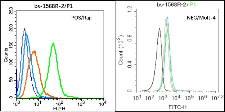 Black line : Positive blank control (Raji); Negative blank control (Molt4)
Green line : Primary Antibody (Rabbit Anti-ENPP3 antibody (SL1568R) )
Orange line:Isotype Control Antibody (Rabbit IgG) .
Blue line : Secondary Antibody (Goat anti-rabbit IgG-PE)/Goat anti-rabbit IgG-AF488)
Raji(Positive)and Molt4(Negative control)cells (black) were incubated in 5% BSA blocking buffer for 30 min at room temperature. Cells were then stained with ENPP3 Antibody(SL1568R)at 1:50 dilution in blocking buffer and incubated for 30 min at room temperature, washed twice with 2% BSA in PBS, followed by secondary antibody(blue) incubation for 40 min at room temperature. Acquisitions of 20,000 events were performed. Cells stained with primary antibody (green), and isotype control (orange).
Cartpieces
Totalgoods,subtotals:¥Checkout
Bought notes(bought amounts latest0)
No one bought this product
User Comment(Total0User Comment Num)
- No comment


 +86 571 56623320
 +86 18668110335

