
Rabbit Anti-phospho-ERK1 + 2 (Thr183/Tyr185)antibody
ERK1 + ERK2 (phospho Thr183/Tyr185); phospho-ERK1/MAPK-1/2(Thr183/Tyr185); ERK 1; ERK 2; ERK-2; ERK1; ERK2; ERT1; ERT2; Extracellular signal regulated kinase 1; Extracellular signal regulated kinase 1; Extracellular signal regulated kinase 2; Extracellula
View History [Clear]
Details
Product Name phospho-ERK1 + 2 (Thr183/Tyr185) Chinese Name 磷酸化丝裂原活化蛋白激酶1/2抗体 Alias ERK1 + ERK2 (phospho Thr183/Tyr185); phospho-ERK1/MAPK-1/2(Thr183/Tyr185); ERK 1; ERK 2; ERK-2; ERK1; ERK2; ERT1; ERT2; Extracellular signal regulated kinase 1; Extracellular signal regulated kinase 1; Extracellular signal regulated kinase 2; Extracellular signal regulated kinase 2; Extracellular signal-regulated kinase 2; HS44KDAP; HUMKER1A; Insulin stimulated MAP2 kinase; MAP kinase 1; MAP kinase 2; MAP kinase isoform p42; MAP kinase isoform p44; MAPK 1; MAPK 2; MAPK1; MAPK2; MGC20180; Microtubule associated protein 2 kinase; Mitogen activated protein kinase 1; Mitogen activated protein kinase 1; Mitogen activated protein kinase 2; Mitogen-activated protein kinase 1; Mitogen-activated protein kinase 2; MK01_MOUSE; p38; p40; p41; p41mapk; p42 MAPK; p42-MAPK; p42MAPK; p42MAPK; p44 ERK1; p44 MAPK; p44ERK1; p44ERK1; p44MAPK; p44MAPK; PRKM 1; PRKM 1; PRKM 2; PRKM 2; PRKM1; PRKM2; Protein kinase mitogen activated 1; Protein kinase mitogen activated 1; Protein kinase mitogen activated 2; Protein kinase mitogen activated 2; Protein tyrosine kinas. literatures Product Type Phosphorylated anti Research Area immunology Neurobiology Signal transduction Stem cells Kinases and Phosphatases Immunogen Species Rabbit Clonality Polyclonal React Species Human, Mouse, (predicted: Rat, Chicken, Dog, Cow, Horse, Rabbit, Guinea Pig, ) Applications WB=1:500-2000 ELISA=1:5000-10000 IHC-P=1:100-500 IHC-F=1:100-500 ICC=1:100 IF=1:100-500 (Paraffin sections need antigen repair)
not yet tested in other applications.
optimal dilutions/concentrations should be determined by the end user.Theoretical molecular weight 42/44kDa Cellular localization The nucleus cytoplasmic Form Liquid Concentration 1mg/ml immunogen KLH conjugated Synthesised phosphopeptide derived from mouse ERK1 around the phosphorylation site of Thr183/Tyr185: FL(p-T)E(p-Y)V Lsotype IgG Purification affinity purified by Protein A Buffer Solution 0.01M TBS(pH7.4) with 1% BSA, 0.03% Proclin300 and 50% Glycerol. Storage Shipped at 4℃. Store at -20 °C for one year. Avoid repeated freeze/thaw cycles. Attention This product as supplied is intended for research use only, not for use in human, therapeutic or diagnostic applications. PubMed PubMed Product Detail Mitogen-activated protein kinase (MAPK) signaling cascades include MAPK or extracellular signal-regulated kinase (ERK), MAPK kinase (MKK or MEK), and MAPK kinase kinase (MAPKKK or MEKK). MAPKK kinase/MEKK phosphorylates and activates its downstream protein kinase, MAPK kinase/MEK, which in turn activates MAPK. The kinases of these signaling cascades are highly conserved, and homologs exist in yeast, Drosophila, and mammalian cells. MAPKKK5 contains 1,374 amino acids with all 11 kinase subdomains. Northern blot analysis shows that MAPKKK5 transcript is abundantly expressed in human heart and pancreas. The MAPKKK5 protein phosphorylates and activates MKK4 (aliases SERK1, MAPKK4) in vitro, and activates c-Jun N-terminal kinase (JNK)/stress-activated protein kinase (SAPK) during transient expression in COS and 293 cells; MAPKKK5 does not activate MAPK/ERK. [provided by RefSeq, Jul 2008]
Function:
Serine/threonine kinase which acts as an essentialcomponent of the MAP kinase signal transduction pathway. MAPK1/ERK2and MAPK3/ERK1 are the 2 MAPKs which play an important role in theMAPK/ERK cascade. They participate also in a signaling cascadeinitiated by activated KIT and KITLG/SCF. Depending on the cellularcontext, the MAPK/ERK cascade mediates diverse biological functionssuch as cell growth, adhesion, survival and differentiation throughthe regulation of transcription, translation, cytoskeletalrearrangements. The MAPK/ERK cascade plays also a role ininitiation and regulation of meiosis, mitosis, and postmitoticfunctions in differentiated cells by phosphorylating a number oftranscription factors. About 160 substrates have already beendiscovered for ERKs. Many of these substrates are localized in thenucleus, and seem to participate in the regulation of transcriptionupon stimulation. However, other substrates are found in thecytosol as well as in other cellular organelles, and those areresponsible for processes such as translation, mitosis andapoptosis. Moreover, the MAPK/ERK cascade is also involved in theregulation of the endosomal dynamics, including lysosome processingand endosome cycling through the perinuclear recycling compartment(PNRC); as well as in the fragmentation of the Golgi apparatusduring mitosis. The substrates include transcription factors (suchas ATF2, BCL6, ELK1, ERF, FOS, HSF4 or SPZ1), cytoskeletal elements(such as CANX, CTTN, GJA1, MAP2, MAPT, PXN, SORBS3 or STMN1),regulators of apoptosis (such as BAD, BTG2, CASP9, DAPK1, IER3,MCL1 or PPARG), regulators of translation (such as EIF4EBP1) and avariety of other signaling-related molecules (like ARHGEF2, DCC,FRS2 or GRB10). Protein kinases (such as RAF1, RPS6KA1/RSK1,RPS6KA3/RSK2, RPS6KA2/RSK3, RPS6KA6/RSK4, SYK, MKNK1/MNK1,MKNK2/MNK2, RPS6KA5/MSK1, RPS6KA4/MSK2, MAPKAPK3 or MAPKAPK5) andphosphatases (such as DUSP1, DUSP4, DUSP6 or DUSP16) are othersubstrates which enable the propagation the MAPK/ERK signal toadditional cytosolic and nuclear targets, thereby extending thespecificity of the cascade. Mediates phosphorylation of TPR inrespons to EGF stimulation. May play a role in the spindle assemblycheckpoint. Phosphorylates PML and promotes its interaction withPIN1, leading to PML degradation (By similarity). [FUNCTION] Acts as a transcriptional repressor. Binds to a[GC]AAA[GC] consensus sequence. Repress the expression ofinterferon gamma-induced genes. Seems to bind to the promoter ofCCL5, DMP1, IFIH1, IFITM1, IRF7, IRF9, LAMP3, OAS1, OAS2, OAS3 andSTAT1. Transcriptional activity is independent of kinase activity(By similarity).
Subunit:
Binds both upstream activators and downstream substratesin multimolecular complexes. Interacts with ADAM15, ARHGEF2, ARRB2,DAPK1 (via death domain), HSF4, IER3, IPO7, DUSP6, NISCH, SGK1, andisoform 1 of NEK2. Interacts (via phosphorylated form) with TPR(via C-terminus region and phosphorylated form); the interactionrequires dimerization of MAPK1/ERK2 and increases following EGFstimulation (By similarity). Interacts (phosphorylated form) withCAV2 ('Tyr-19'-phosphorylated form); the interaction, promoted byinsulin, leads to nuclear location and MAPK1 activation (Bysimilarity). Interacts with DCC (By similarity). Interacts withMORG1, PEA15 and MKNK2. MKNK2 isoform 1 binding prevents fromdephosphorylation and inactivation. The phosphorylated forminteracts with PML (By similarity).
Subcellular Location:
Cytoplasm, cytoskeleton, spindle (Bysimilarity). Nucleus. Cytoplasm, cytoskeleton, centrosome (Bysimilarity). Cytoplasm. Note=Associated with the spindle duringprometaphase and metaphase (By similarity). PEA15-binding andphosphorylated DAPK1 promote its cytoplasmic retention.Phosphorylation at Ser-244 and Ser-246 as well asautophosphorylation at Thr-188 promote nuclear localization (Bysimilarity).
Tissue Specificity:
Widely expressed.
Post-translational modifications:
Dually phosphorylated on Thr-183 and Tyr-185, which activatesthe enzyme. Ligand-activated ALK induces tyrosine phosphorylation(By similarity). Dephosphorylated by PTPRJ at Tyr-185 (Bysimilarity). Phosphorylated upon FLT3 and KIT signaling (Bysimilarity).
Similarity:
Belongs to the protein kinase superfamily. CMGCSer/Thr protein kinase family. MAP kinase subfamily.
Contains 1 protein kinase domain.
SWISS:
P63085
Gene ID:
5595
Database links:Entrez Gene: 5594 Human
Entrez Gene: 5595 Human
Entrez Gene: 26413 Mouse
Entrez Gene: 26417 Mouse
Kinases and Phosphatases(Kinases and Phosphatases)
丝裂原活化蛋白激酶-ERK(Mitogen-activated protein kinase 1, MAPK-1; P42-MAPK;Extracellular signal-regulated kinase 2, ERK-2;MAPK-2曾用名还有:, ERK2,MAPK-α,MAPK1,MAPK2, p42MAPK)是一组可以被多种细胞外信号即获得蛋白丝/苏氨酸激酶,处于胞浆信号传导通路的终末位置,活化后转位到核内,作用于核内转录因子,调节基因表达。它主要参与生长因子、激素、cell factor、应激等各种刺激下细胞的反应、细胞的生长、分化过程。
经研究证实,MAPKSignal transduction通路存在于大多数细胞内,在将细胞外刺激Signal transduction至细胞及其核内,并引起Cell biology学反应(如细胞增殖、分化、转化及凋亡等)的过程中具有至关重要的作用。研究表明,MAPKSignal transduction通路在细胞内具有生物进化的高度保守性,在低等原核细胞和高等哺乳类细胞内,目前均已发现存在着多条并行的MAPK信号通路,不同的细胞外刺激可使用不同的MAPK信号通路,通过其相互调控而介导不同的Cell biology学反应。Product Picture 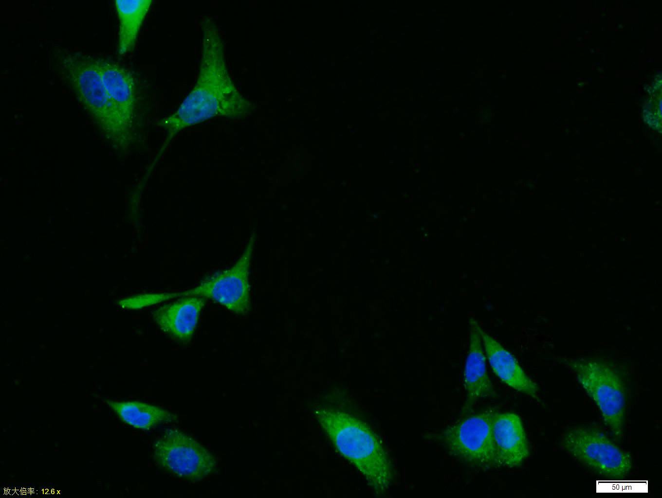 Tissue/cell: HUVEC cell; 4% Paraformaldehyde-fixed; Triton X-100 at room temperature for 20 min; Blocking buffer (normal goat serum, C-0005) at 37°C for 20 min; Antibody incubation with (phospho-ERK1 + 2 (Thr183/Tyr185)) Polyclonal Antibody, Unconjugated (SL1522R) 1:100, 90 minutes at 37°C; followed by a conjugated Goat Anti-Rabbit IgG antibody (SL0295G-FITC) at 37°C for 90 minutes, DAPI (blue, C02-04002) was used to stain the cell nuclei.
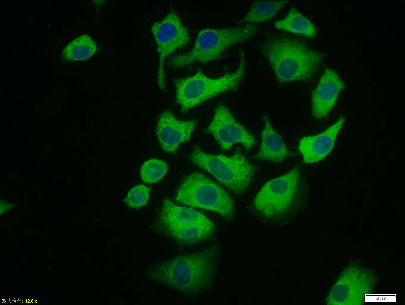 Tissue/cell: Hela cell; 4% Paraformaldehyde-fixed; Triton X-100 at room temperature for 20 min; Blocking buffer (normal goat serum, C-0005) at 37°C for 20 min; Antibody incubation with (phospho-ERK1 + 2 (Thr183/Tyr185)) polyclonal Antibody, Unconjugated (SL1522R) 1:100, 90 minutes at 37°C; followed by a FITC conjugated Goat Anti-Rabbit IgG antibody at 37°C for 90 minutes, DAPI (blue, C02-04002) was used to stain the cell nuclei.
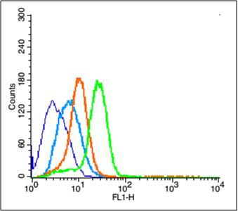 Blank control (blue line): U251 (blue).
Primary Antibody (green line): Rabbit Anti-phospho-ERK1 + 2 (Thr183185) antibody (SL1522R)
Dilution: 3μg /10^6 cells;
Isotype Control Antibody (orange line): Rabbit IgG .
Secondary Antibody (white blue line): Goat anti-rabbit IgG-PE
Dilution: 1μg /test.
Protocol
The cells were fixed with 2% paraformaldehyde (10 min)and then permeabilized with 0.1% PBS-Tween for 20 min at room temperature. Cells stained with Primary Antibody for 30 min at room temperature. The cells were then incubated in 1 X PBS/2%BSA/10% goat serum to block non-specific protein-protein interactions followed by the antibody for 15 min at room temperature. The secondary antibody used for 40 min at room temperature. Acquisition of 20,000 events was performed.
Cartpieces
Totalgoods,subtotals:¥Checkout
Bought notes(bought amounts latest0)
No one bought this product
User Comment(Total0User Comment Num)
- No comment


 +86 571 56623320
 +86 18668110335

