
Rabbit Anti-MUC1 antibody
KL-6; MUC-1; MUC-1/SEC; MUC-1/X; MUC1/ZD; Breast carcinoma associated antigen DF3; CA 15 3; CA15-3; CA15-3 antigen; CA15.3; CA-153; Carcinoma associated mucin; CD 227; CD227; CD227 antigen; DF3 antigen; EMA; Episialin; Epithelial membrane antigen; Epithel
View History [Clear]
Details
Product Name MUC1 Chinese Name 粘蛋白-1抗体 Alias KL-6; MUC-1; MUC-1/SEC; MUC-1/X; MUC1/ZD; Breast carcinoma associated antigen DF3; CA 15 3; CA15-3; CA15-3 antigen; CA15.3; CA-153; Carcinoma associated mucin; CD 227; CD227; CD227 antigen; DF3 antigen; EMA; Episialin; Epithelial membrane antigen; Epithelial mucin tandem repeat sequence; H23 antigen; H23AG; HGNC:7508; MAM6; MUC 1; MUC-1; Mucin 1; Mucin 1 precursor; Mucin1; Peanut reactive urinary mucin; PEM; PEMT; Polymorphic epithelial mucin; PUM; Tumor associated epithelial membrane antigen; Tumor associated mucin. literatures Research Area Tumour immunology Immunogen Species Rabbit Clonality Polyclonal React Species Human, Mouse, Rat, Applications ELISA=1:5000-10000 IHC-P=1:100-500 IHC-F=1:100-500 ICC=1:100-500 IF=1:100-500 (Paraffin sections need antigen repair)
not yet tested in other applications.
optimal dilutions/concentrations should be determined by the end user.Theoretical molecular weight 20/138kDa Cellular localization The nucleus cytoplasmic The cell membrane Secretory protein Form Liquid Concentration 1mg/ml immunogen KLH conjugated synthetic peptide derived from human MUC1: 1101-1255/1255 <Cytoplasmic> Lsotype IgG2b Purification affinity purified by Protein A Buffer Solution 0.01M TBS(pH7.4) with 1% BSA, 0.03% Proclin300 and 50% Glycerol. Storage Shipped at 4℃. Store at -20 °C for one year. Avoid repeated freeze/thaw cycles. Attention This product as supplied is intended for research use only, not for use in human, therapeutic or diagnostic applications. PubMed PubMed Product Detail MUC1 is a large cell surface mucin glycoprotein expressed by most glandular and ductal epithelial cells and some hematopoietic cell lineages. It is expressed on most secretory epithelium, including mammary gland and some hematopoietic cells. It is expressed abundantly in lactating mammary glands and overexpressed abundantly in >90% breast carcinomas and metastases. Transgenic MUC1 has been shown to associate with all four cebB receptors and localize with erbB1 (EGFR) in lactating glands. The MUC1 gene contains seven exons and produces several different alternatively spliced variants. The major expressed form of MUC1 uses all seven exons and is a type 1 transmembrane protein with a large extracellular tandem repeat domain. The tandem repeat domain is highly O glycosylated and alterations in glycosylation have been shown in epithelial cancer cells.
Function:
The alpha subunit has cell adhesive properties. Can act both as an adhesion and an anti-adhesion protein. May provide a protective layer on epithelial cells against bacterial and enzyme attack.
The beta subunit contains a C-terminal domain which is involved in cell signaling, through phosphorylations and protein-protein interactions. Modulates signaling in ERK, SRC and NF-kappa-B pathways. In activated T-cells, influences directly or indirectly the Ras/MAPK pathway. Promotes tumor progression. Regulates TP53-mediated transcription and determines cell fate in the genotoxic stress response. Binds, together with KLF4, the PE21 promoter element of TP53 and represses TP53 activity.
Subunit:
The alpha subunit forms a tight, non-covalent heterodimeric complex with the proteolytically-released beta-subunit.
Subcellular Location:
Apical cell membrane; Single-pass type I membrane protein. Isoform 5: Secreted. Isoform 7: Secreted. Isoform 9: Secreted. Mucin-1 subunit beta: Cell membrane. Cytoplasm. Nucleus.
Tissue Specificity:
Expressed on the apical surface of epithelial cells, especially of airway passages, breast and uterus. Also expressed in activated and unactivated T-cells. Overexpressed in epithelial tumors, such as breast or ovarian cancer and also in non-epithelial tumor cells. Isoform 7 is expressed in tumor cells only.
Post-translational modifications:
Highly glycosylated (N- and O-linked carbohydrates and sialic acid). O-glycosylated to a varying degree on serine and threonine residues within each tandem repeat, ranging from mono- to penta-glycosylation. The average density ranges from about 50% in human milk to over 90% in T47D breast cancer cells. Further sialylation occurs during recycling. Membrane-shed glycoproteins from kidney and breast cancer cells have preferentially sialyated core 1 structures, while secreted forms from the same tissues display mainly core 2 structures. The O-glycosylated content is overlapping in both these tissues with terminal fucose and galactose, 2- and 3-linked galactose, 3- and 3,6-linked GalNAc-ol and 4-linked GlcNAc predominating. Differentially O-glycosylated in breast carcinomas with 3,4-linked GlcNAc. N-glycosylation consists of high-mannose, acidic complex-type and hybrid glycans in the secreted form MUC1/SEC, and neutral complex-type in the transmembrane form, MUC1/TM.
Proteolytic cleavage in the SEA domain occurs in the endoplasmic reticulum by an autoproteolytic mechanism and requires the full-length SEA domain as well as requiring a Ser, Thr or Cys residue at the P + 1 site. Cleavage at this site also occurs on isoform MUC1/X but not on isoform MUC1/Y. Ectodomain shedding is mediated by ADAM17.
Dual palmitoylation on cysteine residues in the CQC motif is required for recycling from endosomes back to the plasma membrane.
Phosphorylated on tyrosines and serine residues in the C-terminal. Phosphorylation on tyrosines in the C-terminal increases the nuclear location of MUC1 and beta-catenin. Phosphorylation by PKC delta induces binding of MUC1 to beta-catenin/CTNNB1 and thus decreases the formation of the beta-catenin/E-cadherin complex. Src-mediated phosphorylation inhibits interaction with GSK3B. Src-and EGFR-mediated phosphorylation on Tyr-1229 increases binding to beta-catenin/CTNNB1. GSK3B-mediated phosphorylation on Ser-1227 decreases this interaction but restores the formation of the beta-cadherin/E-cadherin complex. On T-cell receptor activation, phosphorylated by LCK. PDGFR-mediated phosphorylation increases nuclear colocalization of MUC1CT and CTNNB1.
The N-terminal sequence has been shown to begin at position 24 or 28 (PubMed:11341784).
Similarity:
Contains 1 SEA domain.
SWISS:
P15941
Gene ID:
4582
Database links:Entrez Gene: 4582 Human
Entrez Gene: 17829 Mouse
Omim: 158340 Human
SwissProt: P15941 Human
SwissProt: Q02496 Mouse
Unigene: 89603 Human
Unigene: 16193 Mouse
Unigene: 10779 Rat
CA153为高分子glycoprotein,是乳腺癌Maker之一,其表达的量与乳腺癌的分化程度及雌激素受体高低有关联。近年来,很多学者研究认为:在人的很多种恶性Tumour中都有CA15-3的不同表达。Product Picture 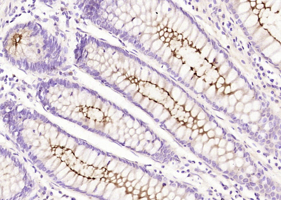 Paraformaldehyde-fixed, paraffin embedded (human colon carcinoma); Antigen retrieval by boiling in sodium citrate buffer (pH6.0) for 15min; Block endogenous peroxidase by 3% hydrogen peroxide for 20 minutes; Blocking buffer (normal goat serum) at 37°C for 30min; Antibody incubation with (MUC1) Polyclonal Antibody, Unconjugated (SL1497R) at 1:200 overnight at 4°C, followed by operating according to SP Kit(Rabbit) (sp-0023) instructionsand DAB staining.
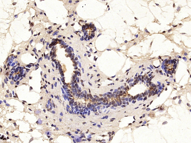 Paraformaldehyde-fixed, paraffin embedded (Rat breast); Antigen retrieval by microwave in sodium citrate buffer (pH6.0) ; Block endogenous peroxidase by 3% hydrogen peroxide for 30 minutes; Blocking buffer (3% BSA) at RT for 30min; Antibody incubation with (MUC1) Polyclonal Antibody, Unconjugated (SL1497R) at 1:400 overnight at 4℃, followed by conjugation to the secondary antibody (labeled with HRP)and DAB staining.
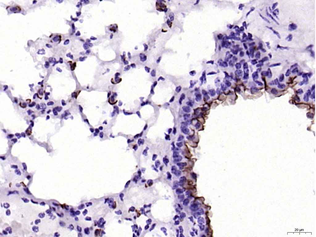 Paraformaldehyde-fixed, paraffin embedded (mouse lung); Antigen retrieval by boiling in sodium citrate buffer (pH6.0) for 15min; Block endogenous peroxidase by 3% hydrogen peroxide for 20 minutes; Blocking buffer (normal goat serum) at 37°C for 30min; Antibody incubation with (MUC1) Polyclonal Antibody, Unconjugated (SL1497R) at 1:200 overnight at 4°C, followed by operating according to SP Kit(Rabbit) (sp-0023) instructionsand DAB staining.
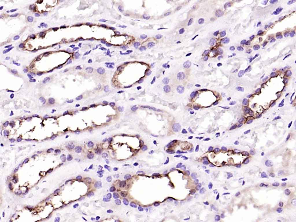 Paraformaldehyde-fixed, paraffin embedded (Human kidney); Antigen retrieval by boiling in sodium citrate buffer (pH6.0) for 15min; Block endogenous peroxidase by 3% hydrogen peroxide for 20 minutes; Blocking buffer (normal goat serum) at 37°C for 30min; Antibody incubation with (MUC1) Polyclonal Antibody, Unconjugated (SL1497R) at 1:200 overnight at 4°C, followed by operating according to SP Kit(Rabbit) (sp-0023) instructionsand DAB staining.
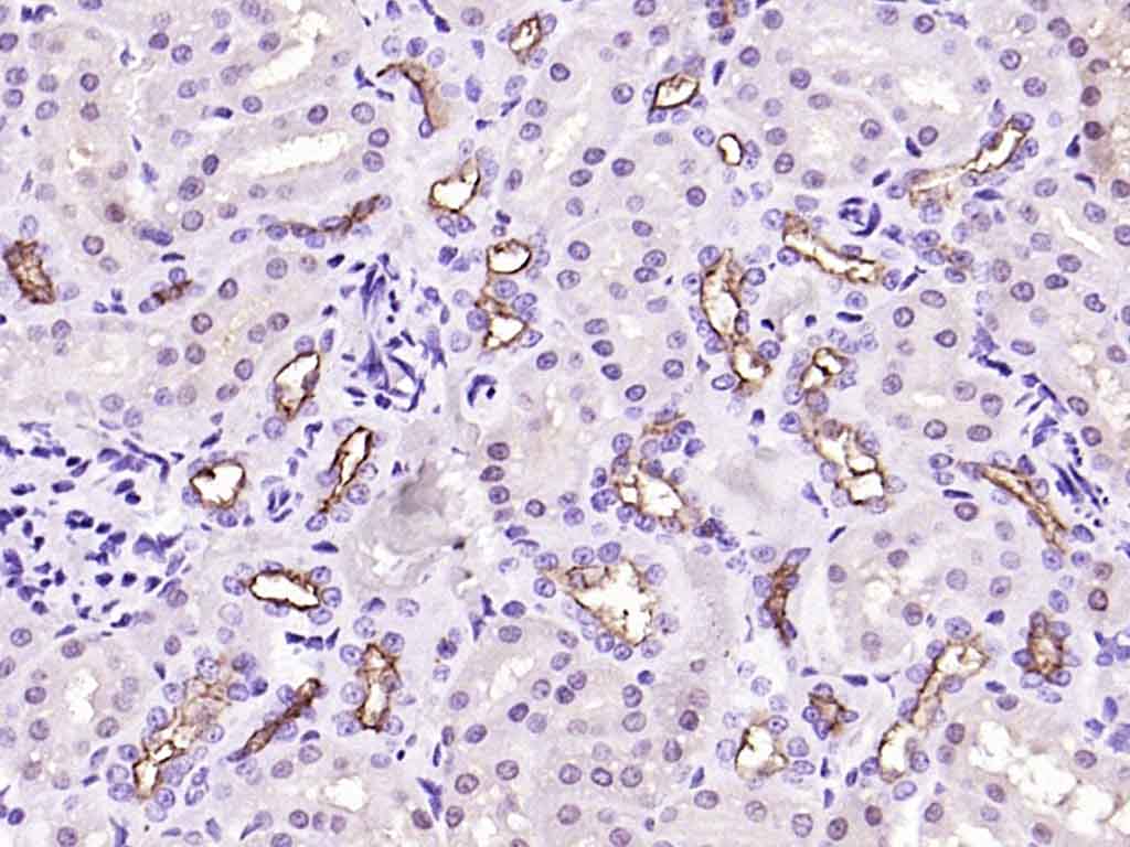 Paraformaldehyde-fixed, paraffin embedded (rat kidney); Antigen retrieval by boiling in sodium citrate buffer (pH6.0) for 15min; Block endogenous peroxidase by 3% hydrogen peroxide for 20 minutes; Blocking buffer (normal goat serum) at 37°C for 30min; Antibody incubation with (MUC1) Polyclonal Antibody, Unconjugated (SL1497R) at 1:200 overnight at 4°C, followed by operating according to SP Kit(Rabbit) (sp-0023) instructionsand DAB staining.
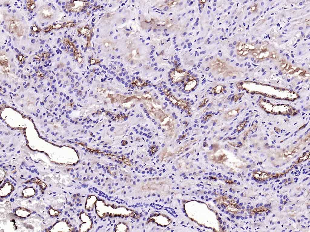 Paraformaldehyde-fixed, paraffin embedded (Human kidney); Antigen retrieval by boiling in sodium citrate buffer (pH6.0) for 15min; Block endogenous peroxidase by 3% hydrogen peroxide for 20 minutes; Blocking buffer (normal goat serum) at 37°C for 30min; Antibody incubation with (MUC1) Polyclonal Antibody, Unconjugated (SL1497R) at 1:200 overnight at 4°C, followed by operating according to SP Kit(Rabbit) (sp-0023) instructionsand DAB staining.
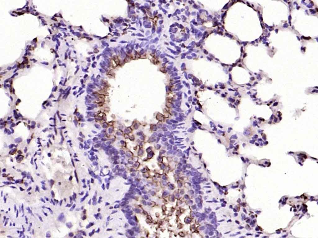 Paraformaldehyde-fixed, paraffin embedded (rat lung); Antigen retrieval by boiling in sodium citrate buffer (pH6.0) for 15min; Block endogenous peroxidase by 3% hydrogen peroxide for 20 minutes; Blocking buffer (normal goat serum) at 37°C for 30min; Antibody incubation with (MUC1) Polyclonal Antibody, Unconjugated (SL1497R) at 1:200 overnight at 4°C, followed by operating according to SP Kit(Rabbit) (sp-0023) instructionsand DAB staining.
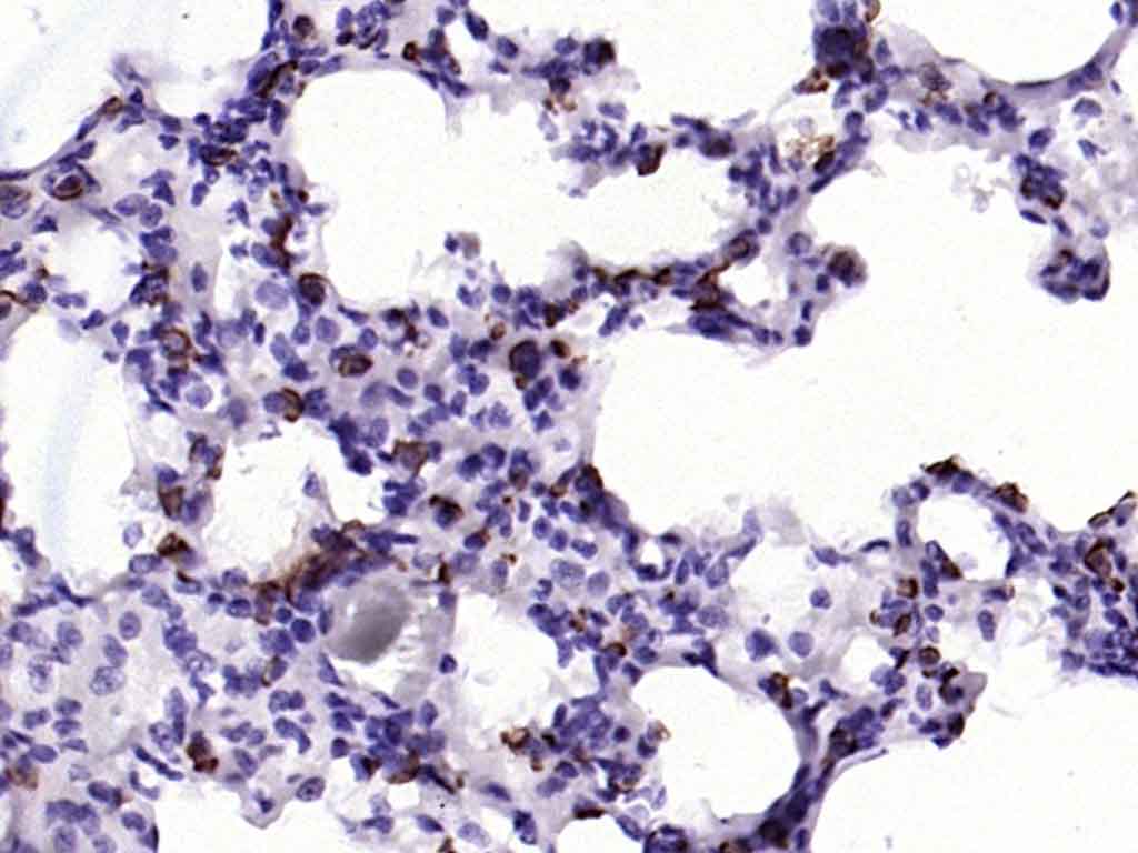 Paraformaldehyde-fixed, paraffin embedded (rat lung); Antigen retrieval by boiling in sodium citrate buffer (pH6.0) for 15min; Block endogenous peroxidase by 3% hydrogen peroxide for 20 minutes; Blocking buffer (normal goat serum) at 37°C for 30min; Antibody incubation with (MUC1) Polyclonal Antibody, Unconjugated (SL1497R) at 1:200 overnight at 4°C, followed by operating according to SP Kit(Rabbit) (sp-0023) instructionsand DAB staining.
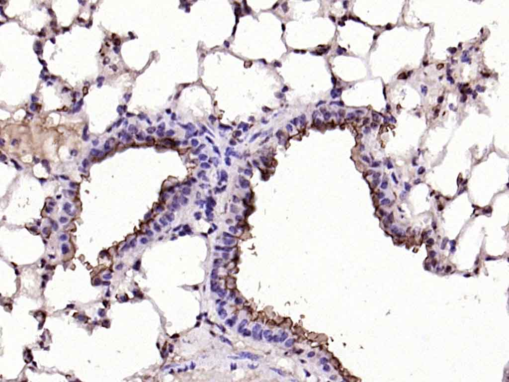 Paraformaldehyde-fixed, paraffin embedded (mouse lung); Antigen retrieval by boiling in sodium citrate buffer (pH6.0) for 15min; Block endogenous peroxidase by 3% hydrogen peroxide for 20 minutes; Blocking buffer (normal goat serum) at 37°C for 30min; Antibody incubation with (MUC1) Polyclonal Antibody, Unconjugated (SL1497R) at 1:200 overnight at 4°C, followed by operating according to SP Kit(Rabbit) (sp-0023) instructionsand DAB staining.
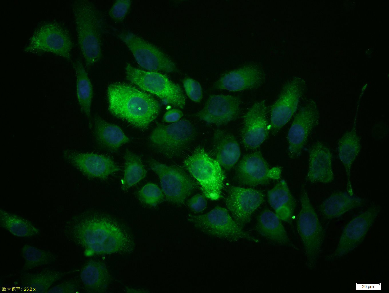 Tissue/cell: MCF-7 cell; 4% Paraformaldehyde-fixed; Triton X-100 at room temperature for 20 min; Blocking buffer (normal goat serum, C-0005) at 37°C for 20 min; Antibody incubation with (MUC1) Polyclonal Antibody, Unconjugated (SL1497R) 1:100, 90 minutes at 37°C; followed by a conjugated Goat Anti-Rabbit IgG antibody (SL0295G-FITC) at 37°C for 90 minutes, DAPI (5ug/ml, blue, C-0033) was used to stain the cell nuclei.
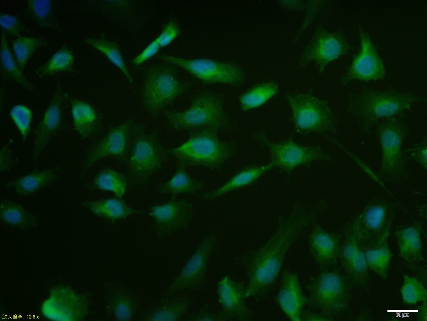 MCF7 cell; 4% Paraformaldehyde-fixed; Triton X-100 at room temperature for 20 min; Blocking buffer (normal goat serum, C-0005) at 37°C for 20 min; Antibody incubation with (MUC1) polyclonal Antibody, Unconjugated (SL1497R) 1:100, 90 minutes at 37°C; followed by a conjugated Goat Anti-Rabbit IgG antibody at 37°C for 90 minutes, DAPI (blue, C02-04002) was used to stain the cell nuclei.
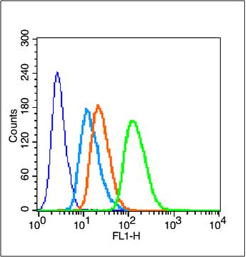 Blank control (blue line): MCF7(fixed with 70% ethanol (Overnight at 4℃) and then permeabilized with 90% ice-cold methanol for 30 min on ice)
Primary Antibody (green line): Rabbit Anti-MUC1 antibody (SL1497R),Dilution: 0.2μg /10^6 cells;
Isotype Control Antibody (orange line): Rabbit IgG .
Secondary Antibody (white blue line): Goat anti-rabbit IgG-FITC,Dilution: 1μg /test.
 Blank control: MCF7.
Primary Antibody (green line): Rabbit Anti-MUC1 antibody (SL1497R)
Dilution: 2μg /10^6 cells;
Isotype Control Antibody (orange line): Rabbit IgG .
Secondary Antibody : Goat anti-rabbit IgG-AF647
Dilution: 1μg /test.
Protocol
The cells were fixed with 4% PFA (10min at room temperature)and then permeabilized with 90% ice-cold methanol for 20 min at-20℃.The cells were then incubated in 5%BSA to block non-specific protein-protein interactions for 30 min at at room temperature .Cells stained with Primary Antibody for 30 min at room temperature. The secondary antibody used for 40 min at room temperature. Acquisition of 20,000 events was performed.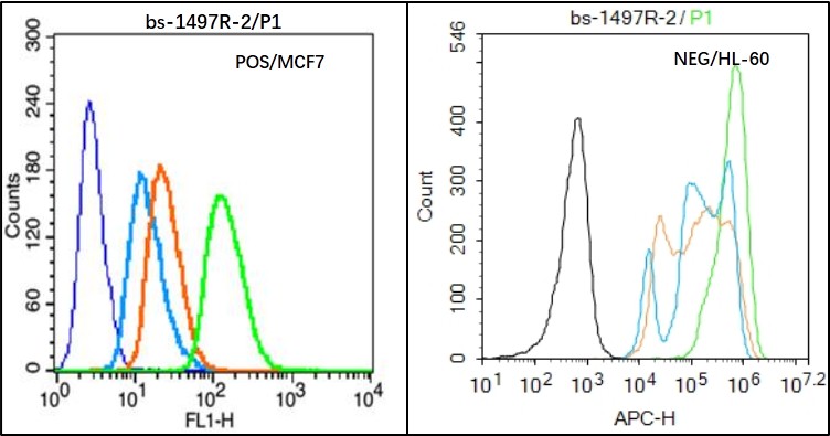 Black line : Positive blank control (MCF7); Negative blank control (HL60)
Green line : Primary Antibody (Rabbit Anti-MUC1 antibody (SL1497R) )
Orange line:Isotype Control Antibody (Rabbit IgG) .
Blue line : Secondary Antibody (Goat anti-rabbit IgG-AF488)/(Goat anti-rabbit IgG-AF488)
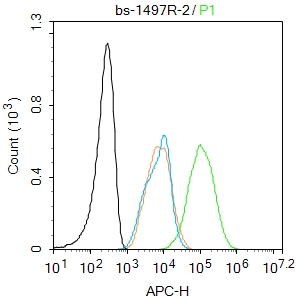
MCF7(Positive)and HL60(Negative control)cells (black) were fixed with 4% PFA for 10min at room temperature, permeabilized with PBST for 20 min at room temperature, and incubated in 5% BSA blocking buffer for 30 min at room temperature. Cells were then stained with MUC1 Antibody(SL1497R)at 1:50 dilution in blocking buffer and incubated for 30 min at room temperature, washed twice with 2% BSA in PBS, followed by secondary antibody(blue) incubation for 40 min at room temperature. Acquisitions of 20,000 events were performed. Cells stained with primary antibody (green), and isotype control (orange).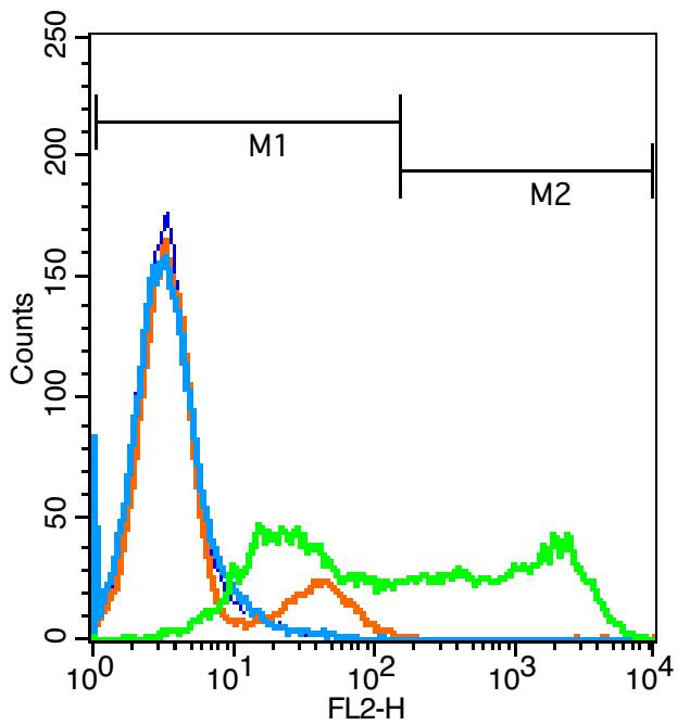 Blank control(blue): Jurkat cells(fixed with 2% paraformaldehyde (10 min)).
Primary Antibody:Rabbit Anti-TSLC1 antibody(SL1497R), Dilution: 5μg in 100 μL 1X PBS containing 0.5% BSA;
Isotype Control Antibody: Rabbit IgG(orange) ,used under the same conditions );
Secondary Antibody: Goat anti-rabbit IgG-PE(white blue), Dilution: 1:200 in 1 X PBS containing 0.5% BSA.
Cartpieces
Totalgoods,subtotals:¥Checkout
Bought notes(bought amounts latest0)
No one bought this product
User Comment(Total0User Comment Num)
- No comment


 +86 571 56623320
 +86 18668110335

