
Rabbit Anti-CD13 antibody
ANPEN; Aminopeptidase N; Alanyl aminopeptidase; Alanyl membrane aminopeptidase; Aminopeptidase M; Aminopeptidase N; ANPEP; APN; CD 13; CD13 antigen; gp150; hAPN; Lap 1; Lap1; Alanyl (membrane) aminopeptidase; AMPN_HUMAN; AP M; AP N; AP-M; AP-N; LAP 1; LAP
View History [Clear]
Details
Product Name CD13 Chinese Name CD13抗体 Alias ANPEN; Aminopeptidase N; Alanyl aminopeptidase; Alanyl membrane aminopeptidase; Aminopeptidase M; Aminopeptidase N; ANPEP; APN; CD 13; CD13 antigen; gp150; hAPN; Lap 1; Lap1; Alanyl (membrane) aminopeptidase; AMPN_HUMAN; AP M; AP N; AP-M; AP-N; LAP 1; LAP1; PEPN; Microsomal aminopeptidase; Myeloid plasma membrane glycoprotein CD13; p150. 氨肽酶N; literatures Research Area Tumour Cell biology Stem cells Cell Surface Molecule Immunogen Species Rabbit Clonality Polyclonal React Species Human, Mouse, (predicted: Rat, ) Applications ELISA=1:5000-10000 IHC-P=1:100-500 IHC-F=1:100-500 Flow-Cyt=1µg/Test IF=1:100-500 (Paraffin sections need antigen repair)
not yet tested in other applications.
optimal dilutions/concentrations should be determined by the end user.Theoretical molecular weight 109kDa Detection molecular weight 150-170kDa Cellular localization The cell membrane Form Liquid Concentration 1mg/ml immunogen KLH conjugated synthetic peptide derived from human CD13: 344-444/444 <Cytoplasmic> Lsotype IgG Purification affinity purified by Protein A Buffer Solution 0.01M TBS(pH7.4) with 1% BSA, 0.03% Proclin300 and 50% Glycerol. Storage Shipped at 4℃. Store at -20 °C for one year. Avoid repeated freeze/thaw cycles. Attention This product as supplied is intended for research use only, not for use in human, therapeutic or diagnostic applications. PubMed PubMed Product Detail Aminopeptidase N is located in the small-intestinal and renal microvillar membrane, and also in other plasma membranes. In the small intestine aminopeptidase N plays a role in the final digestion of peptides generated from hydrolysis of proteins by gastric and pancreatic proteases. Its function in proximal tubular epithelial cells and other cell types is less clear. The large extracellular carboxyterminal domain contains a pentapeptide consensus sequence characteristic of members of the zinc-binding metalloproteinase superfamily. Sequence comparisons with known enzymes of this class showed that CD13 and aminopeptidase N are identical. The latter enzyme was thought to be involved in the metabolism of regulatory peptides by diverse cell types, including small intestinal and renal tubular epithelial cells, macrophages, granulocytes, and synaptic membranes from the CNS. This membrane-bound zinc metalloprotease is known to serve as a receptor for the HCoV-229E alphacoronavirus as well as other non-human coronaviruses. This gene has also been shown to promote angiogenesis, tumor growth, and metastasis and defects in this gene are associated with various types of leukemia and lymphoma. [provided by RefSeq, Apr 2020]
Function:
Broad specificity aminopeptidase. Plays a role in the final digestion of peptides generated from hydrolysis of proteins by gastric and pancreatic proteases. May play a critical role in the pathogenesis of cholesterol gallstone disease. May be involved in the metabolism of regulatory peptides of diverse cell types, responsible for the processing of peptide hormones, such as angiotensin III and IV, neuropeptides, and chemokines. Found to cleave antigen peptides bound to major histocompatibility complex class II molecules of presenting cells and to degrade neurotransmitters at synaptic junctions. Is also implicated as a regulator of IL-8 bioavailability in the endometrium, and therefore may contribute to the regulation of angiogenesis. Is used as a marker for acute myeloid leukemia and plays a role in tumor invasion. In case of human coronavirus 229E (HCoV-229E) infection, serves as receptor for HCoV-229E spike glycoprotein. Mediates as well human cytomegalovirus (HCMV) infection.
Subunit:
Homodimer. Interacts with the S1 domain of HCoV-229E spike protein.
Subcellular Location:
Cell membrane; Single-pass type II membrane protein. Cytoplasm, cytosol (Potential). Note=A soluble form has also been detected.
Tissue Specificity:
Expressed in epithelial cells of the kidney, intestine, and respiratory tract; granulocytes, monocytes, fibroblasts, endothelial cells, cerebral pericytes at the blood-brain barrier, synaptic membranes of cells in the CNS. Also expressed in endometrial stromal cells, but not in the endometrial glandular cells. Found in the vasculature of tissues that undergo angiogenesis and in malignant gliomas and lymph node metastases from multiple tumor types but not in blood vessels of normal tissues. A soluble form has been found in plasma. It is found to be elevated in plasma and effusions of cancer patients.
Post-translational modifications:
Sulfated.
N- and O-glycosylated.
May undergo proteolysis and give rise to a soluble form.
Similarity:
Belongs to the peptidase M1 family.
SWISS:
P15144
Gene ID:
290
Database links:Entrez Gene: 290 Human
Entrez Gene: 16790 Mouse
Omim: 151530 Human
SwissProt: P15144 Human
SwissProt: P97449 Mouse
Unigene: 1239 Human
Unigene: 4487 Mouse
Unigene: 11132 Rat
Unigene: 179371 Rat
氨肽酶N(又称CD13)是氨肽酶系列的一种,氨肽酶是从蛋白质多肽链氨基端催化降解氨基酸残基的水解蛋白酶。是多种冠状病毒的受体,目前在Tumour侵袭、转移、免疫调节和病毒感染等多方面受人们的关注.它在Tumour细胞表面高水平表达,对Tumour细胞外基底膜起到降解作用引发Tumour的侵袭和转移。主要分布于小肠和肾脏,巨噬细胞、粒细胞和中枢神经系统的突触膜也表达CD13.Product Picture 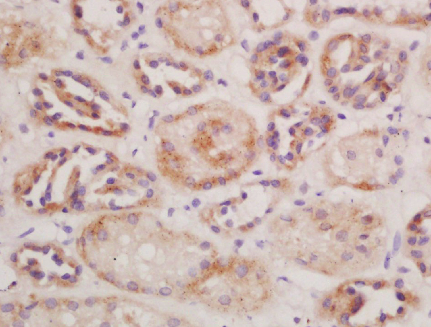 Tissue/cell: mouse kidney tissue; 4% Paraformaldehyde-fixed and paraffin-embedded;
Antigen retrieval: citrate buffer ( 0.01M, pH 6.0 ), Boiling bathing for 15min; Block endogenous peroxidase by 3% Hydrogen peroxide for 30min; Blocking buffer (normal goat serum,C-0005) at 37℃ for 20 min;
Incubation: Anti-CD13/APN/ANPEN Polyclonal Antibody, Unconjugated(SL1383R) 1:200, overnight at 4°C, followed by conjugation to the secondary antibody(SP-0023) and DAB(C-0010) staining
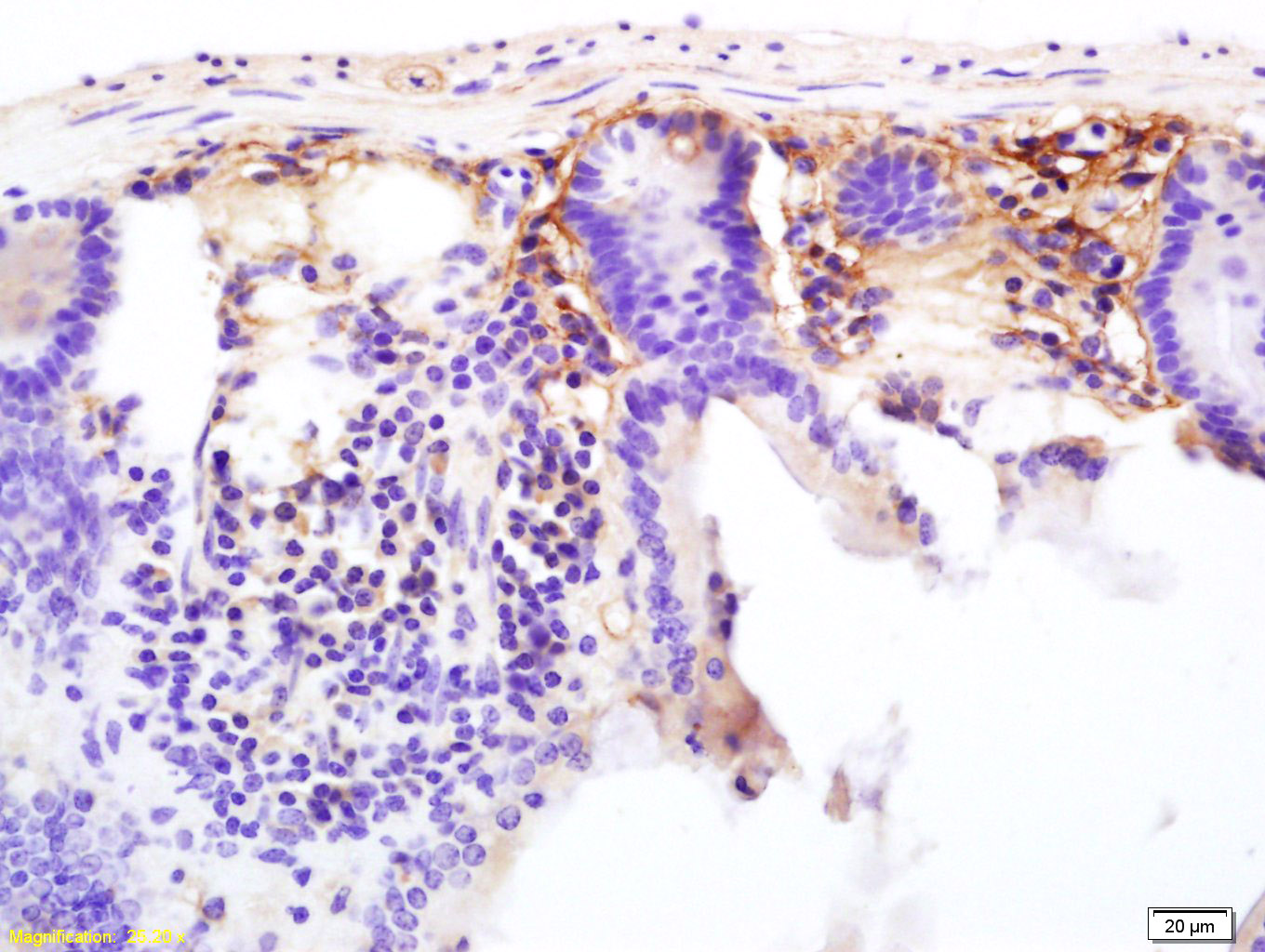 Tissue/cell: mouse colon tissue; 4% Paraformaldehyde-fixed and paraffin-embedded;
Antigen retrieval: citrate buffer ( 0.01M, pH 6.0 ), Boiling bathing for 15min; Block endogenous peroxidase by 3% Hydrogen peroxide for 30min; Blocking buffer (normal goat serum,C-0005) at 37℃ for 20 min;
Incubation: Anti-CD13/APN/ANPEN Polyclonal Antibody, Unconjugated(SL1383R) 1:200, overnight at 4°C, followed by conjugation to the secondary antibody(SP-0023) and DAB(C-0010) staining
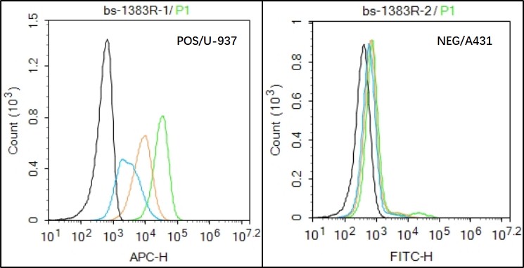 Black line : Positive blank control U937); Negative blank control (A431)
Green line : Primary Antibody (Rabbit Anti- CD13 antibody (SL1383R) )
Orange line:Isotype Control Antibody (Rabbit IgG) .
Blue line : Secondary Antibody (Goat anti-rabbit IgG-AF647)
U937(Positive)and A431 Negative control)cells (black) were incubated in 5% BSA blocking buffer for 30 min at room temperature. Cells were then stained with CD13 Antibody(SL1383R)at 1:100 dilution in blocking buffer and incubated for 30 min at room temperature, washed twice with 2% BSA in PBS, followed by secondary antibody(blue) incubation for 40 min at room temperature. Acquisitions of 20,000 events were performed. Cells stained with primary antibody (green), and isotype control (orange).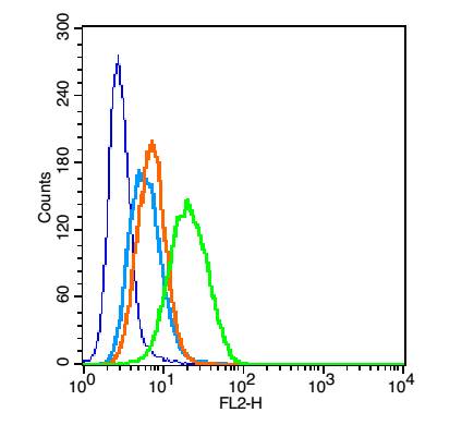 Blank control: U937 (blue).
Primary Antibody:Rabbit Anti- CD13 antibody(SL1383R), Dilution: 1μg in 100 μL 1X PBS containing 0.5% BSA;
Isotype Control Antibody: Rabbit IgG(orange) ,used under the same conditions );
Secondary Antibody: Goat anti-rabbit IgG-PE(white blue), Dilution: 1:200 in 1 X PBS containing 0.5% BSA.
Protocol
The cells were fixed with 2% paraformaldehyde (10 min). Primary antibody (SL1383R, 1μg /1x10^6 cells) were incubated for 30 min on the ice, followed by 1 X PBS containing 0.5% BSA + 1 0% goat serum (15 min) to block non-specific protein-protein interactions. Then the Goat Anti-rabbit IgG/PE antibody was added into the blocking buffer mentioned above to react with the primary antibody at 1/200 dilution for 30 min on ice. Acquisition of 20,000 events was performed.
Cartpieces
Totalgoods,subtotals:¥Checkout
References (0)
No References
Bought notes(bought amounts latest0)
No one bought this product
User Comment(Total0User Comment Num)
- No comment


 +86 571 56623320
 +86 18668110335

