
Rabbit Anti-AKAP12 antibody
A kinase (PRKA) anchor protein (gravin) 12; A kinase Anchor Protein 12; A kinase anchor protein 250kDa; AKAP 12; AKAP 250; AKAP250; DKFZp686M0430; DKFZp686O0331; Gercelin; Gravin; Kinase scaffold protein gravin; Myasthenia gravis autoantigen gravin; Srcs5
View History [Clear]
Details
Product Name AKAP12 Chinese Name 丝氨酸抑制蛋白激酶C底物抗体 Alias A kinase (PRKA) anchor protein (gravin) 12; A kinase Anchor Protein 12; A kinase anchor protein 250kDa; AKAP 12; AKAP 250; AKAP250; DKFZp686M0430; DKFZp686O0331; Gercelin; Gravin; Kinase scaffold protein gravin; Myasthenia gravis autoantigen gravin; Srcs5; SSeCKS; Tsga12. Research Area Neurobiology Signal transduction Kinases and Phosphatases Immunogen Species Rabbit Clonality Polyclonal React Species Human, Mouse, Rat, (predicted: Dog, ) Applications WB=1:500-2000 ELISA=1:5000-10000 IHC-P=1:100-500 IHC-F=1:100-500 IF=1:200-800 (Paraffin sections need antigen repair)
not yet tested in other applications.
optimal dilutions/concentrations should be determined by the end user.Theoretical molecular weight 191kDa Cellular localization cytoplasmic Form Liquid Concentration 1mg/ml immunogen KLH conjugated synthetic peptide derived from human AKAP12: 701-900/1684 Lsotype IgG Purification affinity purified by Protein A Buffer Solution 0.01M TBS(pH7.4) with 1% BSA, 0.03% Proclin300 and 50% Glycerol. Storage Shipped at 4℃. Store at -20 °C for one year. Avoid repeated freeze/thaw cycles. Attention This product as supplied is intended for research use only, not for use in human, therapeutic or diagnostic applications. PubMed PubMed Product Detail The A-kinase anchor proteins (AKAPs) are a group of structurally diverse proteins, which have the common function of binding to the regulatory subunit of protein kinase A (PKA) and confining the holoenzyme to discrete locations within the cell. This gene encodes a member of the AKAP family. The encoded protein is expressed in endothelial cells, cultured fibroblasts, and osteosarcoma cells. It associates with protein kinases A and C and phosphatase, and serves as a scaffold protein in signal transduction. This protein and RII PKA colocalize at the cell periphery. This protein is a cell growth-related protein. Antibodies to this protein can be produced by patients with myasthenia gravis. Alternative splicing of this gene results in two transcript variants encoding different isoforms. [provided by RefSeq, Jul 2008]
Function:
Anchoring protein that mediates the subcellular compartmentation of protein kinase A (PKA) and protein kinase C (PKC).
Subcellular Location:
Cytoplasm
Tissue Specificity:
Expressed in endothelial cells, cultured fibroblasts and osteosarcoma, but not in platelets, leukocytes, monocytic cell lines or peripherical blood cells.
Post-translational modifications:
Phosphorylated upon DNA damage, probably by ATM or ATR.
Similarity:
Contains 3 AKAP domains.
SWISS:
Q02952
Gene ID:
9590
Database links:Entrez Gene: 9590 Human
Omim: 604698 Human
SwissProt: Q02952 Human
Unigene: 197081 Human
Unigene: 371240 Human
Product Picture 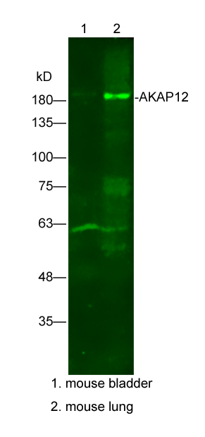 Sample:
Lane1: mouse bladder Lysate at 25 ug
Lane2: mouse lung Lysate at 25 ug
Primary: Anti-AKAP12(SL1381R) at 1/300 dilution
Secondary: IRDye800CW Goat Anti-Rabbit IgG at 1/20000 dilution
Predicted band size: 191kD
Observed band size: 191kD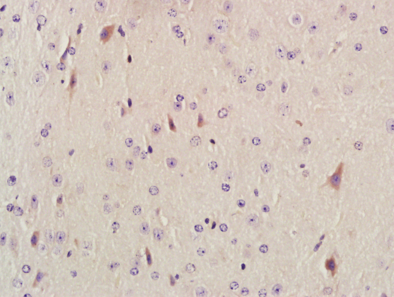 Paraformaldehyde-fixed, paraffin embedded (Mouse brain); Antigen retrieval by boiling in sodium citrate buffer (pH6.0) for 15min; Block endogenous peroxidase by 3% hydrogen peroxide for 20 minutes; Blocking buffer (normal goat serum) at 37°C for 30min; Antibody incubation with (AKAP12) Polyclonal Antibody, Unconjugated (SL1381R) at 1:400 overnight at 4°C, followed by a conjugated secondary antibody (sp-0023) for 20 minutes and DAB staining.
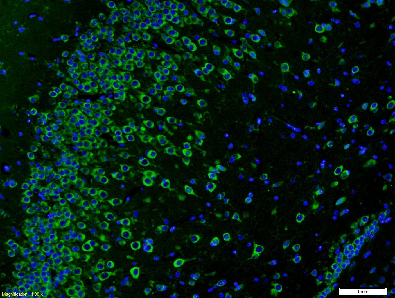 Paraformaldehyde-fixed, paraffin embedded (Mouse brain); Antigen retrieval by boiling in sodium citrate buffer (pH6.0) for 15min; Block endogenous peroxidase by 3% hydrogen peroxide for 20 minutes; Blocking buffer (normal goat serum) at 37°C for 30min; Antibody incubation with (AKAP12) Polyclonal Antibody, Unconjugated (SL1381R) at 1:400 overnight at 4°C, followed by a conjugated secondary antibody (SL0295G-FITC) for 90 minutes, and DAPI for nuclei staining.
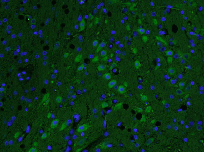 Paraformaldehyde-fixed, paraffin embedded (Rat brain); Antigen retrieval by boiling in sodium citrate buffer (pH6.0) for 15min; Block endogenous peroxidase by 3% hydrogen peroxide for 20 minutes; Blocking buffer (normal goat serum) at 37°C for 30min; Antibody incubation with (AKAP12) Polyclonal Antibody, Unconjugated (SL1381R) at 1:400 overnight at 4°C, followed by a conjugated Goat Anti-Rabbit IgG antibody (SL0295G-FITC) for 90 minutes, and DAPI for nuclei staining.
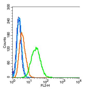 Blank control: RSC 96 (blue).
Primary Antibody:Rabbit Anti-AKAP12 antibody(SL1381R), Dilution: 1μg in 100 μL 1X PBS containing 0.5% BSA;
Isotype Control Antibody: Rabbit IgG(orange) ,used under the same conditions );
Secondary Antibody: Goat anti-rabbit IgG-PE(white blue), Dilution: 1:200 in 1 X PBS containing 0.5% BSA.
Protocol
The cells were fixed with 2% paraformaldehyde (10 min) , then permeabilized with 90% ice-cold methanol for 30 min on ice. Antibody (SL1381R, 1μg /1x10^6 cells) were incubated for 30 min on the ice, followed by 1 X PBS containing 0.5% BSA + 1 0% goat serum (15 min) to block non-specific protein-protein interactions. Then the Goat Anti-rabbit IgG/PE antibody was added into the blocking buffer mentioned above to react with the primary antibody of SL1381R at 1/200 dilution for 30 min on ice. Acquisition of 20,000 events was performed.
Cartpieces
Totalgoods,subtotals:¥Checkout
Bought notes(bought amounts latest0)
No one bought this product
User Comment(Total0User Comment Num)
- No comment


 +86 571 56623320
 +86 18668110335

