
Rabbit Anti-phospho-14-3-3 beta + zeta (Ser186 / Ser184)antibody
14-3-3 beta + zeta (phospho S184 + S186); 14-3-3 beta + zeta (phospho S186); 14-3-3 beta + zeta (phospho Ser186); p-14-3-3 beta + zeta (Ser186) 14 3 3 protein beta; 14 3 3 protein beta/alpha; 14 3 3 protein zeta/delta; 14 3 3 zeta; HS1; KCIP1; YWHAB; YWHA
View History [Clear]
Details
Product Name phospho-14-3-3 beta + zeta (Ser186 / Ser184) Chinese Name 磷酸化14-3-3 β/ζ抗体 Alias 14-3-3 beta + zeta (phospho S184 + S186); 14-3-3 beta + zeta (phospho S186); 14-3-3 beta + zeta (phospho Ser186); p-14-3-3 beta + zeta (Ser186) 14 3 3 protein beta; 14 3 3 protein beta/alpha; 14 3 3 protein zeta/delta; 14 3 3 zeta; HS1; KCIP1; YWHAB; YWHAZ. Product Type Phosphorylated anti Research Area Cell biology Neurobiology Signal transduction Apoptosis Immunogen Species Rabbit Clonality Polyclonal React Species Human, Mouse, Rat, (predicted: Chicken, Dog, Pig, Cow, Horse, ) Applications ELISA=1:5000-10000 IHC-P=1:100-500 IHC-F=1:100-500 Flow-Cyt=0.2μg/Test IF=1:100-500 (Paraffin sections need antigen repair)
not yet tested in other applications.
optimal dilutions/concentrations should be determined by the end user.Theoretical molecular weight 28kDa Cellular localization cytoplasmic The cell membrane Form Liquid Concentration 1mg/ml immunogen KLH conjugated synthesised phosphopeptide derived from human 14-3-3 beta + zeta around the phosphorylation site of Ser186 / Ser184: LN(p-S)PE Lsotype IgG Purification affinity purified by Protein A Buffer Solution 0.01M TBS(pH7.4) with 1% BSA, 0.03% Proclin300 and 50% Glycerol. Storage Shipped at 4℃. Store at -20 °C for one year. Avoid repeated freeze/thaw cycles. Attention This product as supplied is intended for research use only, not for use in human, therapeutic or diagnostic applications. PubMed PubMed Product Detail Members of the 14-3-3 family of proteins are highly conserved proteins, localized in neurons, and are axonally transported to the nerve terminals. They are also present, at lower levels, in various other eukaryotic tissues. 14-3-3 proteins appear to play important roles in a variety of signal transduction pathways, including those involved in cell cycle regulation and cell survival. Because 14-3-3 proteins bind to specific phosphoserine-containing sequences they are likely to have an important role in signaling pathways mediated by serine/threonine protein kinases. Evidence indicates 14-3-3 is required for Raf 1 kinase activity and phosphorylation amoung many other functions.
Function:
14-3-3 proteins are highly conserved proteins which play a role in both signal transduction and progression through the cell cycle by binding to and regulating several different proteins. 14-3-3 proteins activate tyrosine and tryptophan hydroxylases and protein kinase C. They mediate signal transduction by binding to phosphoserine-containing proteins. There are at least 7 mammalian isoforms: alpha, beta, gamma, delta, epsilon, zeta, and eta. An eighth subtype, termed theta has been found in rat brain. The 14-3-3 proteins exists in vitro and in vivo as either homo- or heterodimers which interact via their N-terminal domains and are subject to phosphorylation by protein kinase C. 14-3-3 proteins are localized in the cytoplasm of neurons in the cerebral cortex and are axonally transported to the nerve terminals. They may be present at lower levels in various other eukaryotic tissues. Northern blot analysis has shown expression of the eta chain in cultured cell lines derived from various tumors.
Subunit:
Interacts with CDK16 and BSPRY. Interacts with WEE1 (C-terminal). Interacts with SAMSN1. Interacts with MLF1 (phosphorylated form); the interaction retains it in the cytoplasm. Interacts with Thr-phosphorylated ITGB2. Interacts with BCL2L11. Homodimer. Heterodimerizes with YWHAE. Homo- and hetero-dimerization is inhibited by phosphorylation on Ser-58. Interacts with FOXO4, NOXA1, SSH1 and ARHGEF2. Interacts with Pseudomonas aeruginosa exoS (unphosphorylated form). Interacts with BAX; the interaction occurs in the cytoplasm. Under stress conditions, MAPK8-mediated phosphorylation releases BAX to mitochondria. Interacts with phosphorylated RAF1; the interaction is inhibited when YWHAZ is phosphorylated on Thr-232. Interacts with TP53; the interaction enhances p53 transcriptional activity. The Ser-58 phosphorylated form inhibits this interaction and p53 transcriptional activity. Interacts with ABL1 (phosphorylated form); the interaction retains ABL1 in the cytoplasm. Interacts with PKA-phosphorylated AANAT; the interaction modulates AANAT enzymatic activity by increasing affinity for arylalkylamines and acetyl-CoA and protecting the enzyme from dephosphorylation and proteasomal degradation. It may also prevent thiol-dependent inactivation. Interacts with AKT1; the interaction phosphorylates YWHAZ and modulates dimerization. Interacts with GAB2 and TLK2.
Subcellular Location:
Cytoplasmic. Melanosome (fractions from stage I to stage IV)
Post-translational modifications:
The delta, brain-specific form differs from the zeta form in being phosphorylated (By similarity). Phosphorylation on Ser-184 by MAPK8; promotes dissociation of BAX and translocation of BAX to mitochondria. Phosphorylation on Ser-58 by PKA; disrupts homodimerization and heterodimerization with YHAE and TP53. This phosphorylation appears to be activated by sphingosine. Phosphorylation on Thr-232; inhibits binding of RAF1.
Similarity:
Belongs to the 14-3-3 family.
SWISS:
P31946
Gene ID:
7529
Database links:Entrez Gene: 7529 Human
Entrez Gene: 7534 Human
Entrez Gene: 22631 Mouse
Entrez Gene: 54401 Mouse
Omim: 601288 Human
Omim: 601289 Human
SwissProt: P31946 Human
SwissProt: P63104 Human
SwissProt: P35215 Mouse
SwissProt: Q9CQV8 Mouse
Unigene: 492407 Human
Unigene: 643544 Human
Unigene: 1292 Rat
Unigene: 8653 Rat
Product Picture 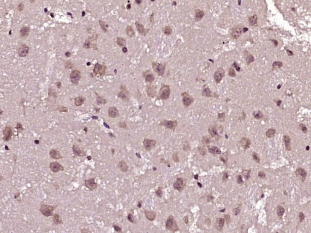 Paraformaldehyde-fixed, paraffin embedded (mouse brain tissue); Antigen retrieval by boiling in sodium citrate buffer (pH6.0) for 15min; Block endogenous peroxidase by 3% hydrogen peroxide for 20 minutes; Blocking buffer (normal goat serum) at 37°C for 30min; Antibody incubation with (YWHAB) Polyclonal Antibody, Unconjugated (SL13773R) at 1:400 overnight at 4°C, followed by operating according to SP Kit(Rabbit) (sp-0023) instructionsand DAB staining.
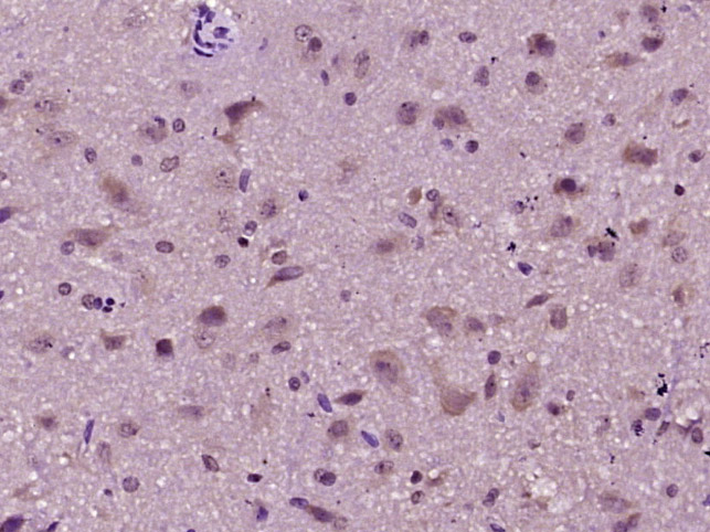 Paraformaldehyde-fixed, paraffin embedded (rat brain tissue); Antigen retrieval by boiling in sodium citrate buffer (pH6.0) for 15min; Block endogenous peroxidase by 3% hydrogen peroxide for 20 minutes; Blocking buffer (normal goat serum) at 37°C for 30min; Antibody incubation with (YWHAB) Polyclonal Antibody, Unconjugated (SL13773R) at 1:400 overnight at 4°C, followed by operating according to SP Kit(Rabbit) (sp-0023) instructionsand DAB staining.
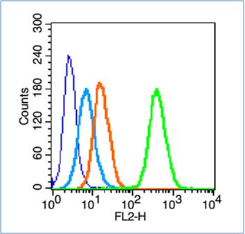 Blank control (blue line): HL60 (fixed with 70% ethanol (overninght at 4℃) and then permeabilized with 0.1% PBS-Tween for 20 min at room temperature).
Primary Antibody (green line): Rabbit Anti-phospho-14-3-3 beta + zeta (Ser186) antibody (SL13773R),Dilution: 0.2μg /10^6 cells;
Isotype Control Antibody (orange line): Rabbit IgG .
Secondary Antibody (white blue line): Goat anti-rabbit IgG-PE,Dilution: 1μg /test.
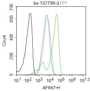 Blank control:A431.
Primary Antibody (green line): Rabbit Anti-phospho-14-3-3 beta+zeta (Ser186/Ser184) antibody (SL13773R)
Dilution: 2μg /10^6 cells;
Isotype Control Antibody (orange line): Rabbit IgG .
Secondary Antibody : Goat anti-rabbit IgG-AF647
Dilution: 1μg /test.
Protocol
The cells were fixed with 4% PFA (10min at room temperature)and then permeabilized with 0.1% PBST for 20 min at room temperature. The cells were then incubated in 5%BSA to block non-specific protein-protein interactions for 30 min at room temperature .Cells stained with Primary Antibody for 30 min at room temperature. The secondary antibody used for 40 min at room temperature. Acquisition of 20,000 events was performed.
Cartpieces
Totalgoods,subtotals:¥Checkout
Bought notes(bought amounts latest0)
No one bought this product
User Comment(Total0User Comment Num)
- No comment


 +86 571 56623320
 +86 18668110335

