
Rabbit Anti-MAP2 antibody
Microtubule-associated protein 2; DKFZp686I2148; Dendrite specific MAP; DKFZp686I2148; MAP 2; MAP-2; MAP2A; MAP2B; MAP2C; Microtubule associated protein 2; Mtap 2; MAP2_HUMAN.
View History [Clear]
Details
Product Name MAP2 Chinese Name 微管相关蛋白2抗体 Alias Microtubule-associated protein 2; DKFZp686I2148; Dendrite specific MAP; DKFZp686I2148; MAP 2; MAP-2; MAP2A; MAP2B; MAP2C; Microtubule associated protein 2; Mtap 2; MAP2_HUMAN. literatures Research Area Tumour Cell biology immunology Neurobiology Signal transduction Apoptosis Cytoskeleton Immunogen Species Rabbit Clonality Polyclonal React Species Human, Mouse, Rat, Applications WB=1:500-2000 ELISA=1:5000-10000 IHC-P=1:100-500 IHC-F=1:100-500 Flow-Cyt=1ug/Test IF=1:200-800 (Paraffin sections need antigen repair)
not yet tested in other applications.
optimal dilutions/concentrations should be determined by the end user.Theoretical molecular weight 70/201kDa Cellular localization cytoplasmic Form Liquid Concentration 1mg/ml immunogen KLH conjugated synthetic peptide derived from human MAP2: 1-120/1827 Lsotype IgG Purification affinity purified by Protein A Buffer Solution 0.01M TBS(pH7.4) with 1% BSA, 0.03% Proclin300 and 50% Glycerol. Storage Shipped at 4℃. Store at -20 °C for one year. Avoid repeated freeze/thaw cycles. Attention This product as supplied is intended for research use only, not for use in human, therapeutic or diagnostic applications. PubMed PubMed Product Detail MAP2 is the major microtubule associated protein of brain tissue. There are three forms of MAP2; two are similarily sized with apparent molecular weights of 280 kDa (MAP2a and MAP2b) and the third with a lower molecular weight of 70 kDa (MAP2c). In the newborn rat brain, MAP2b and MAP2c are present, while MAP2a is absent. Between postnatal days 10 and 20, MAP2a appears. At the same time, the level of MAP2c drops by 10-fold. This change happens during the period when dendrite growth is completed and when neurons have reached their mature morphology. MAP2 is degraded by a Cathepsin D-like protease in the brain of aged rats. There is some indication that MAP2 is expressed at higher levels in some types of neurons than in other types. MAP2 is known to promote microtubule assembly and to form side-arms on microtubules. It also interacts with neurofilaments, actin, and other elements of the cytoskeleton.
Function:
The exact function of MAP2 is unknown but MAPs may stabilize the microtubules against depolymerization. They also seem to have a stiffening effect on microtubules.
Subcellular Location:
Cytoplasm, cytoskeleton (Probable).
Post-translational modifications:
Phosphorylated at serine residues in K-X-G-S motifs by MAP/microtubule affinity-regulating kinase (MARK1 or MARK2), causing detachment from microtubules, and their disassembly. MAP2A/c is phosphorylated. Phosphorylated upon DNA damage, probably by ATM or ATR. Isoform MAP2c is phosphorylated by FYN at Tyr-67.
Similarity:
Contains 3 Tau/MAP repeats.
SWISS:
P11137
Gene ID:
4133
Database links:Entrez Gene: 4133 Human
Omim: 157130 Human
SwissProt: P11137 Human
SwissProt: P20357 Mouse
Unigene: 368281 Human
Unigene: 256966 Mouse
微管相关蛋白 2(MAP-2)是一种磷蛋白质,在正常脑组织中,主要存在于神经元的胞体、树突和树突棘,是脑内最丰富的蛋白之一。MAP-2在其调节微管的聚合作用和微管的稳定性以及对神经元轴突和树突的发生、延长、稳定和突触可塑性调节具有重要作用。Product Picture 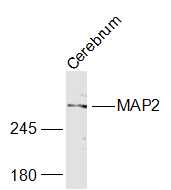 Sample:
Cerebrum (Mouse) Lysate at 40 ug
Primary: Anti-MAP2 (SL1369R) at 1/1000 dilution
Secondary: IRDye800CW Goat Anti-Rabbit IgG at 1/20000 dilution
Predicted band size: 201 kD
Observed band size: 280 kD
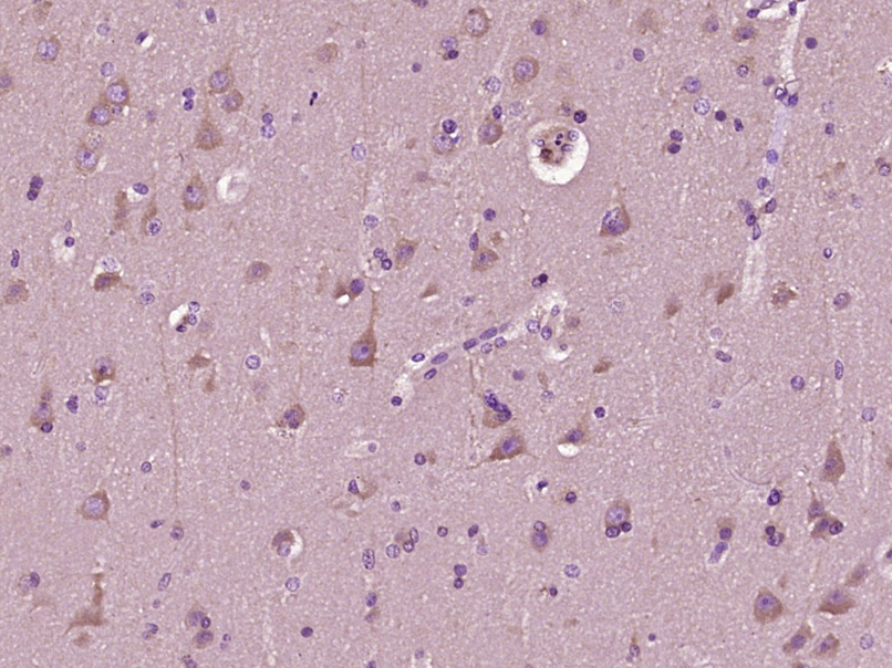 Paraformaldehyde-fixed, paraffin embedded (Human brain glioma); Antigen retrieval by boiling in sodium citrate buffer (pH6.0) for 15min; Block endogenous peroxidase by 3% hydrogen peroxide for 20 minutes; Blocking buffer (normal goat serum) at 37°C for 30min; Antibody incubation with (MAP2) Polyclonal Antibody, Unconjugated (SL1369R) at 1:400 overnight at 4°C, followed by operating according to SP Kit(Rabbit) (sp-0023) instructionsand DAB staining.
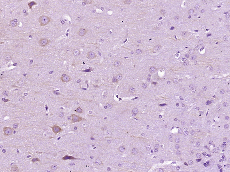 Paraformaldehyde-fixed, paraffin embedded (Rat brain); Antigen retrieval by boiling in sodium citrate buffer (pH6.0) for 15min; Block endogenous peroxidase by 3% hydrogen peroxide for 20 minutes; Blocking buffer (normal goat serum) at 37°C for 30min; Antibody incubation with (MAP2) Polyclonal Antibody, Unconjugated (SL1369R) at 1:400 overnight at 4°C, followed by operating according to SP Kit(Rabbit) (sp-0023) instructionsand DAB staining.
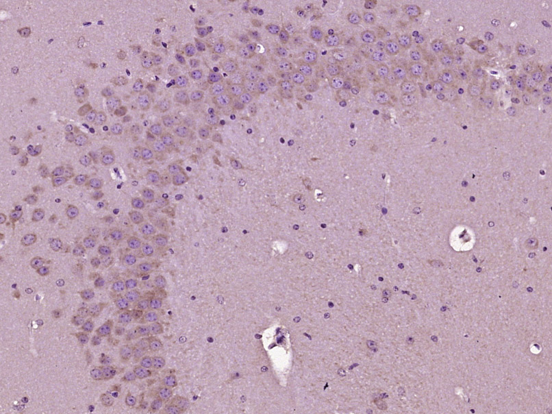 Paraformaldehyde-fixed, paraffin embedded (Mouse brain); Antigen retrieval by boiling in sodium citrate buffer (pH6.0) for 15min; Block endogenous peroxidase by 3% hydrogen peroxide for 20 minutes; Blocking buffer (normal goat serum) at 37°C for 30min; Antibody incubation with (MAP2) Polyclonal Antibody, Unconjugated (SL1369R) at 1:400 overnight at 4°C, followed by operating according to SP Kit(Rabbit) (sp-0023) instructionsand DAB staining.
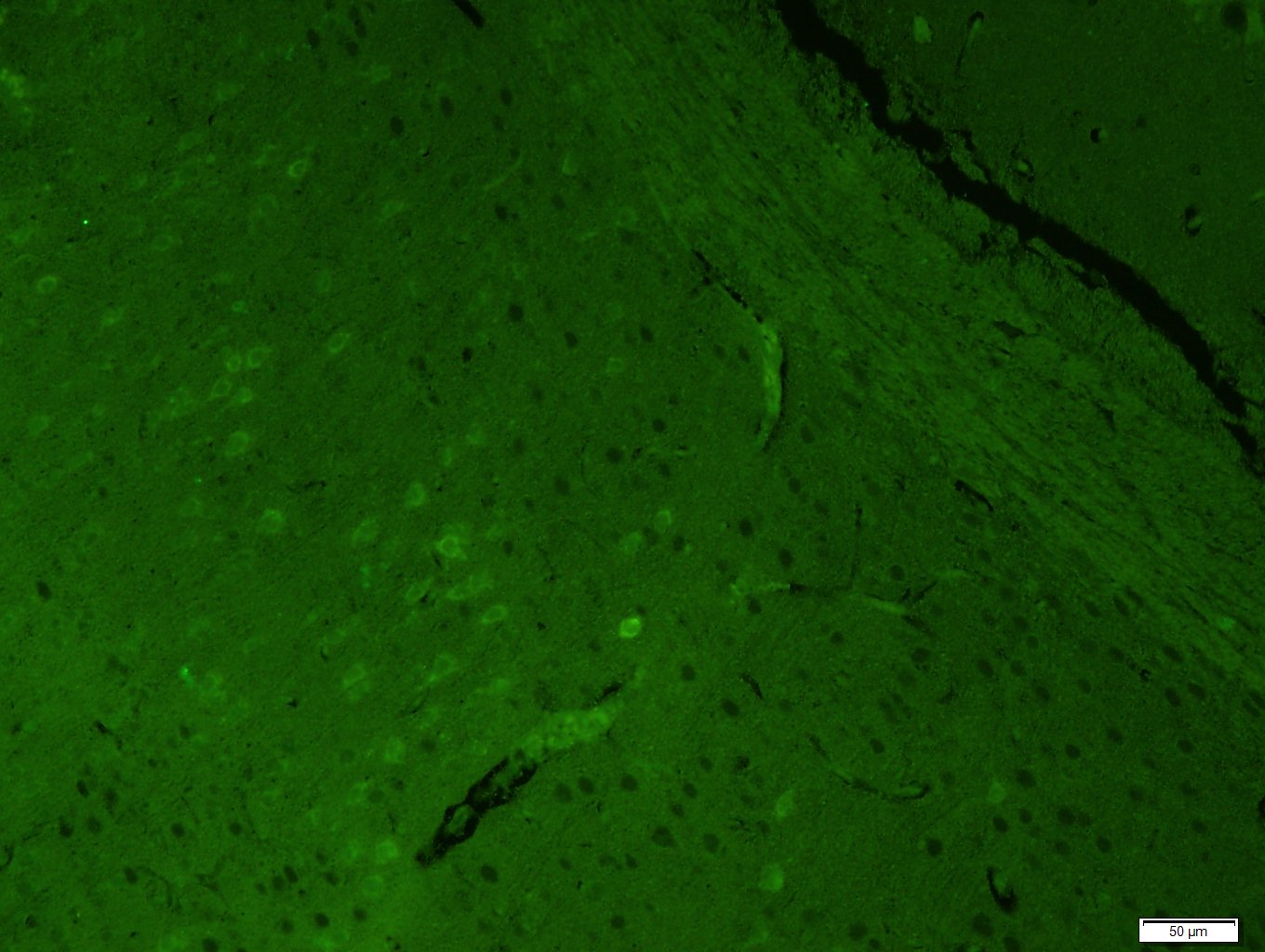 Paraformaldehyde-fixed, paraffin embedded (Mouse brain); Antigen retrieval by boiling in sodium citrate buffer (pH6.0) for 15min; Block endogenous peroxidase by 3% hydrogen peroxide for 20 minutes; Blocking buffer (normal goat serum) at 37°C for 30min; Antibody incubation with (MAP2) Polyclonal Antibody, Unconjugated (SL1369R) at 1:400 overnight at 4°C, followed by a conjugated Goat Anti-Rabbit IgG antibody (SL0295G-FITC) for 90 minutes, and DAPI for nuclei staining.
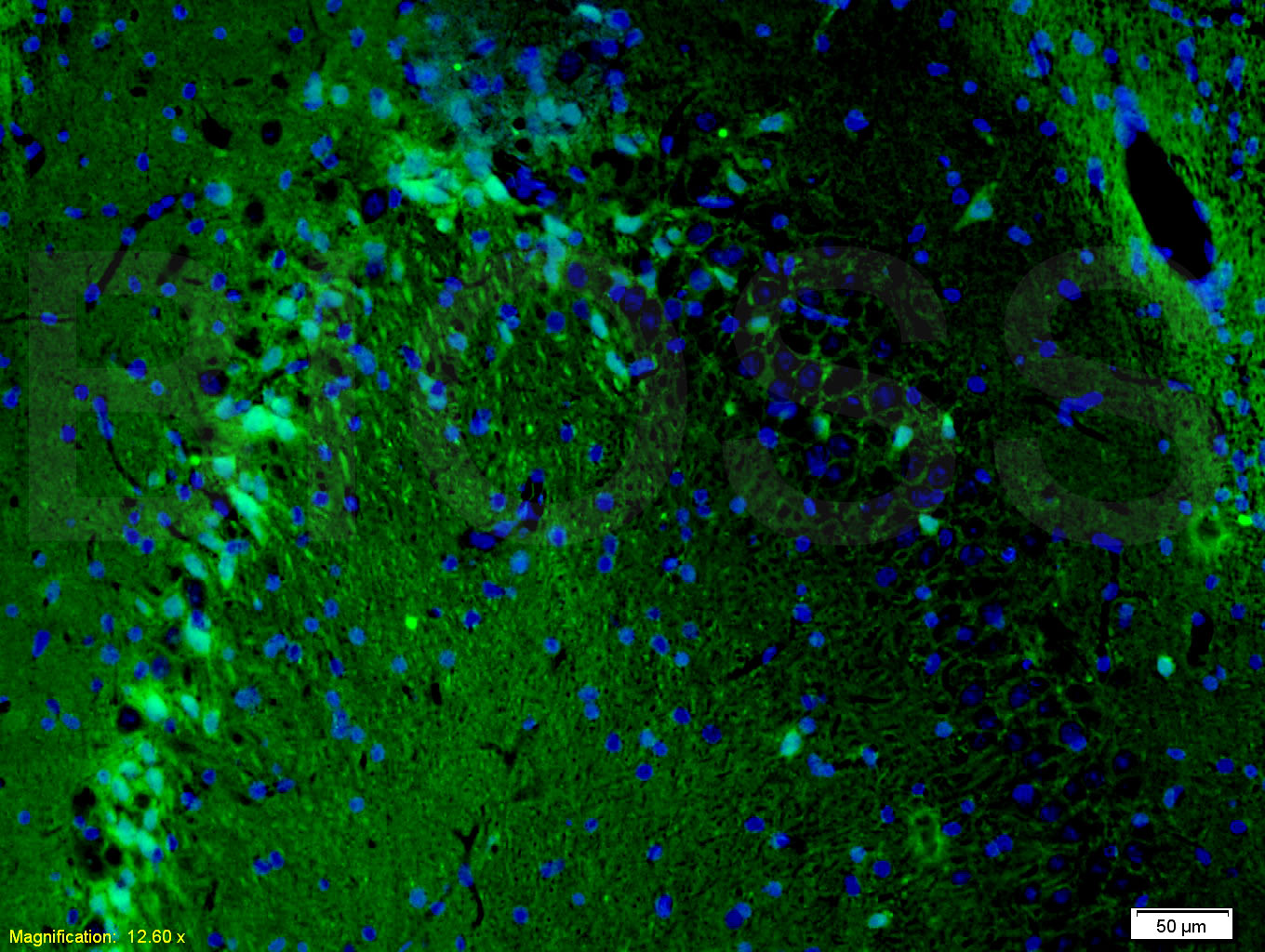 Tissue/cell: rat brain tissue;4% Paraformaldehyde-fixed and paraffin-embedded;
Antigen retrieval: citrate buffer ( 0.01M, pH 6.0 ), Boiling bathing for 15min; Blocking buffer (normal goat serum,C-0005) at 37℃ for 20 min;
Incubation: Anti-MAP2/MAP-2a.b.c Polyclonal Antibody, Unconjugated(SL1369R) 1:200, overnight at 4°C; The secondary antibody was Goat Anti-Rabbit IgG, FITC conjugated (SL0295G-FITC)used at 1:200 dilution for 40 minutes at 37°C. DAPI(5ug/ml,blue,C-0033) was used to stain the cell nuclei
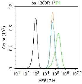 Blank control: SH-SY5Y.
Primary Antibody (green line): Rabbit Anti-MAP2 antibody (SL1369R)
Dilution: 1μg /10^6 cells;
Isotype Control Antibody (orange line): Rabbit IgG .
Secondary Antibody : Goat anti-rabbit IgG-AF647
Dilution: 1μg /test.
Protocol
The cells were fixed with 4% PFA (10min at room temperature)and then permeabilized with 90% ice-cold methanol for 20 min at-20℃.The cells were then incubated in 5%BSA to block non-specific protein-protein interactions for 30 min at room temperature .Cells stained with Primary Antibody for 30 min at room temperature. The secondary antibody used for 40 min at room temperature. Acquisition of 20,000 events was performed.
Cartpieces
Totalgoods,subtotals:¥Checkout
Bought notes(bought amounts latest0)
No one bought this product
User Comment(Total0User Comment Num)
- No comment


 +86 571 56623320
 +86 18668110335

