
Rabbit Anti-VHL antibody
VHL_HUMAN; von Hippel-Lindau disease tumor suppressor; Protein G7; pVHL; RCA1; VHL1; HRCA1;
View History [Clear]
Details
Product Name VHL Chinese Name VHL抗体 Alias VHL_HUMAN; von Hippel-Lindau disease tumor suppressor; Protein G7; pVHL; RCA1; VHL1; HRCA1; Research Area Tumour Cell biology immunology Chromatin and nuclear signals transcriptional regulatory factor Cell differentiation The new supersedes the old Epigenetics Ubiquitin Immunogen Species Rabbit Clonality Polyclonal React Species Human, Rat, (predicted: Mouse, Dog, Cow, ) Applications ELISA=1:5000-10000 IHC-P=1:100-500 IHC-F=1:100-500 Flow-Cyt=1ug/test IF=1:100-500 (Paraffin sections need antigen repair)
not yet tested in other applications.
optimal dilutions/concentrations should be determined by the end user.Theoretical molecular weight 24kDa Cellular localization The nucleus cytoplasmic The cell membrane Form Liquid Concentration 1mg/ml immunogen KLH conjugated synthetic peptide derived from human VHL: 101-213/213 Lsotype IgG Purification affinity purified by Protein A Buffer Solution 0.01M TBS(pH7.4) with 1% BSA, 0.03% Proclin300 and 50% Glycerol. Storage Shipped at 4℃. Store at -20 °C for one year. Avoid repeated freeze/thaw cycles. Attention This product as supplied is intended for research use only, not for use in human, therapeutic or diagnostic applications. PubMed PubMed Product Detail This gene encodes a component of a ubiquitination complex. The encoded protein is involved in the ubiquitination and degradation of hypoxia-inducible-factor (HIF), which is a transcription factor that plays a central role in the regulation of gene expression by oxygen. In addition to oxygen-related gene expression, this protein plays a role in many other cellular processes including cilia formation, cytokine signaling, regulation of senescence, and formation of the extracellular matrix. Variants of this gene are associated with von Hippel-Lindau syndrome, pheochromocytoma, erythrocytosis, renal cell carcinoma, and cerebellar hemangioblastoma. [provided by RefSeq, Jun 2022]
Function:
Involved in the ubiquitination and subsequent proteasomal degradation via the von Hippel-Lindau ubiquitination complex. Seems to act as target recruitment subunit in the E3 ubiquitin ligase complex and recruits hydroxylated hypoxia-inducible factor (HIF) under normoxic conditions. Involved in transcriptional repression through interaction with HIF1A, HIF1AN and histone deacetylases. Ubiquitinates, in an oxygen-responsive manner, ADRB2.
Subunit:
Component of the VCB (VHL-Elongin BC-CUL2) complex; this complex acts as a ubiquitin-ligase E3 and directs proteasome-dependent degradation of targeted proteins. Interacts with CUL2; this interaction is dependent on the integrity of the trimeric VBC complex. Interacts (via the beta domain) with HIF1A (via the NTAD domain); this interaction mediates degradation of HIF1A in normoxia and, in hypoxia, prevents ubiqitination and degradation of HIF1A by mediating hypoxia-induced translocation to the nucleus, a process which requires a hypoxia-dependent regulatory signal. Interacts with ADRB2; the interaction, in normoxia, is dependent on hydroxylation of ADRB2 and the subsequent VCB-mediated ubiquitination and degradation of ADRB2. Under hypoxia, hydroxylation, interaction with VHL, ubiquitination and subsequent degradation of ADRB2 are dramatically decreased. Interacts with RNF139, USP33 and PHF17.
Subcellular Location:
Isoform 1: Cytoplasm. Membrane; Peripheral membrane protein. Nucleus. Note=Found predominantly in the cytoplasm and with less amounts nuclear or membrane-associated. Colocalizes with ADRB2 at the cell membrane.
Isoform 3: Cytoplasm. Nucleus. Note=Equally distributed between the nucleus and the cytoplasm but not membrane-associated.
Tissue Specificity:
Expressed in the adult and fetal brain and kidney.
DISEASE:
Defects in VHL are a cause of susceptibility to pheochromocytoma (PCC) [MIM:171300]. A catecholamine-producing tumor of chromaffin tissue of the adrenal medulla or sympathetic paraganglia. The cardinal symptom, reflecting the increased secretion of epinephrine and norepinephrine, is hypertension, which may be persistent or intermittent.
Defects in VHL are the cause of von Hippel-Lindau disease (VHLD) [MIM:193300]. VHLD is a dominantly inherited familial cancer syndrome characterized by the development of retinal angiomatosis, cerebellar and spinal hemangioblastoma, renal cell carcinoma (RCC), phaeochromocytoma and pancreatic tumors. VHL type 1 is without pheochromocytoma, type 2 is with pheochromocytoma. VHL type 2 is further subdivided into types 2A (pheochromocytoma, retinal angioma, and hemangioblastomas without renal cell carcinoma and pancreatic cyst) and 2B (pheochromocytoma, retinal angioma, and hemangioblastomas with renal cell carcinoma and pancreatic cyst). VHL type 2C refers to patients with isolated pheochromocytoma without hemangioblastoma or renal cell carcinoma. The estimated incidence is 3/100000 births per year and penetrance is 97% by age 60 years.
Defects in VHL are the cause of familial erythrocytosis type 2 (ECYT2) [MIM:263400]; also called VHL-dependent polycythemia or Chuvash type polycythemia. ECYT2 is an autosomal recessive disorder characterized by an increase in serum red blood cell mass, hypersensitivity of erythroid progenitors to erythropoietin, increased erythropoietin serum levels, and normal oxygen affinity. Patients with ECYT2 carry a high risk for peripheral thrombosis and cerebrovascular events.
Defects in VHL are a cause of renal cell carcinoma (RCC) [MIM:144700]. Renal cell carcinoma is a heterogeneous group of sporadic or hereditary carcinoma derived from cells of the proximal renal tubular epithelium. It is subclassified into clear cell renal carcinoma (non-papillary carcinoma), papillary renal cell carcinoma, chromophobe renal cell carcinoma, collecting duct carcinoma with medullary carcinoma of the kidney, and unclassified renal cell carcinoma.
SWISS:
P40337
Gene ID:
7428
Database links:Entrez Gene: 7428 Human
Entrez Gene: 22346 Mouse
Omim: 608537 Human
SwissProt: P40337 Human
SwissProt: P40338 Mouse
Unigene: 517792 Human
Unigene: 607789 Human
Unigene: 29407 Mouse
von Hippel-Lindau是一种Tumour抑制因子,在细胞对氧的感受过程中发挥关键作用,VHL蛋白除了调节血管生成外还在调节细胞的生长和生存、对调节细胞周期、Apoptosis和Extracellular matrix方面起重要作用。Product Picture 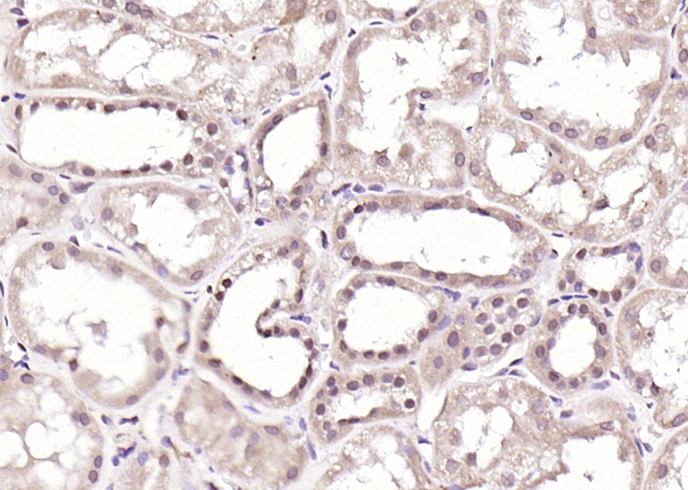 Paraformaldehyde-fixed, paraffin embedded (Human kidney); Antigen retrieval by boiling in sodium citrate buffer (pH6.0) for 15min; Block endogenous peroxidase by 3% hydrogen peroxide for 20 minutes; Blocking buffer (normal goat serum) at 37°C for 30min; Antibody incubation with (VHL) Polyclonal Antibody, Unconjugated (SL1367R) at 1:200 overnight at 4°C, followed by operating according to SP Kit(Rabbit) (sp-0023) instructionsand DAB staining.
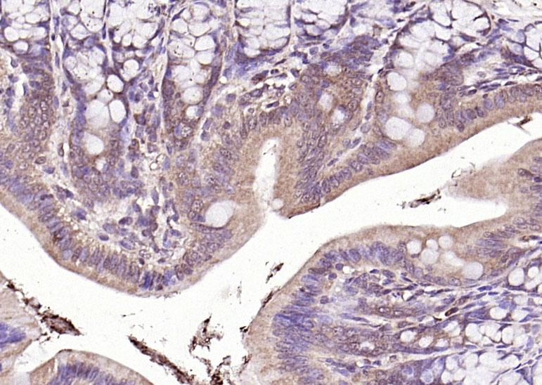 Paraformaldehyde-fixed, paraffin embedded (rat colon); Antigen retrieval by boiling in sodium citrate buffer (pH6.0) for 15min; Block endogenous peroxidase by 3% hydrogen peroxide for 20 minutes; Blocking buffer (normal goat serum) at 37°C for 30min; Antibody incubation with (VHL) Polyclonal Antibody, Unconjugated (SL1367R) at 1:200 overnight at 4°C, followed by operating according to SP Kit(Rabbit) (sp-0023) instructionsand DAB staining.
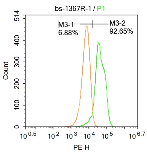 Blank control:Molt-4.
Primary Antibody (green line): Rabbit Anti-VHL antibody (SL1367R)
Dilution: 1μg /10^6 cells;
Isotype Control Antibody (orange line): Rabbit IgG .
Secondary Antibody : Goat anti-rabbit IgG-AF647
Dilution: 1μg /test.
Protocol
The cells were fixed with 4% PFA (10min at room temperature)and then permeabilized with 90% ice-cold methanol for 20 min at-20℃. The cells were then incubated in 5%BSA to block non-specific protein-protein interactions for 30 min at at room temperature .Cells stained with Primary Antibody for 30 min at room temperature. The secondary antibody used for 40 min at room temperature. Acquisition of 20,000 events was performed.
Cartpieces
Totalgoods,subtotals:¥Checkout
References (0)
No References
Bought notes(bought amounts latest0)
No one bought this product
User Comment(Total0User Comment Num)
- No comment


 +86 571 56623320
 +86 18668110335

