
Rabbit Anti-P73 antibody
P73_HUMAN; Tumor protein p73; p53-like transcription factor; p53-related protein; TP73; p53 Related Protein; CILD47;
View History [Clear]
Details
Product Name P73 Chinese Name Tumour蛋白P73抗体 Alias P73_HUMAN; Tumor protein p73; p53-like transcription factor; p53-related protein; TP73; p53 Related Protein; CILD47; literatures Research Area Tumour Cell biology immunology Signal transduction Apoptosis transcriptional regulatory factor Immunogen Species Rabbit Clonality Polyclonal React Species Human, Mouse, Rat, Applications ELISA=1:5000-10000 IHC-P=1:100-500 IHC-F=1:100-500 ICC=1:100 IF=1:100-500 (Paraffin sections need antigen repair)
not yet tested in other applications.
optimal dilutions/concentrations should be determined by the end user.Theoretical molecular weight 70kDa Cellular localization The nucleus cytoplasmic Form Liquid Concentration 1mg/ml immunogen KLH conjugated synthetic peptide derived from mouse P73 protein: 501-631/631 Lsotype IgG Purification affinity purified by Protein A Buffer Solution 0.01M TBS(pH7.4) with 1% BSA, 0.03% Proclin300 and 50% Glycerol. Storage Shipped at 4℃. Store at -20 °C for one year. Avoid repeated freeze/thaw cycles. Attention This product as supplied is intended for research use only, not for use in human, therapeutic or diagnostic applications. PubMed PubMed Product Detail P73 protein is a structural and functional homologue of p53, a tumor suppressor gene. In this study, The p73 protein, p19ras, by the yeast two-hybrid screening method. Alternative splicing of the proto-oncogene H-ras pre-mRNA has led to two distinct transcripts, Ras proteins are known to be small membrane-localized guanine nucleotide-binding proteins. However, unlike other Ras proteins, p19ras is localized in the nucleus and the cytosol and its interaction with P73 protein occurred exclusively in the nucleus. Oncogenic MDM2 (mouse double minutes 2) is a known repressor of p73 transcriptional activity. In this study, when p19ras was bound to MDM2, it further inhibited the association of MDM2 to the p73 protein. Therefore, this study presents a novel pathway of Ras signaling that occurs in the nucleus, involving p19ras and p73.
Function:
Participates in the apoptotic response to DNA damage. Isoforms containing the transactivation domain are pro-apoptotic, isoforms lacking the domain are anti-apoptotic and block the function of p53 and transactivating p73 isoforms. May be a tumor suppressor protein. [COFACTOR] Binds 1 zinc ion per subunit
Subunit:
Found in a complex with p53/TP53 and CABLES1. The C-terminal oligomerization domain binds to the ABL1 tyrosine kinase SH3 domain. Interacts with HECW2. Isoform Beta interacts homotypically and with p53/TP53, whereas isoform Alpha does not. Isoform Gamma interacts homotypically and with all p73 isoforms. Isoform Delta interacts with isoform Gamma, isoform Alpha, and homotypically. Isoforms Alpha and Beta interact with HIPK2. Isoform Alpha interacts with RANBP9. Isoform Beta interacts with WWOX. Interacts (via SAM domain) with FBXO45 (via B30.2/SPRY domain). Interacts with YAP1 (phosphorylated form). Interacts with HCK (via SH3 domain); this inhibits TP73 activity and degradation.
Subcellular Location:
Nucleus. Cytoplasm. Note=Accumulates in the nucleus in response to DNA damage.
Tissue Specificity:
Expressed in striatal neurons of patients with Huntington disease (at protein level). Brain, kidney, placenta, colon, heart, liver, spleen, skeletal muscle, prostate, thymus and pancreas. Highly expressed in fetal tissue.
Post-translational modifications:
Isoform alpha (but not isoform beta) is sumoylated on Lys-627, which potentiates proteasomal degradation but does not affect transcriptional activity. Phosphorylation by PLK1 and PLK3 inhibits the transcription regulator activity and pro-apoptotic function.
Higher levels of phosphorylation seen in the brain from patients with Huntington disease.
Polyubiquitinated by RCHY1/PIRH2; leading to its degradation by the proteasome.
Similarity:
Belongs to the p53 family.
Contains 1 SAM (sterile alpha motif) domain.
SWISS:
Q9JJP2
Gene ID:
22062
Database links:Entrez Gene: 7161 Human
Entrez Gene: 22062 Mouse
Omim: 601990 Human
SwissProt: O15350 Human
SwissProt: Q9JJP2 Mouse
Unigene: 697294 Human
Unigene: 706990 Human
Unigene: 78015 Mouse
P73是p53基因家族的第一个成员p53基因是一个经典的抑癌基因,与p53在结构和功能方面具有很高的相似性。Product Picture 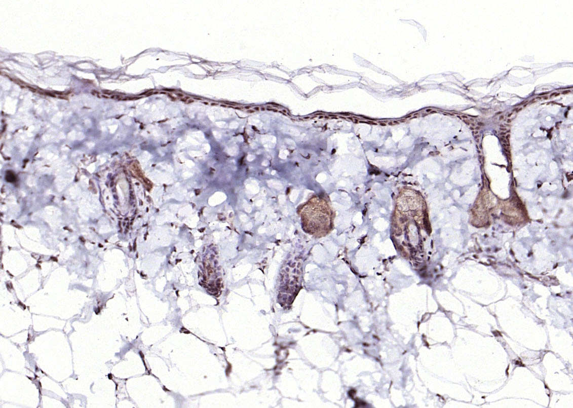 Paraformaldehyde-fixed, paraffin embedded (rat skin); Antigen retrieval by boiling in sodium citrate buffer (pH6.0) for 15min; Block endogenous peroxidase by 3% hydrogen peroxide for 20 minutes; Blocking buffer (normal goat serum) at 37°C for 30min; Antibody incubation with (P73) Polyclonal Antibody, Unconjugated (SL1346R) at 1:200 overnight at 4°C, followed by operating according to SP Kit(Rabbit) (sp-0023) instructionsand DAB staining.
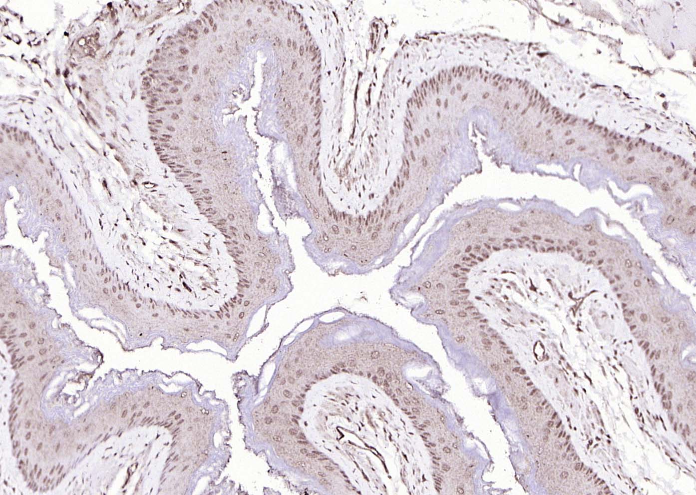 Paraformaldehyde-fixed, paraffin embedded (mouse esophagus); Antigen retrieval by boiling in sodium citrate buffer (pH6.0) for 15min; Block endogenous peroxidase by 3% hydrogen peroxide for 20 minutes; Blocking buffer (normal goat serum) at 37°C for 30min; Antibody incubation with (P73) Polyclonal Antibody, Unconjugated (SL1346R) at 1:200 overnight at 4°C, followed by operating according to SP Kit(Rabbit) (sp-0023) instructionsand DAB staining.
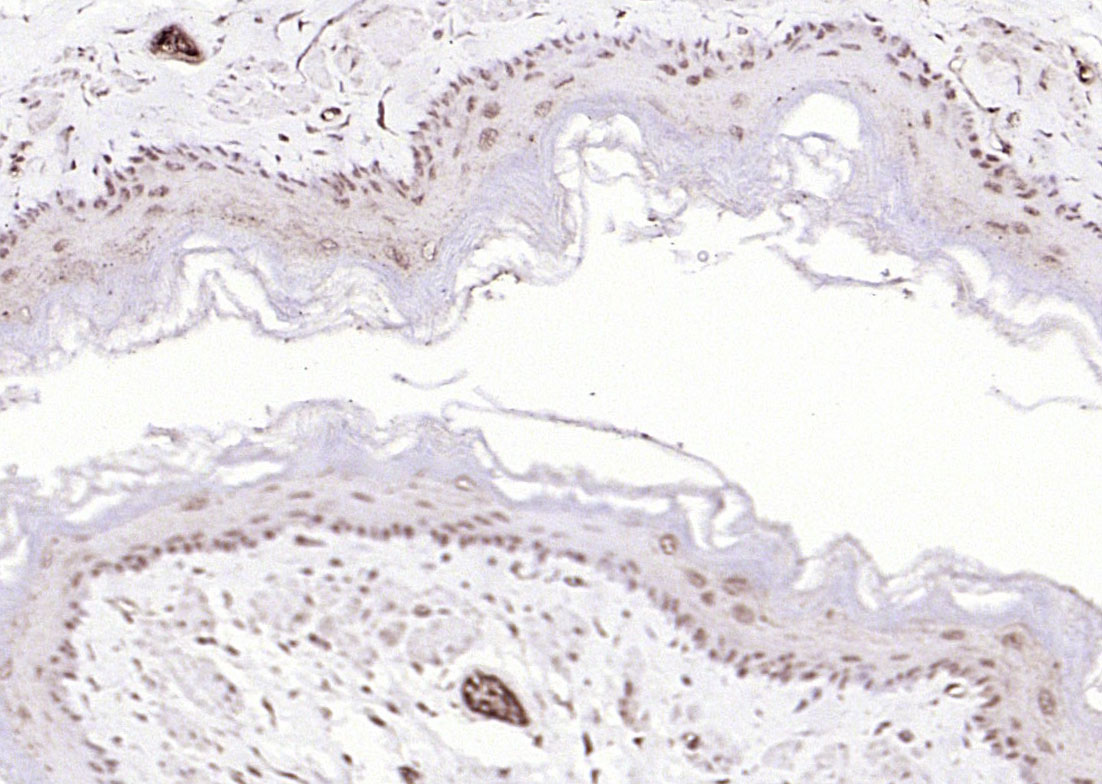 Paraformaldehyde-fixed, paraffin embedded (Rat esophagus); Antigen retrieval by boiling in sodium citrate buffer (pH6.0) for 15min; Block endogenous peroxidase by 3% hydrogen peroxide for 20 minutes; Blocking buffer (normal goat serum) at 37°C for 30min; Antibody incubation with (P73) Polyclonal Antibody, Unconjugated (SL1346R) at 1:200 overnight at 4°C, followed by operating according to SP Kit(Rabbit) (sp-0023) instructionsand DAB staining.
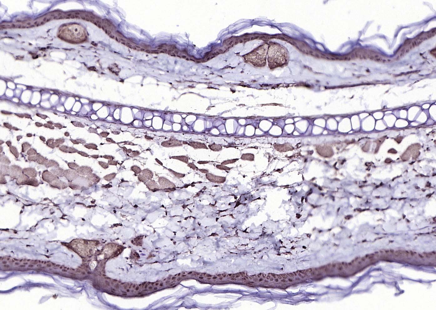 Paraformaldehyde-fixed, paraffin embedded (mouse skin); Antigen retrieval by boiling in sodium citrate buffer (pH6.0) for 15min; Block endogenous peroxidase by 3% hydrogen peroxide for 20 minutes; Blocking buffer (normal goat serum) at 37°C for 30min; Antibody incubation with (P73) Polyclonal Antibody, Unconjugated (SL1346R) at 1:200 overnight at 4°C, followed by operating according to SP Kit(Rabbit) (sp-0023) instructionsand DAB staining.
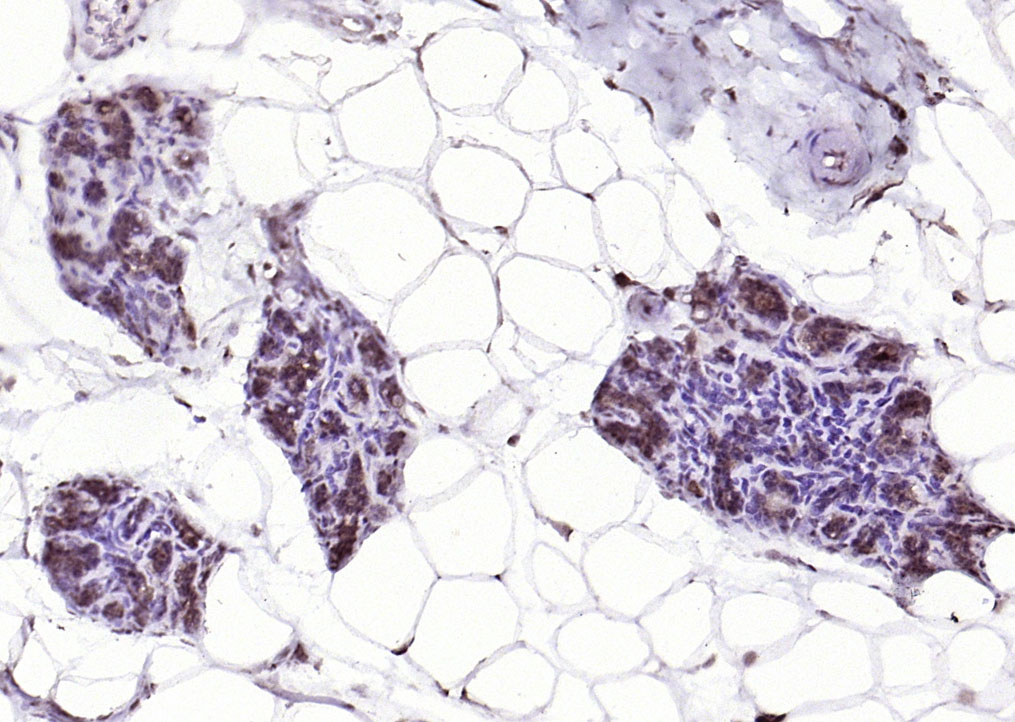 Paraformaldehyde-fixed, paraffin embedded (Rat mammary gland); Antigen retrieval by boiling in sodium citrate buffer (pH6.0) for 15min; Block endogenous peroxidase by 3% hydrogen peroxide for 20 minutes; Blocking buffer (normal goat serum) at 37°C for 30min; Antibody incubation with (P73) Polyclonal Antibody, Unconjugated (SL1346R) at 1:200 overnight at 4°C, followed by operating according to SP Kit(Rabbit) (sp-0023) instructionsand DAB staining.
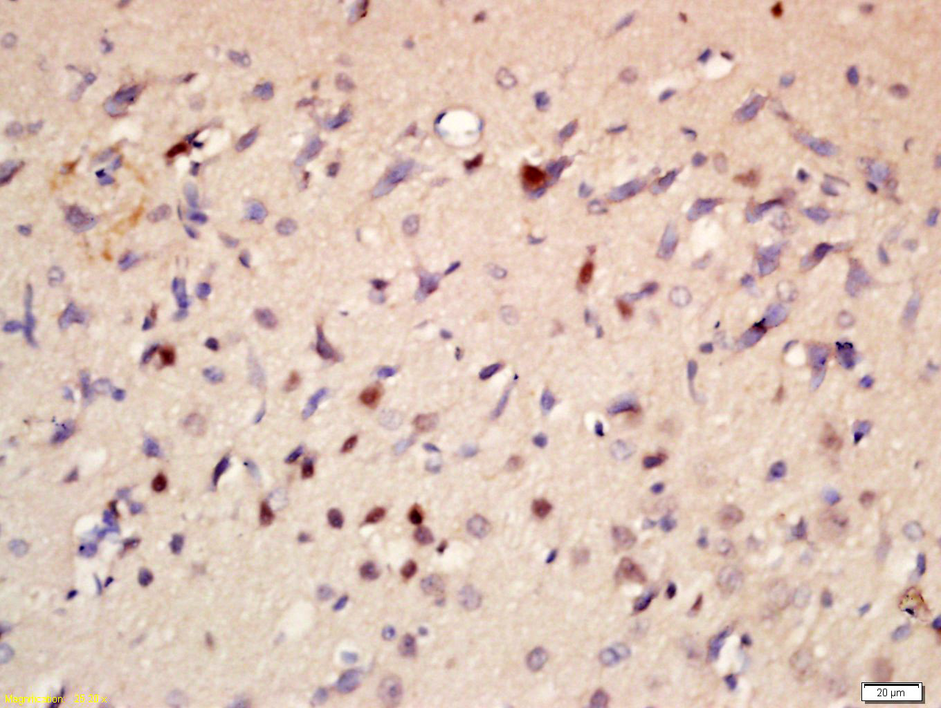 Tissue/cell: rat brain tissue; 4% Paraformaldehyde-fixed and paraffin-embedded;
Antigen retrieval: citrate buffer ( 0.01M, pH 6.0 ), Boiling bathing for 15min; Block endogenous peroxidase by 3% Hydrogen peroxide for 30min; Blocking buffer (normal goat serum,C-0005) at 37℃ for 20 min;
Incubation: Anti-P73 protein Polyclonal Antibody, Unconjugated(SL1346R) 1:200, overnight at 4°C, followed by conjugation to the secondary antibody(SP-0023) and DAB(C-0010) staining
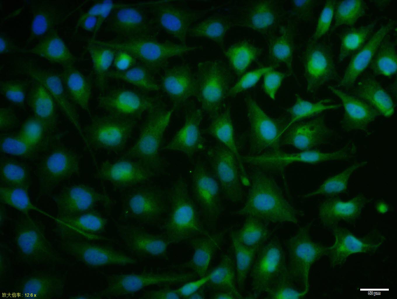 MCF7 cell; 4% Paraformaldehyde-fixed; Triton X-100 at room temperature for 20 min; Blocking buffer (normal goat serum, C-0005) at 37°C for 20 min; Antibody incubation with (P73) polyclonal Antibody, Unconjugated (SL1346R) 1:100, 90 minutes at 37°C; followed by a conjugated Goat Anti-Rabbit IgG antibody at 37°C for 90 minutes, DAPI (blue, C02-04002) was used to stain the cell nuclei.
Cartpieces
Totalgoods,subtotals:¥Checkout
Bought notes(bought amounts latest0)
No one bought this product
User Comment(Total0User Comment Num)
- No comment


 +86 571 56623320
 +86 18668110335

