
Rabbit Anti-GCAP1 antibody
COD3; GCAP 1; GCAP; Guanylate Cyclase Activating Protein Photoreceptor 1; Guanylate Cyclase Activating Protein Photoreceptor 1; Guanylate Cyclase Activator 1A; Guanylate Cyclase Activator 1A; Guanylin 1; Guanylin 1; guanylyl cyclase activating protein 1;
View History [Clear]
Details
Product Name GCAP1 Chinese Name 鸟苷酸环化酶激活蛋白1抗体 Alias COD3; GCAP 1; GCAP; Guanylate Cyclase Activating Protein Photoreceptor 1; Guanylate Cyclase Activating Protein Photoreceptor 1; Guanylate Cyclase Activator 1A; Guanylate Cyclase Activator 1A; Guanylin 1; Guanylin 1; guanylyl cyclase activating protein 1; Guanylyl cyclase-activating protein 1; GUC1A_HUMAN; GUCA; GUCA1; GUCA1A; GUCA1A. Research Area Cell biology Neurobiology Signal transduction Immunogen Species Rabbit Clonality Polyclonal React Species Mouse, Rat, (predicted: Human, Dog, Cow, Horse, Sheep, ) Applications WB=1:500-2000 ELISA=1:5000-10000 IHC-P=1:100-500 IHC-F=1:100-500 ICC=1:100-500 IF=1:100-500 (Paraffin sections need antigen repair)
not yet tested in other applications.
optimal dilutions/concentrations should be determined by the end user.Theoretical molecular weight 23kDa Cellular localization The cell membrane Form Liquid Concentration 1mg/ml immunogen KLH conjugated synthetic peptide derived from human GCAP1: 51-150/201 Lsotype IgG Purification affinity purified by Protein A Buffer Solution 0.01M TBS(pH7.4) with 1% BSA, 0.03% Proclin300 and 50% Glycerol. Storage Shipped at 4℃. Store at -20 °C for one year. Avoid repeated freeze/thaw cycles. Attention This product as supplied is intended for research use only, not for use in human, therapeutic or diagnostic applications. PubMed PubMed Product Detail intracellular stimulation of guanylate cyclase (GC) by calcium, a key event in the recovery of the dark state of rod photoreceptors after exposure to light, is mediated by guanylate cyclase-activating protein (GCAP1). GCAPs are calcium-The binding proteins belonging to the calmodulin superfamily. GCAP1 is a calcium-binding protein that stimulates synthesis of c-GMP in photoreceptors. GCAP1 is present in rod and cone photoreceptor outer segments where phototransduction occurs. In contrast to other calcium-binding proteins from the calmodulin superfamily, the calcium-free form of GCAP1 stimulates the effector enzyme. By molecular cloning of human and mouse GCAP cDNA, the known mammalian GCAPs are found to be more than 90% similar, consisting of 201 to 205 amino acids, and containing three identically conserved calcium-binding sites. A related protein, GCAP2, is detectable only in the retina and results from a gene duplication event.
Function:
Stimulates guanylyl cyclase 1 (GC1) when free calcium ions concentration is low and inhibits GC1 when free calcium ions concentration is elevated. This Ca(2+)-sensitive regulation of GC is a key event in recovery of the dark state of rod photoreceptors following light exposure.
Subcellular Location:
Membrane.
Tissue Specificity:
Retina; cone outer and inner segments, in particular, in disk membrane regions, and to a lesser extent rod inner and outer segments.
DISEASE:
Defects in GUCA1A are the cause of cone dystrophy type 3 (COD3) [MIM:602093]. COD3 is an autosomal dominant cone dystrophy. Cone dystrophies are retinal dystrophies characterized by progressive degeneration of the cone photoreceptors with preservation of rod function, as indicated by electroretinogram. However, some rod involvement may be present in some cone dystrophies, particularly at late stage. Affected individuals suffer from photophobia, loss of visual acuity, color vision and central visual field. Another sign is the absence of macular lesions for many years. Cone dystrophies are distinguished from the cone-rod dystrophies, in which some loss of peripheral vision also occurs.
Similarity:
Contains 4 EF-hand domains.
SWISS:
P43080
Gene ID:
2978
Database links:Entrez Gene: 2978 Human
Omim: 600364 Human
SwissProt: P43080 Human
Unigene: 92858 Human
Product Picture 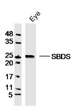 Sample:Eye (Mouse)Lysate at 40 ug
Primary: Anti-GCAP1(SL13306R)at 1/300 dilution
Secondary: IRDye800CW Goat Anti-RabbitIgG at 1/20000 dilution
Predicted band size: 23kD
Observed band size: 25kD
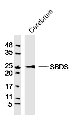 Sample:Cerebrum (Rat)Lysate at 40 ug
Primary: Anti-GCAP1(SL13306R)at 1/300 dilution
Secondary: IRDye800CW Goat Anti-RabbitIgG at 1/20000 dilution
Predicted band size: 23kD
Observed band size: 23kD
Cartpieces
Totalgoods,subtotals:¥Checkout
References (0)
No References
Bought notes(bought amounts latest0)
No one bought this product
User Comment(Total0User Comment Num)
- No comment


 +86 571 56623320
 +86 18668110335

