
Rabbit Anti-ZO-1/TJP1 antibody
ZO1_HUMAN; Tight junction protein ZO-1; TJP1; ZO1; Tight junction protein 1; Zona occludens protein 1; Zonula occludens protein 1; ZO1 tight junction protein;
View History [Clear]
Details
Product Name ZO-1/TJP1 Chinese Name 胞质紧密粘连蛋白1/闭锁小带蛋白1抗体 Alias ZO1_HUMAN; Tight junction protein ZO-1; TJP1; ZO1; Tight junction protein 1; Zona occludens protein 1; Zonula occludens protein 1; ZO1 tight junction protein; literatures Research Area Cell biology immunology Signal transduction Cell adhesion molecule Cell Surface Molecule Immunogen Species Rabbit Clonality Polyclonal React Species Human, Pig, (predicted: Mouse, Rat, Chicken, Dog, Cow, Rabbit, Guinea Pig, ) Applications WB=1:500-2000 ELISA=1:5000-10000 IHC-P=1:100-500 IHC-F=1:100-500 Flow-Cyt=1μg/Test IF=1:100-500 (Paraffin sections need antigen repair)
not yet tested in other applications.
optimal dilutions/concentrations should be determined by the end user.Theoretical molecular weight 191kDa Cellular localization cytoplasmic The cell membrane Form Liquid Concentration 1mg/ml immunogen KLH conjugated synthetic peptide derived from human ZO-1: 1551-1702/1733 Lsotype IgG Purification affinity purified by Protein A Buffer Solution 0.01M TBS(pH7.4) with 1% BSA, 0.03% Proclin300 and 50% Glycerol. Storage Shipped at 4℃. Store at -20 °C for one year. Avoid repeated freeze/thaw cycles. Attention This product as supplied is intended for research use only, not for use in human, therapeutic or diagnostic applications. PubMed PubMed Product Detail This gene encodes a member of the membrane-associated guanylate kinase (MAGUK) family of proteins, and acts as a tight junction adaptor protein that also regulates adherens junctions. Tight junctions regulate the movement of ions and macromolecules between endothelial and epithelial cells. The multidomain structure of this scaffold protein, including a postsynaptic density 95/disc-large/zona occludens (PDZ) domain, a Src homology (SH3) domain, a guanylate kinase (GuK) domain and unique (U) motifs all help to co-ordinate binding of transmembrane proteins, cytosolic proteins, and F-actin, which are required for tight junction function. Alternative splicing results in multiple transcript variants encoding different isoforms. [provided by RefSeq, Aug 2017]
Function:
The N-terminal may be involved in transducing a signal required for tight junction assembly, while the C-terminal may have specific properties of tight junctions. The alpha domain might be involved in stabilizing junctions. Plays a role in the regulation of cell migration by targeting CDC42BPB to the leading edge of migrating cells.
Subunit:
Interacts with BVES (via the C-terminus cytoplasmic tail). Interacts with HSPA4 and KIRREL1. Homodimer, and heterodimer with TJP2/ZO-2 and TJP3/ZO-3. Interacts with OCLN, CALM, claudins, CGN/cingulin, CXADR, GJA12, GJD3 and UBN1. Interacts (via ZU5 domain) with CDC42BPB and MYZAP. Interacts (via PDZ domain) with GJA1.
Subcellular Location:
Cell membrane; Peripheral membrane protein; Cytoplasmic side. Cell junction, tight junction. Cell junction. Cell junction, gap junction. Note=Moves from the cytoplasm to the cell membrane concurrently with cell-cell contact. Detected at the leading edge of migrating and wounded cells.
Tissue Specificity:
The alpha-containing isoform is found in most epithelial cell junctions. The short isoform is found both in endothelial cells and the highly specialized epithelial junctions of renal glomeruli and Sertoli cells of the seminiferous tubules.
Post-translational modifications:
Phosphorylated. Dephosphorylated by PTPRJ.
Similarity:
Belongs to the MAGUK family.
Contains 1 guanylate kinase-like domain.
Contains 3 PDZ (DHR) domains.
Contains 1 SH3 domain.
Contains 1 ZU5 domain.
SWISS:
Q07157
Gene ID:
7082
Database links:Entrez Gene: 7082 Human
Entrez Gene: 21872 Mouse
Omim: 601009 Human
SwissProt: Q07157 Human
SwissProt: P39447 Mouse
Unigene: 510833 Human
Unigene: 4342 Mouse
胞质紧密粘连蛋白1(ZO-1)是多结构域蛋白家族膜结合鸟苷酸激酶的家族成员,在紧密连接蛋白的组成成分中起到对组织分化和器官形成方面起较重要的作用。ZO-1在包括肾、胎盘、血脑屏障等许多组织都有不同的表达,可与紧密连接上的很多Transmembrane protein相互作用。也有学者认为:ZO-1的改变与细胞通透性的增加有关。Product Picture 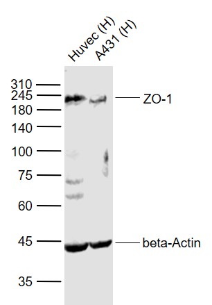 Sample:
Lane 1: Huvec (Human) Cell Lysate at 30 ug
Lane 2: A431 (Human) Cell Lysate at 30 ug
Primary:
Anti-ZO-1 (SL1329R) at 1/1000 dilution
Anti-beta-Actin (SL0061R) at 1/2000 dilution
Secondary: IRDye800CW Goat Anti-Rabbit IgG at 1/20000 dilution
Predicted band size: 220 kD
Observed band size: 220 kD
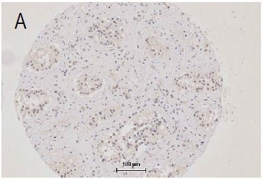 Independently Validated Antibody, image provided by Science Direct, badge number 029577:Formalin-fixed and paraffin embedded human testis labeled with Anti-ZO-1 Polyclonal Antibody, Unconjugated (SL1329R) at 1:250 followed by conjugation to the secondary antibody.
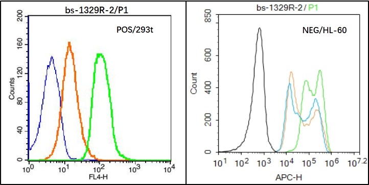 Black line : Positive blank control (293T); Negative blank control (HL60)
Green line : Primary Antibody (Rabbit Anti-ZO-1 antibody (SL11320R) )
Orange line:Isotype Control Antibody (Rabbit IgG) .
Blue line : Secondary Antibody (Goat anti-rabbit IgG-AF647)
293T(Positive)and HL60(Negative control)cells (black) were fixed with 4% PFA for 10min at room temperature, permeabilized with PBST for 20 min at room temperature, and incubated in 5% BSA blocking buffer for 30 min at room temperature. Cells were then stained with ZO-1 Antibody(SL1329R)at 1:50 dilution in blocking buffer and incubated for 30 min at room temperature, washed twice with 2% BSA in PBS, followed by secondary antibody(blue) incubation for 40 min at room temperature. Acquisitions of 20,000 events were performed. Cells stained with primary antibody (green), and isotype control (orange).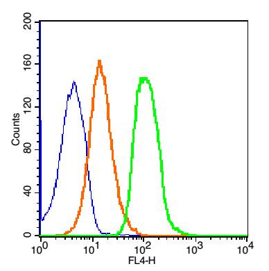 Blank control: 293T Cells(blue).
Primary Antibody: Rabbit Anti-ZO-1/AF647 Conjugated antibody (SL1329R-AF647), Dilution: 1μg in 100 μL 1X PBS containing 0.5% BSA;
Isotype Control Antibody: Rabbit IgG/AF647(orange) ,used under the same conditions.
Protocol
The cells were washed twice with phosphate-buffered saline (PBS). The cells were incubated in 1 X PBS containing 0.5% BSA + 1 0% goat serum (15 min) to block non-specific protein-protein interactions followed by the antibody (SL1329R-AF647, 1μg /1x10^6 cells) for 30 min on ice. Acquisition of 20,000 events was performed.
Cartpieces
Totalgoods,subtotals:¥Checkout
Bought notes(bought amounts latest0)
No one bought this product
User Comment(Total0User Comment Num)
- No comment


 +86 571 56623320
 +86 18668110335

