
Rabbit Anti-FHOD1 antibody
FH1/FH2 domain containing protein; FH1/FH2 domain-containing protein 1; Fhod1; FHOD1_HUMAN; FHOS; FHOS1; Formin homolog overexpressed in spleen 1; Formin homology 2 domain containing 1; Formin homology 2 domain containing protein 1; Formin homology 2 doma
View History [Clear]
Details
Product Name FHOD1 Chinese Name 肢体畸形相关蛋白FHOD1抗体 Alias FH1/FH2 domain containing protein; FH1/FH2 domain-containing protein 1; Fhod1; FHOD1_HUMAN; FHOS; FHOS1; Formin homolog overexpressed in spleen 1; Formin homology 2 domain containing 1; Formin homology 2 domain containing protein 1; Formin homology 2 domain-containing protein 1. Research Area Cell biology Developmental biology Signal transduction Cytoskeleton Immunogen Species Rabbit Clonality Polyclonal React Species Human, Mouse, Rat, (predicted: Dog, Pig, Horse, Rabbit, ) Applications WB=1:500-2000 ELISA=1:5000-10000 IHC-P=1:100-500 IHC-F=1:100-500 Flow-Cyt=2ug/Test IF=1:100-500 (Paraffin sections need antigen repair)
not yet tested in other applications.
optimal dilutions/concentrations should be determined by the end user.Theoretical molecular weight 126kDa Cellular localization cytoplasmic Form Liquid Concentration 1mg/ml immunogen KLH conjugated synthetic peptide derived from human FHOD1: 601-700/1164 Lsotype IgG Purification affinity purified by Protein A Buffer Solution 0.01M TBS(pH7.4) with 1% BSA, 0.03% Proclin300 and 50% Glycerol. Storage Shipped at 4℃. Store at -20 °C for one year. Avoid repeated freeze/thaw cycles. Attention This product as supplied is intended for research use only, not for use in human, therapeutic or diagnostic applications. PubMed PubMed Product Detail The limb deformity (ld) locus influences normal limb development and gives rise to alternative mRNAs that can translate into a family of protein products known as formins. Formins play a crucial role in cytoskeletal reorganization by influencing actin filament assembly. The temporal genetic hierarchy influencing normal limb development can deregulate and mediate mammalian developmental syndromes. FHOD1 induces the formation of and associates with bundled actin stress fibers in response to the activity of the Rho-ROCK cascade. It influences several cellular activities including cell migration, cytoskeletal arrangement, signal transduction and gene expression.
Function:
Required for the assembly of F-actin structures, such as stress fibers. Depends on the Rho-ROCK cascade for its activity. Contributes to the coordination of microtubules with actin fibers and plays a role in cell elongation.
Subunit:
Self-associates via the FH2 domain. Binds to F-actin via its N-terminus. Binds to the cytoplasmic domain of CD21 via its C-terminus. Interacts with ROCK1 in a Src-dependent manner.
Subcellular Location:
Cytoplasm. Cytoplasm; cytoskeleton. Predominantly cytoplasmic.
Tissue Specificity:
Ubiquitous. Highly expressed in spleen.
Post-translational modifications:
Phosphorylated by ROCK1.
Similarity:
Belongs to the formin homology family.
Contains 1 FH1 (formin homology 1) domain.
Contains 1 FH2 (formin homology 2) domain.
Contains 1 GBD/FH3 (Rho GTPase-binding/formin homology 3) domain.
SWISS:
Q9Y613
Gene ID:
29109
Database links:Entrez Gene: 29109 Human
Omim: 606881 Human
SwissProt: Q9Y613 Human
Unigene: 95231 Human
Product Picture 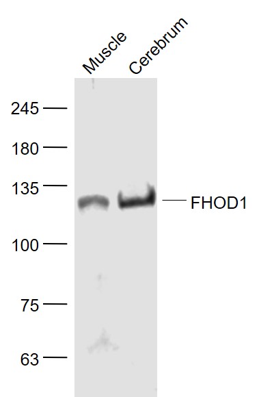 Sample:
Muscle (Mouse) Lysate at 40 ug
Cerebrum (Rat) Lysate at 40 ug
Primary: Anti- FHOD1 (SL13158R) at 1/1000 dilution
Secondary: IRDye800CW Goat Anti-Rabbit IgG at 1/20000 dilution
Predicted band size: 126 kD
Observed band size: 126 kD
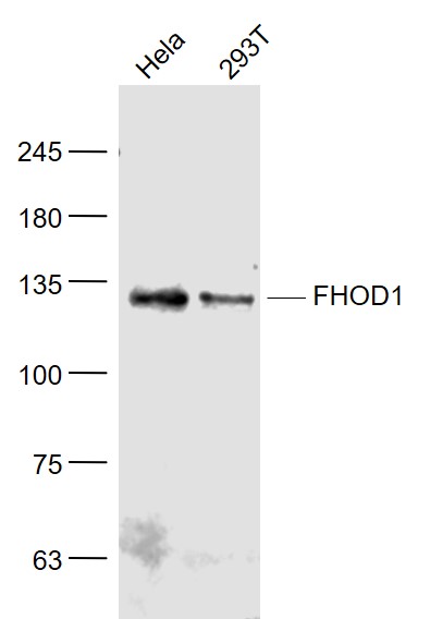 Sample:
Hela(Human) Cell Lysate at 30 ug
293T(Human) Cell Lysate at 30 ug
Primary: Anti- FHOD1 (SL13158R) at 1/1000 dilution
Secondary: IRDye800CW Goat Anti-Rabbit IgG at 1/20000 dilution
Predicted band size: 126 kD
Observed band size: 126 kD
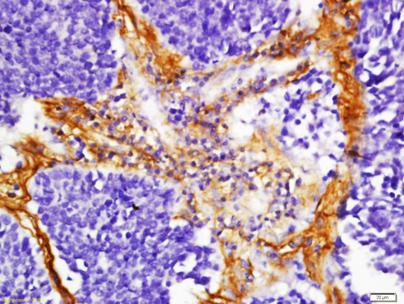 Tissue/cell: human lung carcinoma; 4% Paraformaldehyde-fixed and paraffin-embedded;
Antigen retrieval: citrate buffer ( 0.01M, pH 6.0 ), Boiling bathing for 15min; Block endogenous peroxidase by 3% Hydrogen peroxide for 30min; Blocking buffer (normal goat serum,C-0005) at 37℃ for 20 min;
Incubation: Anti-FHOD1 Polyclonal Antibody, Unconjugated(SL13158R) 1:200, overnight at 4°C, followed by conjugation to the secondary antibody(SP-0023) and DAB(C-0010) staining
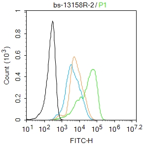 Blank control:MCF7.
Primary Antibody (green line): Rabbit Anti-FHOD1 antibody (SL13158R)
Dilution: 2μg /10^6 cells;
Isotype Control Antibody (orange line): Rabbit IgG .
Secondary Antibody : Goat anti-rabbit IgG-AF488
Dilution: 1μg /test.
Protocol
The cells were fixed with 4% PFA (10min at room temperature)and then permeabilized with 0.1% PBST for 20 min at room temperature.The cells were then incubated in 5%BSA to block non-specific protein-protein interactions for 30 min at room temperature .Cells stained with Primary Antibody for 30 min at room temperature. The secondary antibody used for 40 min at room temperature. Acquisition of 20,000 events was performed.
Cartpieces
Totalgoods,subtotals:¥Checkout
Bought notes(bought amounts latest0)
No one bought this product
User Comment(Total0User Comment Num)
- No comment


 +86 571 56623320
 +86 18668110335

