
Rabbit Anti-Bak antibody
Apoptosis Regulator Bak; BAK 1; BAK; BAK like; Bak NT; BAK1; Bcl 2 homologous antagonist/killer; Bcl 2 like 7 protein; Bcl2 homologous antagonist killer; Bcl2 like 7 Protein; BCL2-antagonist/killer 1; BCL2L7; CDN 1; CDN1; Cell death inhibitor 1; MGC117255
View History [Clear]
Details
Product Name Bak Chinese Name Bcl-2同源拮抗剂抗体 Alias Apoptosis Regulator Bak; BAK 1; BAK; BAK like; Bak NT; BAK1; Bcl 2 homologous antagonist/killer; Bcl 2 like 7 protein; Bcl2 homologous antagonist killer; Bcl2 like 7 Protein; BCL2-antagonist/killer 1; BCL2L7; CDN 1; CDN1; Cell death inhibitor 1; MGC117255; MGC3887; NBak; Pro apoptotic protein BAK; BAK_HUMAN; BAK_MOUSE. literatures Research Area immunology Apoptosis Immunogen Species Rabbit Clonality Polyclonal React Species Human, Mouse, (predicted: Rat, ) Applications WB=1:500-2000 ELISA=1:5000-10000 IHC-P=1:100-500 IHC-F=1:100-500 Flow-Cyt=1μg/Test IF=1:100-500 (Paraffin sections need antigen repair)
not yet tested in other applications.
optimal dilutions/concentrations should be determined by the end user.Theoretical molecular weight 23kDa Cellular localization cytoplasmic The cell membrane Form Liquid Concentration 1mg/ml immunogen KLH conjugated synthetic peptide derived from mouse Bak: 21-120/209 Lsotype IgG Purification affinity purified by Protein A Buffer Solution 0.01M TBS(pH7.4) with 1% BSA, 0.03% Proclin300 and 50% Glycerol. Storage Shipped at 4℃. Store at -20 °C for one year. Avoid repeated freeze/thaw cycles. Attention This product as supplied is intended for research use only, not for use in human, therapeutic or diagnostic applications. PubMed PubMed Product Detail The protein encoded by this gene belongs to the BCL2 protein family. BCL2 family members form oligomers or heterodimers and act as anti- or pro-apoptotic regulators that are involved in a wide variety of cellular activities. This protein localizes to mitochondria, and functions to induce apoptosis. It interacts with and accelerates the opening of the mitochondrial voltage-dependent anion channel, which leads to a loss in membrane potential and the release of cytochrome c. This protein also interacts with the tumor suppressor P53 after exposure to cell stress. [provided by RefSeq, Jul 2008]
Function:
In the presence of an appropriate stimulus, accelerates programmed cell death by binding to, and antagonizing the anti-apoptotic action of BCL2 or its adenovirus homolog E1B 19k protein. Low micromolar levels of zinc ions inhibit the promotion of apoptosis.
Subunit:
Belongs to the Bcl-2 family.
Subcellular Location:
Mitochondrion membrane.
Tissue Specificity:
Expressed in a wide variety of tissues.
Similarity:
Belongs to the Bcl-2 family.
SWISS:
O08734
Gene ID:
12018
Database links:
Entrez Gene: 578 Human
Entrez Gene: 12018 Mouse
Omim: 600516 Human
SwissProt: Q16611 Human
SwissProt: Q6I9T6 Human
SwissProt: O08734 Mouse
Unigene: 485139 Human
Unigene: 2443 Mouse
Bak属于bcl-2基因家族中的成员,具有抑制凋亡的作用。BAK基因是新发现的Apoptosis促进因子,BAK含BH1和BH2结构域,其功能类似于BAX;拮抗BCL2、BCL-XL的抗凋亡效应。Product Picture 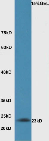 Sample:Lung (Mouse) Lysate at 30 ug
Primary: Anti-Bak(SL1284R) at 1:300 dilution;
Secondary: HRP conjugated Goat-Anti-Rabbit IgG(bse-0295G) at 1: 5000 dilution;
Predicted band size : 23kD
Observed band size : 23kD
 Tissue/cell: human lung carcinoma; 4% Paraformaldehyde-fixed and paraffin-embedded;
Antigen retrieval: citrate buffer ( 0.01M, pH 6.0 ), Boiling bathing for 15min; Block endogenous peroxidase by 3% Hydrogen peroxide for 30min; Blocking buffer (normal goat serum,C-0005) at 37℃ for 20 min;
Incubation: Anti-Bak Polyclonal Antibody, Unconjugated(SL1284R) 1:500, overnight at 4°C, followed by conjugation to the secondary antibody(SP-0023) and DAB(C-0010) staining
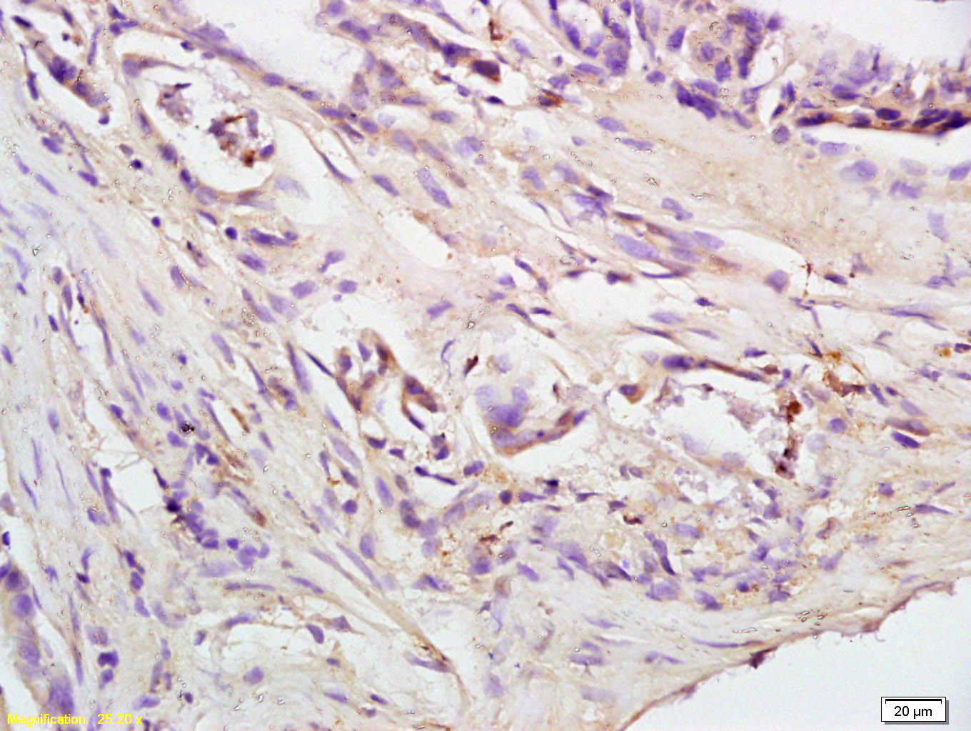 Tissue/cell: human colon carcinoma; 4% Paraformaldehyde-fixed and paraffin-embedded;
Antigen retrieval: citrate buffer ( 0.01M, pH 6.0 ), Boiling bathing for 15min; Block endogenous peroxidase by 3% Hydrogen peroxide for 30min; Blocking buffer (normal goat serum,C-0005) at 37℃ for 20 min;
Incubation: Anti-Bak Polyclonal Antibody, Unconjugated(SL1284R) 1:200, overnight at 4°C, followed by conjugation to the secondary antibody(SP-0023) and DAB(C-0010) staining
 Blank control: THP-1.
Primary Antibody (green line): Rabbit Anti-Bak antibody (SL1284R)
Dilution: 2μg /10^6 cells;
Isotype Control Antibody (orange line): Rabbit IgG .
Secondary Antibody : Goat anti-rabbit IgG-FITC
Dilution: 1μg /test.
Protocol
The cells were fixed with 4% PFA (10min at room temperature)and then permeabilized with 0.1% PBST methanol for 20 min at room temperature.The cells were then incubated in 5%BSA to block non-specific protein-protein interactions for 30 min at room temperature .Cells stained with Primary Antibody for 30 min at room temperature.The secondary antibody used for 40 min at room temperature. Acquisition of 20,000 events was performed. Blank control: Hela(blue).
Primary Antibody:Rabbit Anti- Bak antibody(SL1638R), Dilution: 1μg in 100 μL 1X PBS containing 0.5% BSA;
Isotype Control Antibody: Rabbit IgG(orange) ,used under the same conditions );
Secondary Antibody: Goat anti-rabbit IgG-PE(white blue), Dilution: 1:200 in 1 X PBS containing 0.5% BSA.
Protocol
The cells were fixed with 2% paraformaldehyde (10 min) , then permeabilized with 90% ice-cold methanol for 30 min on ice. Antibody (SL1284R, 1μg /1x10^6 cells) were incubated for 30 min on the ice, followed by 1 X PBS containing 0.5% BSA + 1 0% goat serum (15 min) to block non-specific protein-protein interactions. Then the Goat Anti-rabbit IgG/PE antibody was added into the blocking buffer mentioned above to react with the primary antibody of SL1284R at 1/200 dilution for 30 min on ice. Acquisition of 20,000 events was performed.
Cartpieces
Totalgoods,subtotals:¥Checkout
Bought notes(bought amounts latest0)
No one bought this product
User Comment(Total0User Comment Num)
- No comment


 +86 571 56623320
 +86 18668110335

