
Rabbit Anti-ATP6V0D2 antibody
V-ATPase D2; ATP6D2; ATPase H+ transporting lysosomal 38kDa V0 subunit D; ATPase H+ transporting lysosomal 38kDa V0 subunit D isoform 2; ATPase H+ transporting lysosomal 38kDa V0 subunit D2; FLJ38708; V ATPase subunit d 2; Vacuolar ATP synthase subunit d
View History [Clear]
Details
Product Name ATP6V0D2 Chinese Name ATP6V0D2蛋白抗体 Alias V-ATPase D2; ATP6D2; ATPase H+ transporting lysosomal 38kDa V0 subunit D; ATPase H+ transporting lysosomal 38kDa V0 subunit D isoform 2; ATPase H+ transporting lysosomal 38kDa V0 subunit D2; FLJ38708; V ATPase subunit d 2; Vacuolar ATP synthase subunit d 2; Vacuolar proton pump subunit d 2; VMA 6; VMA6; VA0D2_HUMAN. literatures Research Area Tumour Cell biology Signal transduction The new supersedes the old Immunogen Species Rabbit Clonality Polyclonal React Species Human, (predicted: Mouse, Rat, Dog, Pig, Sheep, ) Applications WB=1:500-2000 ELISA=1:5000-10000
not yet tested in other applications.
optimal dilutions/concentrations should be determined by the end user.Theoretical molecular weight 40kDa Cellular localization cytoplasmic The cell membrane Form Liquid Concentration 1mg/ml immunogen KLH conjugated synthetic peptide derived from human ATP6V0D2/V-ATPase D2: 251-350/350 Lsotype IgG Purification affinity purified by Protein A Buffer Solution 0.01M TBS(pH7.4) with 1% BSA, 0.03% Proclin300 and 50% Glycerol. Storage Shipped at 4℃. Store at -20 °C for one year. Avoid repeated freeze/thaw cycles. Attention This product as supplied is intended for research use only, not for use in human, therapeutic or diagnostic applications. PubMed PubMed Product Detail Vacuolar-type H+-ATPase (V-ATPase) is a multisubunit enzyme responsible for acidification of eukaryotic intracellular organelles. V-ATPases pump protons against an electrochemical gradient, while F-ATPases reverse the process, thereby synthesizing ATP. A peripheral V1 domain, which is responsible for ATP hydrolysis, and a integral V0 domain, which is responsible for proton translocation, compose V-ATPase. Nine subunits (A–H) make up the V1 domain and five subunits (a, d, c, c' and c") make up the V0 domain. Like F-ATPase, V-ATPase most likely operates through a rotary mechanism. V-ATPase D2 is a 350 amino acid protein that is expressed in kidney, lung and osteoclast. V-ATPase D2 has been implicated as a regulator of urine acidification, osteoclast fusion and bone formation. Furthermore, V-ATPase D2 has been identified as a dendritic cell marker.
Function:
Subunit of the integral membrane V0 complex of vacuolar ATPase. Vacuolar ATPase is responsible for acidifying a variety of intracellular compartments in eukaryotic cells, thus providing most of the energy required for transport processes in the vacuolar system. May play a role in coupling of proton transport and ATP hydrolysis (By similarity).
Subunit:
V-ATPase is a heteromultimeric enzyme composed of a peripheral catalytic V1 complex (components A to H) attached to an integral membrane V0 proton pore complex (components: a, c, c', c'' and d).
Subcellular Location:
Apical plasma membrane.
Tissue Specificity:
Kidney, osteoclast and lung.
Similarity:
Belongs to the V-ATPase V0D/AC39 subunit family.
SWISS:
Q8N8Y2
Gene ID:
245972
Database links:Entrez Gene: 245972 Human
Entrez Gene: 242341 Mouse
SwissProt: Q8N8Y2 Human
SwissProt: Q80SY3 Mouse
Unigene: 436360 Human
Product Picture 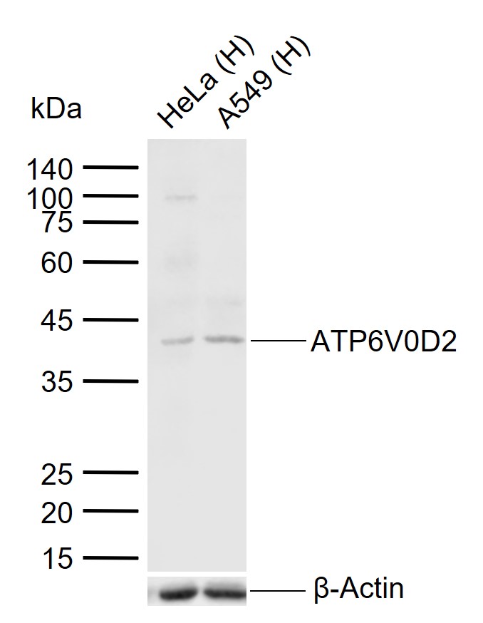 Sample:
Lane 1: Human HeLa cell lysates
Lane 2: Human A549 cell lysates
Primary: Anti-ATP6V0D2 (SL12548R) at 1/1000 dilution
Secondary: IRDye800CW Goat Anti-Rabbit IgG at 1/20000 dilution
Predicted band size: 40 kDa
Observed band size: 40 kDa
Cartpieces
Totalgoods,subtotals:¥Checkout
Bought notes(bought amounts latest0)
No one bought this product
User Comment(Total0User Comment Num)
- No comment


 +86 571 56623320
 +86 18668110335

