
Rabbit Anti-Cytokeratin 1 antibody
Cytokeratin-1; 67 kDa cytokeratin; CK 1; CK1; Cytokeratin1; EHK1; Hair alpha protein; K 1; K1; Keratin 1; Keratin type II cytoskeletal 1; Keratin1; KRT 1; KRT1A; K2C1_HUMAN; Keratin, type II cytoskeletal 1; Cytokeratin-1; CK-1; Keratin-1; Type-II keratin
View History [Clear]
Details
Product Name Cytokeratin 1 Chinese Name 细胞角蛋白1抗体 Alias Cytokeratin-1; 67 kDa cytokeratin; CK 1; CK1; Cytokeratin1; EHK1; Hair alpha protein; K 1; K1; Keratin 1; Keratin type II cytoskeletal 1; Keratin1; KRT 1; KRT1A; K2C1_HUMAN; Keratin, type II cytoskeletal 1; Cytokeratin-1; CK-1; Keratin-1; Type-II keratin Kb1. literatures Research Area Tumour Signal transduction Stem cells Kinases and Phosphatases Immunogen Species Rabbit Clonality Polyclonal React Species Human, Mouse, Rabbit, (predicted: Rat, Dog, Cow, Horse, ) Applications ELISA=1:5000-10000 IHC-P=1:100-500 IHC-F=1:100-500 Flow-Cyt=1μg/Test IF=1:100-500 (Paraffin sections need antigen repair)
not yet tested in other applications.
optimal dilutions/concentrations should be determined by the end user.Theoretical molecular weight 70kDa Cellular localization cytoplasmic The cell membrane Form Liquid Concentration 1mg/ml immunogen KLH conjugated synthetic peptide derived from human Cytokeratin 1: 301-400/644 Lsotype IgG Purification affinity purified by Protein A Buffer Solution 0.01M TBS(pH7.4) with 1% BSA, 0.03% Proclin300 and 50% Glycerol. Storage Shipped at 4℃. Store at -20 °C for one year. Avoid repeated freeze/thaw cycles. Attention This product as supplied is intended for research use only, not for use in human, therapeutic or diagnostic applications. PubMed PubMed Product Detail The protein encoded by this gene is a member of the keratin gene family. The type II cytokeratins consist of basic or neutral proteins which are arranged in pairs of heterotypic keratin chains coexpressed during differentiation of simple and stratified epithelial tissues. This type II cytokeratin is specifically expressed in the spinous and granular layers of the epidermis with family member KRT10 and mutations in these genes have been associated with bullous congenital ichthyosiform erythroderma. The type II cytokeratins are clustered in a region of chromosome 12q12-q13. [provided by RefSeq].
Function:
May regulate the activity of kinases such as PKC and SRC via binding to integrin beta-1 (ITB1) and the receptor of activated protein kinase C (RACK1/GNB2L1). In complex with C1QBP is a high affinty receptor for kininogen-1/HMWK.
Subunit:
Heterotetramer of two type I and two type II keratins. Keratin-1 is generally associated with keratin-10. Interacts with ITGB1 in the presence of GNB2L1 and SRC, and with GNB2L1. Interacts with C1QBP; the association represents a cell surface kininogen receptor.
Subcellular Location:
Cell membrane. Note=Located on plasma membrane of neuroblastoma NMB7 cells.
Tissue Specificity:
The source of this protein is neonatal foreskin. The 67-kDa type II keratins are expressed in terminally differentiating epidermis.
Post-translational modifications:
Undergoes deimination of some arginine residues (citrullination).
DISEASE:
Defects in KRT1 are a cause of epidermolytic hyperkeratosis (EHK) [MIM:113800]. An autosomal dominant skin disorder characterized by widespread blistering and an ichthyotic erythroderma at birth that persist into adulthood. Histologically there is a diffuse epidermolytic degeneration in the lower spinous layer of the epidermis. Within a few weeks from birth, erythroderma and blister formation diminish and hyperkeratoses develop.
Defects in KRT1 are the cause of ichthyosis hystrix Curth-Macklin type (IHCM) [MIM:146590]. IHCM is a genodermatosis with severe verrucous hyperkeratosis. Affected individuals manifest congenital verrucous black scale on the scalp, neck, and limbs with truncal erythema, palmoplantar keratoderma and keratoses on the lips, ears, nipples and buttocks.
Defects in KRT1 are a cause of palmoplantar keratoderma non-epidermolytic (NEPPK) [MIM:600962]. NEPKK is a dermatological disorder characterized by focal palmoplantar keratoderma with oral, genital, and follicular lesions.
Defects in KRT1 are a cause of ichthyosis annular epidermolytic (AEI) [MIM:607602]; also known as cyclic ichthyosis with epidermolytic hyperkeratosis. AEI is a skin disorder resembling bullous congenital ichthyosiform erythroderma. Affected individuals present with bullous ichthyosis in early childhood and hyperkeratotic lichenified plaques in the flexural areas and extensor surfaces at later ages. The feature that distinguishes AEI from BCIE is dramatic episodes of flares of annular polycyclic plaques with scale, which coalesce to involve most of the body surface and can persist for several weeks or even months.
Defects in KRT1 are the cause of palmoplantar keratoderma striate type 3 (SPPK3) [MIM:607654]; also known as keratosis palmoplantaris striata III. SPPK3 is a dermatological disorder affecting palm and sole skin where stratum corneum and epidermal layers are thickened. There is no involvement of non-palmoplantar skin, and both hair and nails are normal.
Similarity:
Belongs to the intermediate filament family.
SWISS:
P04264
Gene ID:
3848
Database links:Entrez Gene: 3848 Human
Entrez Gene: 16678 Mouse
Omim: 139350 Human
SwissProt: P04264 Human
SwissProt: P04104 Mouse
Unigene: 80828 Human
Unigene: 183137 Mouse
Unigene: 31789 Rat
结构蛋白(Structural Proteins)
细胞角蛋白常用于Tumour细胞的分化、增殖及转移方面的研究。有学者认为:在TumourCell differentiation过程中有细胞角蛋白的表达,把细胞角蛋白作为TumourStem cells的Maker。阳性部位:主要在胞浆。CK119, CK8, CK19同源.Product Picture 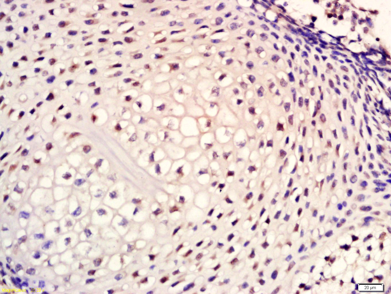 Tissue/cell: mouse embryos tissue; 4% Paraformaldehyde-fixed and paraffin-embedded;
Antigen retrieval: citrate buffer ( 0.01M, pH 6.0 ), Boiling bathing for 15min; Block endogenous peroxidase by 3% Hydrogen peroxide for 30min; Blocking buffer (normal goat serum,C-0005) at 37℃ for 20 min;
Incubation: Anti-Cytokeratin 1 Polyclonal Antibody, Unconjugated(SL1244R) 1:200, overnight at 4°C, followed by conjugation to the secondary antibody(SP-0023) and DAB(C-0010) staining
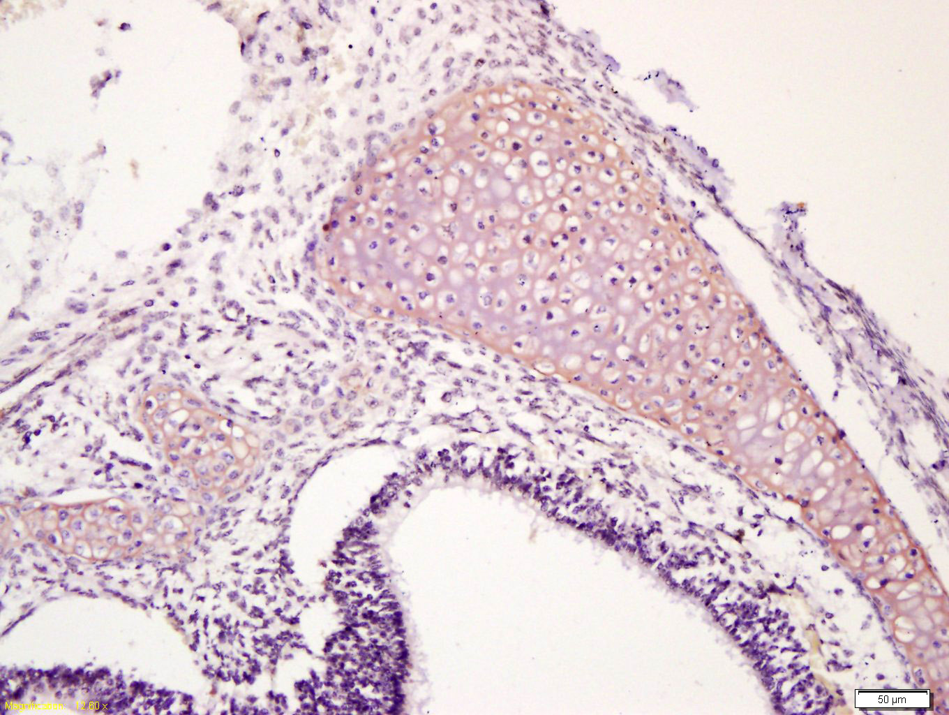 Tissue/cell: mouse embryo tissue; 4% Paraformaldehyde-fixed and paraffin-embedded;
Antigen retrieval: citrate buffer ( 0.01M, pH 6.0 ), Boiling bathing for 15min; Block endogenous peroxidase by 3% Hydrogen peroxide for 30min; Blocking buffer (normal goat serum,C-0005) at 37℃ for 20 min;
Incubation: Anti-Cytokeratin 1 Polyclonal Antibody, Unconjugated(SL1244R) 1:200, overnight at 4°C, followed by conjugation to the secondary antibody(SP-0023) and DAB(C-0010) staining
 Blank control (blue line): Hela cells(blue).
Primary Antibody (green line): Rabbit Anti-Cytokeratin 1/FITC Conjugated antibody (SL1244R-PE)
Dilution: 1μg /10^6 cells;
Isotype Control Antibody (orange line): Rabbit IgG-PE.
Protocol
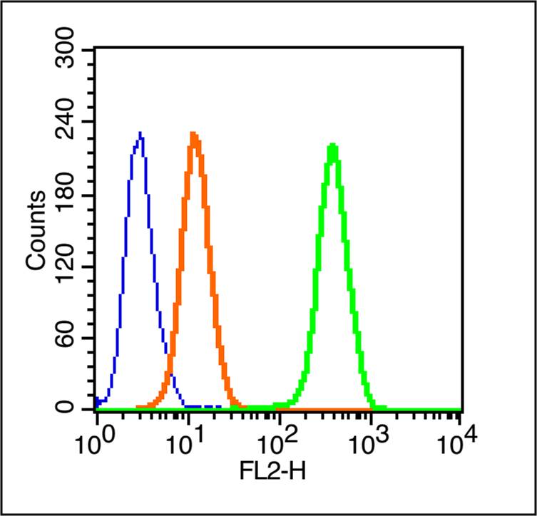
The cells were fixed with 70% ice-cold methanol overnight at 4℃ . The cells were then incubated in 1 X PBS/2%BSA/10% goat serum to block non-specific protein-protein interactions followed by the antibody for 15 min at room temperature. Cells stained with Primary Antibody for 30 min at room temperature.Acquisition of 20,000 events was performed.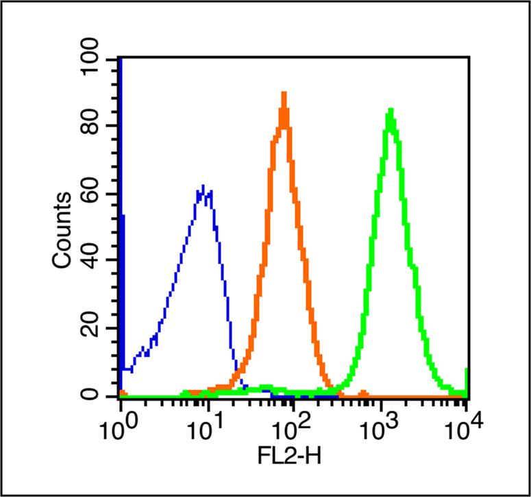 Blank control (blue line): Rabbit spleen cells (blue).
Primary Antibody (green line): Rabbit Anti-Cytokeratin 1/PE Conjugated antibody (SL1244R-PE)
Dilution: 0.2μg /10^6 cells;
Isotype Control Antibody (orange line): Rabbit IgG-PE .
Protocol
The cells were fixed with 70% ice-cold methanol overnight at 4℃ . The cells were then incubated in 1 X PBS/2%BSA/10% goat serum to block non-specific protein-protein interactions followed by the antibody for 15 min at room temperature. Cells stained with Primary Antibody for 30 min at room temperature.Acquisition of 20,000 events was performed.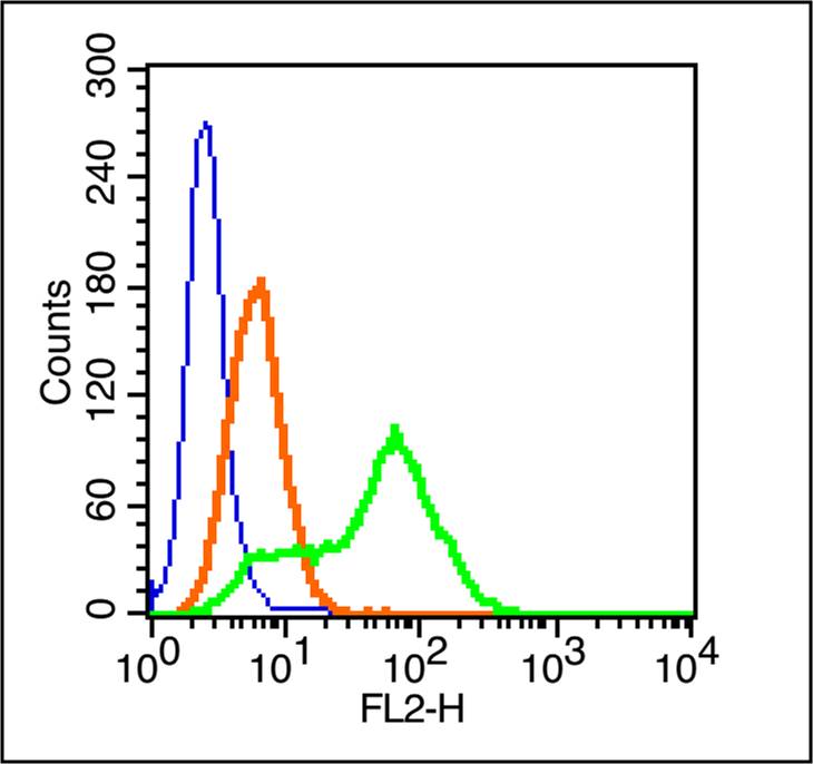 Blank control (blue line): MCF7 cells(blue).
Primary Antibody (green line): Rabbit Anti-Cytokeratin 1/FITC Conjugated antibody (SL1244R-FITC)
Dilution: 1μg /10^6 cells;
Isotype Control Antibody (orange line): Rabbit IgG-FITC.
Protocol
The cells were fixed with 70% ice-cold methanol overnight at 4℃ . The cells were then incubated in 1 X PBS/2%BSA/10% goat serum to block non-specific protein-protein interactions followed by the antibody for 15 min at room temperature. Cells stained with Primary Antibody for 30 min at room temperature.Acquisition of 20,000 events was performed.
Cartpieces
Totalgoods,subtotals:¥Checkout
Bought notes(bought amounts latest0)
No one bought this product
User Comment(Total0User Comment Num)
- No comment


 +86 571 56623320
 +86 18668110335

