
Rabbit Anti-ASCL2 antibody
Achaete scute complex like 2; Achaete scute homolog 2; achaete-scute complex-like 2; Achaete-scute homolog 2; ASCL2; ASCL2_HUMAN; ASH-2; Ash2; bHLHa45; Class A basic helix-loop-helix protein 45; HASH2; Mash2.
View History [Clear]
Details
Product Name ASCL2 Chinese Name ASCL2蛋白抗体 Alias Achaete scute complex like 2; Achaete scute homolog 2; achaete-scute complex-like 2; Achaete-scute homolog 2; ASCL2; ASCL2_HUMAN; ASH-2; Ash2; bHLHa45; Class A basic helix-loop-helix protein 45; HASH2; Mash2. literatures Research Area Cell biology Developmental biology Signal transduction Stem cells Epigenetics Immunogen Species Rabbit Clonality Polyclonal React Species Human, Mouse, (predicted: Rat, Chicken, Dog, Pig, Cow, ) Applications ELISA=1:5000-10000 IHC-P=1:100-500 IHC-F=1:100-500 Flow-Cyt=2ug/Test ICC=1:100 IF=1:100-500 (Paraffin sections need antigen repair)
not yet tested in other applications.
optimal dilutions/concentrations should be determined by the end user.Theoretical molecular weight 22kDa Cellular localization The nucleus Form Liquid Concentration 1mg/ml immunogen KLH conjugated synthetic peptide derived from Human ASCL2: 51-120/193 Lsotype IgG Purification affinity purified by Protein A Buffer Solution 0.01M TBS(pH7.4) with 1% BSA, 0.03% Proclin300 and 50% Glycerol. Storage Shipped at 4℃. Store at -20 °C for one year. Avoid repeated freeze/thaw cycles. Attention This product as supplied is intended for research use only, not for use in human, therapeutic or diagnostic applications. PubMed PubMed Product Detail Members of the myogenic determination family are basic helix-loop-helix (bHLH) proteins that can be separated into two classes, both of which work together to activate DNA transcription. Class A proteins include the ubiquitously expressed E-box binding factors, namely E2A, ITF-2 and HEB, while class B proteins, such as MyoD, myogenin and Neuro D (BETA2), are transiently expressed and exhibit a much more limited tissue distribution. Working in opposition to these positively acting factors are a specialized group of basic helix-loop-helix (bHLH) transcription factors that function as dominant negative regulators and are involved in cell lineage determination and differentiation. ASCL2 is a 193 amino acid protein that localizes to the nucleus and contains one bHLH domain. Expressed in developing placental tissue, ASCL2 binds to DNA and functions as a transcriptional regulator that is involved in the maturation of neuronal precursors in the peripheral and central nervous systems.
Function:
AS-C proteins are involved in the determination of the neuronal precursors in the peripheral nervous system and the central nervous system.
Subunit:
Efficient DNA binding requires dimerization with another bHLH protein.
Subcellular Location:
Nucleus.
Tissue Specificity:
Expressed specifically in the extravillous trophoblasts of the developing placenta.
Similarity:
Contains 1 basic helix-loop-helix (bHLH) domain.
SWISS:
Q99929
Gene ID:
430
Database links:Entrez Gene: 430 Human
Entrez Gene: 17173 Mouse
SwissProt: Q99929 Human
SwissProt: O35885 Mouse
Unigene: 152475 Human
Unigene: 196417 Mouse
Unigene: 10486 Rat
Product Picture 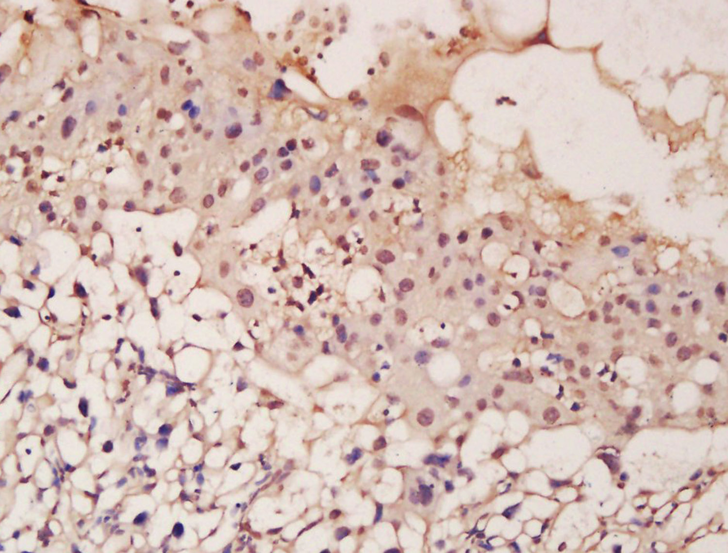 Tissue/cell: mouse placenta tissue; 4% Paraformaldehyde-fixed and paraffin-embedded;
Antigen retrieval: citrate buffer ( 0.01M, pH 6.0 ), Boiling bathing for 15min; Block endogenous peroxidase by 3% Hydrogen peroxide for 30min; Blocking buffer (normal goat serum,C-0005) at 37℃ for 20 min;
Incubation: Anti-ASCL2 Polyclonal Antibody, Unconjugated(SL12349R) 1:200, overnight at 4°C, followed by conjugation to the secondary antibody(SP-0023) and DAB(C-0010) staining
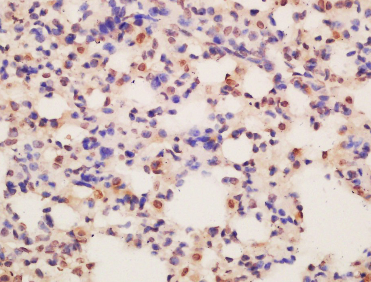 Tissue/cell: mouse lung tissue; 4% Paraformaldehyde-fixed and paraffin-embedded;
Antigen retrieval: citrate buffer ( 0.01M, pH 6.0 ), Boiling bathing for 15min; Block endogenous peroxidase by 3% Hydrogen peroxide for 30min; Blocking buffer (normal goat serum,C-0005) at 37℃ for 20 min;
Incubation: Anti-ASCL2 Polyclonal Antibody, Unconjugated(SL12349R) 1:200, overnight at 4°C, followed by conjugation to the secondary antibody(SP-0023) and DAB(C-0010) staining
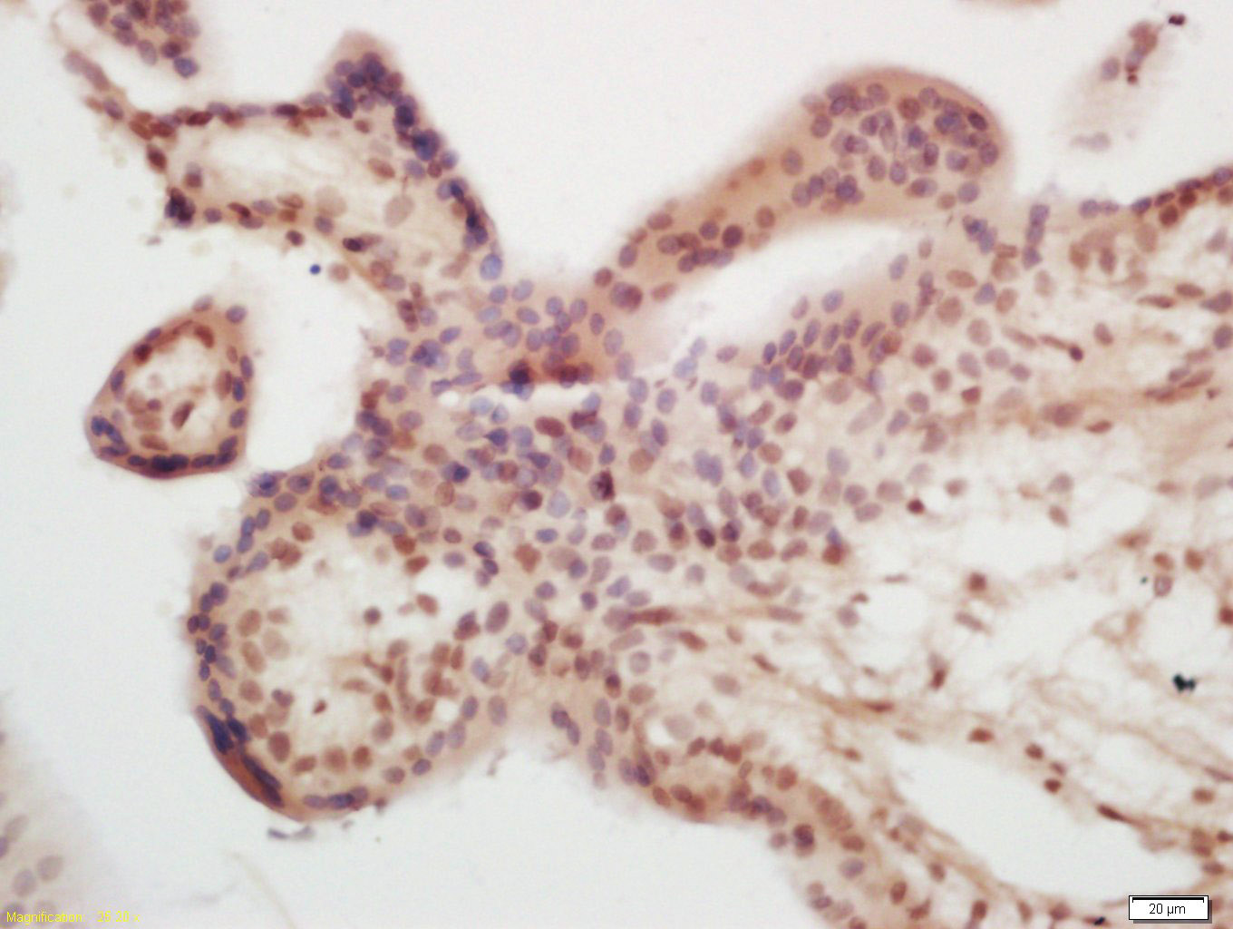 Tissue/cell: human placenta tissue; 4% Paraformaldehyde-fixed and paraffin-embedded;
Antigen retrieval: citrate buffer ( 0.01M, pH 6.0 ), Boiling bathing for 15min; Block endogenous peroxidase by 3% Hydrogen peroxide for 30min; Blocking buffer (normal goat serum,C-0005) at 37℃ for 20 min;
Incubation: Anti-ASCL2 Polyclonal Antibody, Unconjugated(SL12349R) 1:200, overnight at 4°C, followed by conjugation to the secondary antibody(SP-0023) and DAB(C-0010) staining
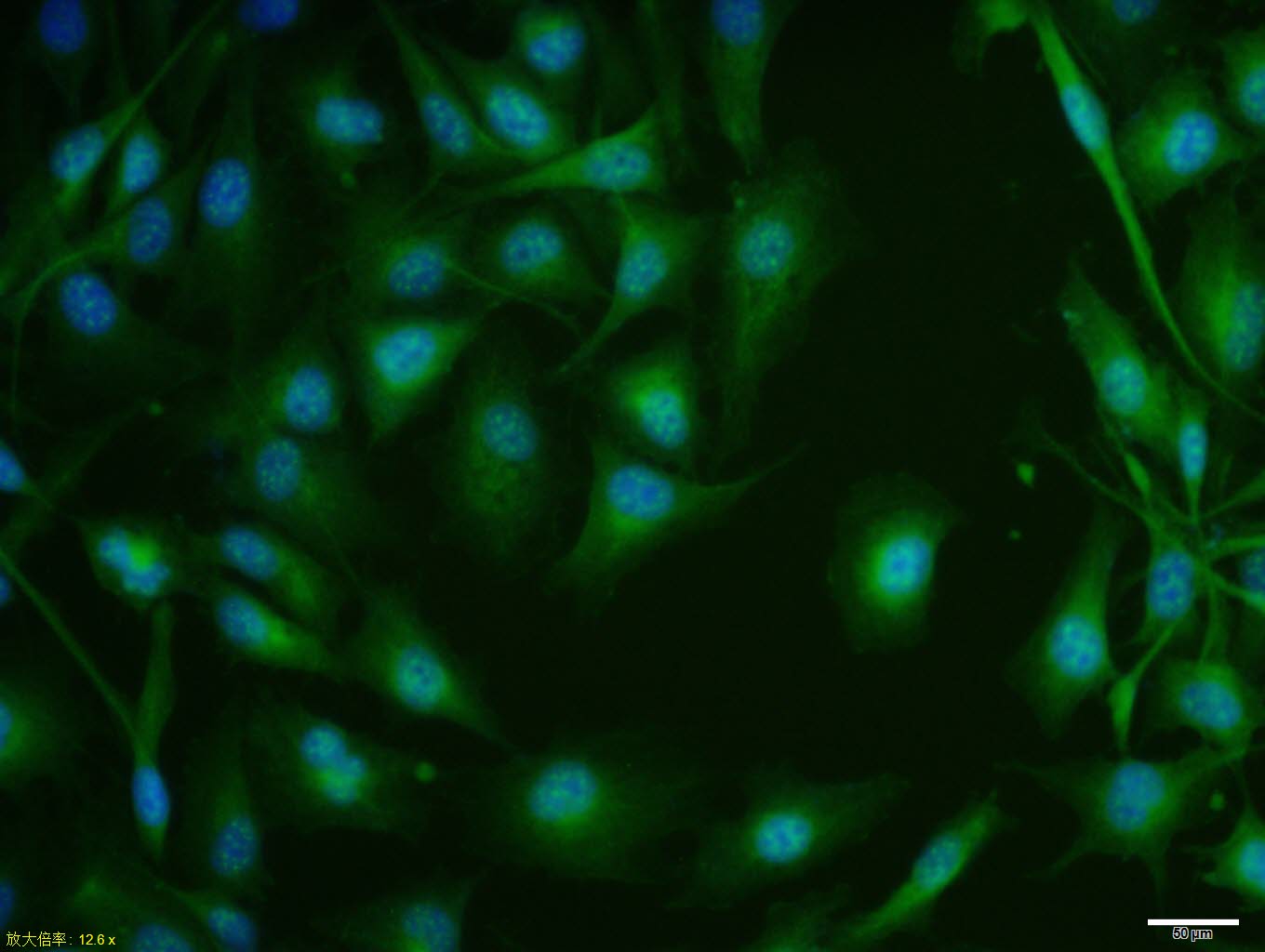 MCF7 cell; 4% Paraformaldehyde-fixed; Triton X-100 at room temperature for 20 min; Blocking buffer (normal goat serum, C-0005) at 37°C for 20 min; Antibody incubation with (ASCL2) polyclonal Antibody, Unconjugated (SL12349R) 1:100, 90 minutes at 37°C; followed by a conjugated Goat Anti-Rabbit IgG antibody at 37°C for 90 minutes, DAPI (blue, C02-04002) was used to stain the cell nuclei.
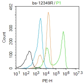 Blank control:K562.
Primary Antibody (green line): Rabbit Anti-ASCL2 antibody (SL12349R)
Dilution: 2μg /10^6 cells;
Isotype Control Antibody (orange line): Rabbit IgG .
Secondary Antibody : Goat anti-rabbit IgG-PE
Dilution: 1μg /test.
Protocol
The cells were fixed with 4% PFA (10min at room temperature)and then permeabilized with 90% ice-cold methanol for 20 min at-20℃. The cells were then incubated in 5%BSA to block non-specific protein-protein interactions for 30 min at room temperature .Cells stained with Primary Antibody for 30 min at room temperature. The secondary antibody used for 40 min at room temperature. Acquisition of 20,000 events was performed.
Cartpieces
Totalgoods,subtotals:¥Checkout
Bought notes(bought amounts latest0)
No one bought this product
User Comment(Total0User Comment Num)
- No comment


 +86 571 56623320
 +86 18668110335

