
Rabbit Anti-GLI1 antibody
Gli 1; Gli1; Gli-1; GLI; GLI Kruppel family member 1; Glioma associated oncogene; Glioma associated oncogene homolog 1 (zinc finger protein); Oncogene GLI; Zfp 5; Zfp5; Zinc finger protein GLI 1; Zinc finger protein GLI1; GLI1_HUMAN.
View History [Clear]
Details
Product Name GLI1 Chinese Name 脑胶质瘤相关蛋白抗体(Zinc finger protein5) Alias Gli 1; Gli1; Gli-1; GLI; GLI Kruppel family member 1; Glioma associated oncogene; Glioma associated oncogene homolog 1 (zinc finger protein); Oncogene GLI; Zfp 5; Zfp5; Zinc finger protein GLI 1; Zinc finger protein GLI1; GLI1_HUMAN. literatures Research Area Tumour Cell biology Neurobiology Signal transduction Epigenetics Immunogen Species Rabbit Clonality Polyclonal React Species Human, Mouse, Rat, (predicted: Dog, Cow, Horse, Rabbit, ) Applications WB=1:500-2000 ELISA=1:5000-10000 IHC-P=1:100-500 IHC-F=1:100-500 Flow-Cyt=1ug/Test IF=1:100-500 (Paraffin sections need antigen repair)
not yet tested in other applications.
optimal dilutions/concentrations should be determined by the end user.Theoretical molecular weight 118kDa Cellular localization The nucleus cytoplasmic Form Liquid Concentration 1mg/ml immunogen KLH conjugated synthetic peptide derived from human GLI1/Zfp5: 601-700/1106 Lsotype IgG Purification affinity purified by Protein A Buffer Solution 0.01M TBS(pH7.4) with 1% BSA, 0.03% Proclin300 and 50% Glycerol. Storage Shipped at 4℃. Store at -20 °C for one year. Avoid repeated freeze/thaw cycles. Attention This product as supplied is intended for research use only, not for use in human, therapeutic or diagnostic applications. PubMed PubMed Product Detail This gene encodes a member of the Kruppel family of zinc finger proteins. The encoded transcription factor is activated by the sonic hedgehog signal transduction cascade and regulates stem cell proliferation. The activity and nuclear localization of this protein is negatively regulated by p53 in an inhibitory loop. Multiple transcript variants encoding different isoforms have been found for this gene. [provided by RefSeq, May 2009]
Function:
Acts as a transcriptional activator. May regulate the transcription of specific genes during normal development. May play a role in craniofacial development and digital development, as well as development of the central nervous system and gastrointestinal tract. Mediates SHH signaling and thus cell proliferation and differentiation.
Subcellular Location:
Cytoplasm. Nucleus. Tethered in the cytoplasm by binding to SUFU. Activation and translocation to the nucleus is promoted by interaction with STK36. Phosphorylation by ULK3 may promote nuclear localization. Translocation to the nucleus is promoted by interaction with ZIC1.
Tissue Specificity:
Testis, myometrium and fallopian tube. Also expressed in the brain with highest expression in the cerebellum, optic nerve and olfactory tract.
SWISS:
P08151
Gene ID:
2735
Database links:Entrez Gene: 2735 Human
Entrez Gene: 14632 Mouse
Omim: 165220 Human
SwissProt: P08151 Human
SwissProt: P47806 Mouse
Unigene: 632702 Human
Unigene: 391450 Mouse
Unigene: 219157 Rat
GLI1是一种具有很强活性的转录激活因子,GLI1诱导G1/S细胞周期调节蛋白的表达从而促进细胞的增殖; 直接诱导抗凋亡因子Bcl-2的表达以抑制凋亡; 直接激活促进上皮组织向间质转化因子的转录从而加重了Tumour的侵袭性。目前主要用于Tumour及神经方面的研究。Product Picture 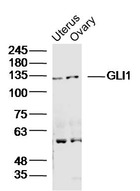 Sample:
Uterus(Mouse)Lysate at 40 ug
Ovary (Mouse)Lysate at 40 ug
Primary: Anti-GLI1(SL1206R)at 1/300 dilution
Secondary: IRDye800CW Goat Anti-RabbitIgG at 1/20000 dilution
Predicted band size: 118kD
Observed band size: 130kD
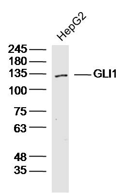 Sample:HepG2 (Human)Cell Lysate at 40 ug
Primary: Anti-GLI1(SL1206R)at 1/300 dilution
Secondary: IRDye800CW Goat Anti-RabbitIgG at 1/20000 dilution
Predicted band size: 118kD
Observed band size: 130kD
 Paraformaldehyde-fixed, paraffin embedded (Rat testis); Antigen retrieval by boiling in sodium citrate buffer (pH6.0) for 15min; Block endogenous peroxidase by 3% hydrogen peroxide for 20 minutes; Blocking buffer (normal goat serum) at 37°C for 30min; Antibody incubation with (GLI1) Polyclonal Antibody, Unconjugated (SL1206R) at 1:400 overnight at 4°C, followed by operating according to SP Kit(Rabbit) (sp-0023) instructionsand DAB staining.
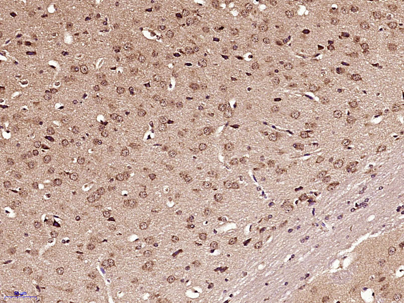 Paraformaldehyde-fixed, paraffin embedded (Mouse brain); Antigen retrieval by boiling in sodium citrate buffer (pH6.0) for 15min; Block endogenous peroxidase by 3% hydrogen peroxide for 20 minutes; Blocking buffer (normal goat serum) at 37°C for 30min; Antibody incubation with (GLI1) Polyclonal Antibody, Unconjugated (SL1206R) at 1:400 overnight at 4°C, followed by operating according to SP Kit(Rabbit) (sp-0023) instructionsand DAB staining.
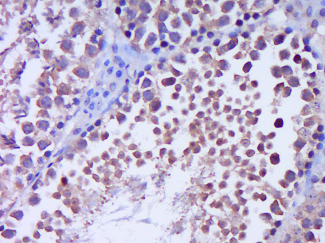
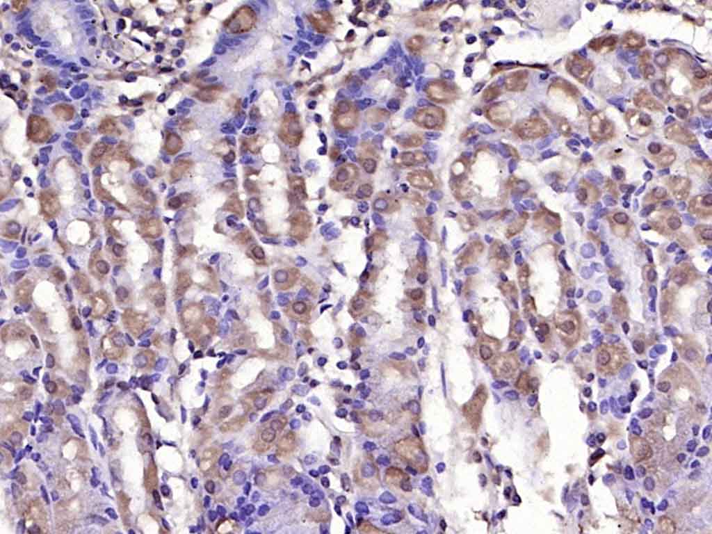 Paraformaldehyde-fixed, paraffin embedded (human gastric carcinoma); Antigen retrieval by boiling in sodium citrate buffer (pH6.0) for 15min; Block endogenous peroxidase by 3% hydrogen peroxide for 20 minutes; Blocking buffer (normal goat serum) at 37°C for 30min; Antibody incubation with (GLI1) Polyclonal Antibody, Unconjugated (SL1206R) at 1:200 overnight at 4°C, followed by operating according to SP Kit(Rabbit) (sp-0023) instructionsand DAB staining.
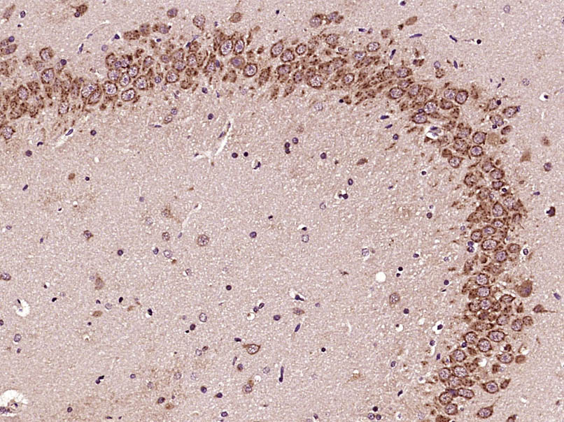 Paraformaldehyde-fixed, paraffin embedded (Rat brain); Antigen retrieval by boiling in sodium citrate buffer (pH6.0) for 15min; Block endogenous peroxidase by 3% hydrogen peroxide for 20 minutes; Blocking buffer (normal goat serum) at 37°C for 30min; Antibody incubation with (GLI1) Polyclonal Antibody, Unconjugated (SL1206R) at 1:400 overnight at 4°C, followed by operating according to SP Kit(Rabbit) (sp-0023) instructionsand DAB staining.
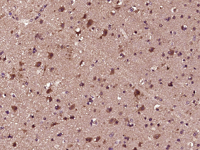 Paraformaldehyde-fixed, paraffin embedded (Human brain glioma); Antigen retrieval by boiling in sodium citrate buffer (pH6.0) for 15min; Block endogenous peroxidase by 3% hydrogen peroxide for 20 minutes; Blocking buffer (normal goat serum) at 37°C for 30min; Antibody incubation with (GLI1) Polyclonal Antibody, Unconjugated (SL1206R) at 1:400 overnight at 4°C, followed by operating according to SP Kit(Rabbit) (sp-0023) instructionsand DAB staining.
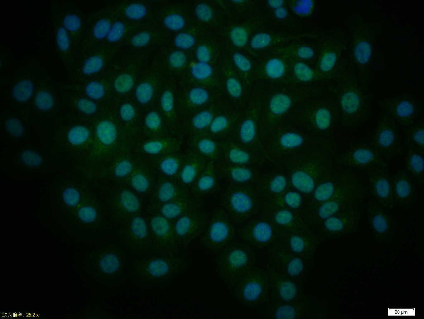 Tissue/cell: HepG2 cell; 4% Paraformaldehyde-fixed; Triton X-100 at room temperature for 20 min; Blocking buffer (normal goat serum, C-0005) at 37°C for 20 min; Antibody incubation with (GLI1) Polyclonal Antibody, Unconjugated (SL1206R) 1:50, 90 minutes at 37°C; followed by a conjugated Goat Anti-Rabbit IgG antibody (SL0295G-FITC) at 37°C for 90 minutes, DAPI (5ug/ml, blue, C-0033) was used to stain the cell nuclei.
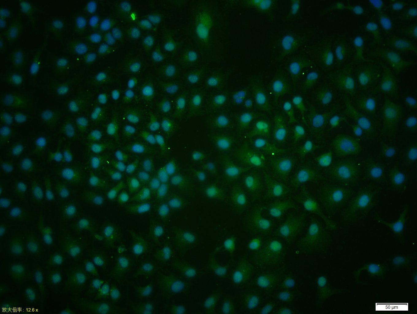 Tissue/cell: HeLa cell; 4% Paraformaldehyde-fixed; Triton X-100 at room temperature for 20 min; Blocking buffer (normal goat serum, C-0005) at 37°C for 20 min; Antibody incubation with (GLI1) Polyclonal Antibody, Unconjugated (SL1206R) 1:50, 90 minutes at 37°C; followed by a conjugated Goat Anti-Rabbit IgG antibody (SL0295G-FITC) at 37°C for 90 minutes, DAPI (5ug/ml, blue, C-0033) was used to stain the cell nuclei.
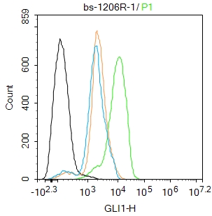 Blank control:Hela.
Primary Antibody (green line): Rabbit Anti-GLI1 antibody (SL1206R)
Dilution: 1ug/Test;
Secondary Antibody : Goat anti-rabbit IgG-FITC
Dilution: 0.5ug/Test.
Protocol
The cells were fixed with 4% PFA (10min at room temperature)and then permeabilized with 90% ice-cold methanol for 20 min at -20℃.The cells were then incubated in 5%BSA to block non-specific protein-protein interactions for 30 min at room temperature .Cells stained with Primary Antibody for 30 min at room temperature. The secondary antibody used for 40 min at room temperature. Acquisition of 20,000 events was performed.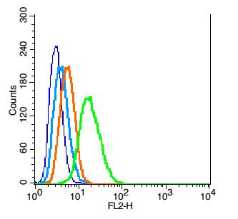 Blank control: RSC96(blue), the cells were fixed with 2% paraformaldehyde (10 min) and then permeabilized with ice-cold 90% methanol for 30 min on ice.
Isotype Control Antibody: Rabbit IgG(orange) ;
Secondary Antibody: Goat anti-rabbit IgG-PE(white blue), Dilution: 1:200 in 1 X PBS containing 0.5% BSA .
Primary Antibody Dilution: 1μg in 100 μL1X PBS containing 0.5% BSA(green).
Cartpieces
Totalgoods,subtotals:¥Checkout
Bought notes(bought amounts latest0)
No one bought this product
User Comment(Total0User Comment Num)
- No comment


 +86 571 56623320
 +86 18668110335

