
Rabbit Anti-HLA-DRA antibody
DR alpha chain precursor; DRB1; DRB4; HLA class II histocompatibility antigen; HLA class II histocompatibility antigen DR alpha chain; HLA DR1B; HLA DR3B; HLA DRA; HLA DRA1; HLA DRB3; HLA DRB4; HLA DRB5; HLADR4B; HLADRA1; HLADRB; Major histocompatibility
View History [Clear]
Details
Product Name HLA-DRA Chinese Name HLA-DR抗体 Alias DR alpha chain precursor; DRB1; DRB4; HLA class II histocompatibility antigen; HLA class II histocompatibility antigen DR alpha chain; HLA DR1B; HLA DR3B; HLA DRA; HLA DRA1; HLA DRB3; HLA DRB4; HLA DRB5; HLADR4B; HLADRA1; HLADRB; Major histocompatibility complex class II DR alpha; Major histocompatibility complex class II DR beta 1; Major histocompatibility complex class II DR beta 3; Major histocompatibility complex class II DR beta 4; Major histocompatibility complex class II DR beta 5; MGC117330; MHC cell surface glycoprotein; MHC class II antigen DRA; MHC II; DRA_HUMAN. literatures Research Area Cell biology immunology Immunogen Species Rabbit Clonality Polyclonal React Species Human, Mouse, Rat, (predicted: Dog, Pig, Cow, Horse, Sheep, ) Applications ELISA=1:5000-10000 IHC-P=1:100-500 Flow-Cyt=1μg/Test (Paraffin sections need antigen repair)
not yet tested in other applications.
optimal dilutions/concentrations should be determined by the end user.Theoretical molecular weight 26kDa Cellular localization cytoplasmic The cell membrane Form Liquid Concentration 1mg/ml immunogen KLH conjugated synthetic peptide derived from human HLA-DRA: 1-100/254 <Extracellular> Lsotype IgG Purification affinity purified by Protein A Buffer Solution 0.01M TBS(pH7.4) with 1% BSA, 0.03% Proclin300 and 50% Glycerol. Storage Shipped at 4℃. Store at -20 °C for one year. Avoid repeated freeze/thaw cycles. Attention This product as supplied is intended for research use only, not for use in human, therapeutic or diagnostic applications. PubMed PubMed Product Detail HLA-DRA is one of the HLA class II alpha chain paralogues. This class II molecule is a heterodimer consisting of an alpha and a beta chain, both anchored in the membrane. It plays a central role in the immune system by presenting peptides derived from extracellular proteins. Class II molecules are expressed in antigen presenting cells (APC: B lymphocytes, dendritic cells, macrophages). The alpha chain is approximately 33-35 kDa and its gene contains 5 exons. Exon 1 encodes the leader peptide, exons 2 and 3 encode the two extracellular domains, and exon 4 encodes the transmembrane domain and the cytoplasmic tail. DRA does not have polymorphisms in the peptide binding part and acts as the sole alpha chain for DRB1, DRB3, DRB4 and DRB5. [provided by RefSeq]
Function:
Binds peptides derived from antigens that access the endocytic route of antigen presenting cells (APC) and presents them on the cell surface for recognition by the CD4 T-cells. The peptide binding cleft accommodates peptides of 10-30 residues. The peptides presented by MHC class II molecules are generated mostly by degradation of proteins that access the endocytic route, where they are processed by lysosomal proteases and other hydrolases. Exogenous antigens that have been endocytosed by the APC are thus readily available for presentation via MHC II molecules, and for this reason this antigen presentation pathway is usually referred to as exogenous. As membrane proteins on their way to degradation in lysosomes as part of their normal turn-over are also contained in the endosomal/lysosomal compartments, exogenous antigens must compete with those derived from endogenous components. Autophagy is also a source of endogenous peptides, autophagosomes constitutively fuse with MHC class II loading compartments.
Subunit:
Heterodimer of an alpha and a beta subunit; also referred as MHC class II molecule. In the endoplasmic reticulum (ER) it forms an heterononamer; 3 MHC class II molecules bind to a CD74 homotrimer (also known as invariant chain or HLA class II histocompatibility antigen gamma chain).
Subcellular Location:
Cell membrane; Single-pass type I membrane protein. Endoplasmic reticulum membrane; Single-pass type I membrane protein. Golgi apparatus, trans-Golgi network membrane; Single-pass type I membrane protein. Endosome membrane; Single-pass type I membrane protein. Lysosome membrane; Single-pass type I membrane protein. Late endosome membrane; Single-pass type I membrane protein. Note=The MHC class II complex transits through a number of intracellular compartments in the endocytic pathway until it reaches the cell membrane for antigen presentation.
Post-translational modifications:
Ubiquitinated by MARCH1 or MARCH8 at Lys-244 leading to down-regulation of MHC class II. When associated with ubiquitination of the beta subunit of HLA-DR: HLA-DRB4 'Lys-254', the down-regulation of MHC class II may be highly effective.
Similarity:
Belongs to the MHC class II family.
Contains 1 Ig-like C1-type (immunoglobulin-like) domain.
SWISS:
P01903
Gene ID:
3122
Database links:Entrez Gene: 3122 Human
Omim: 142860 Human
SwissProt: P01903 Human
Unigene: 520048 Human
HLA-DR抗原主要存在于endothelial cells、Blymphocyte、单核细胞。HLA-DR分子为二聚体结构,由非多态性的DRα链和多态性的DRβ链组成.Product Picture 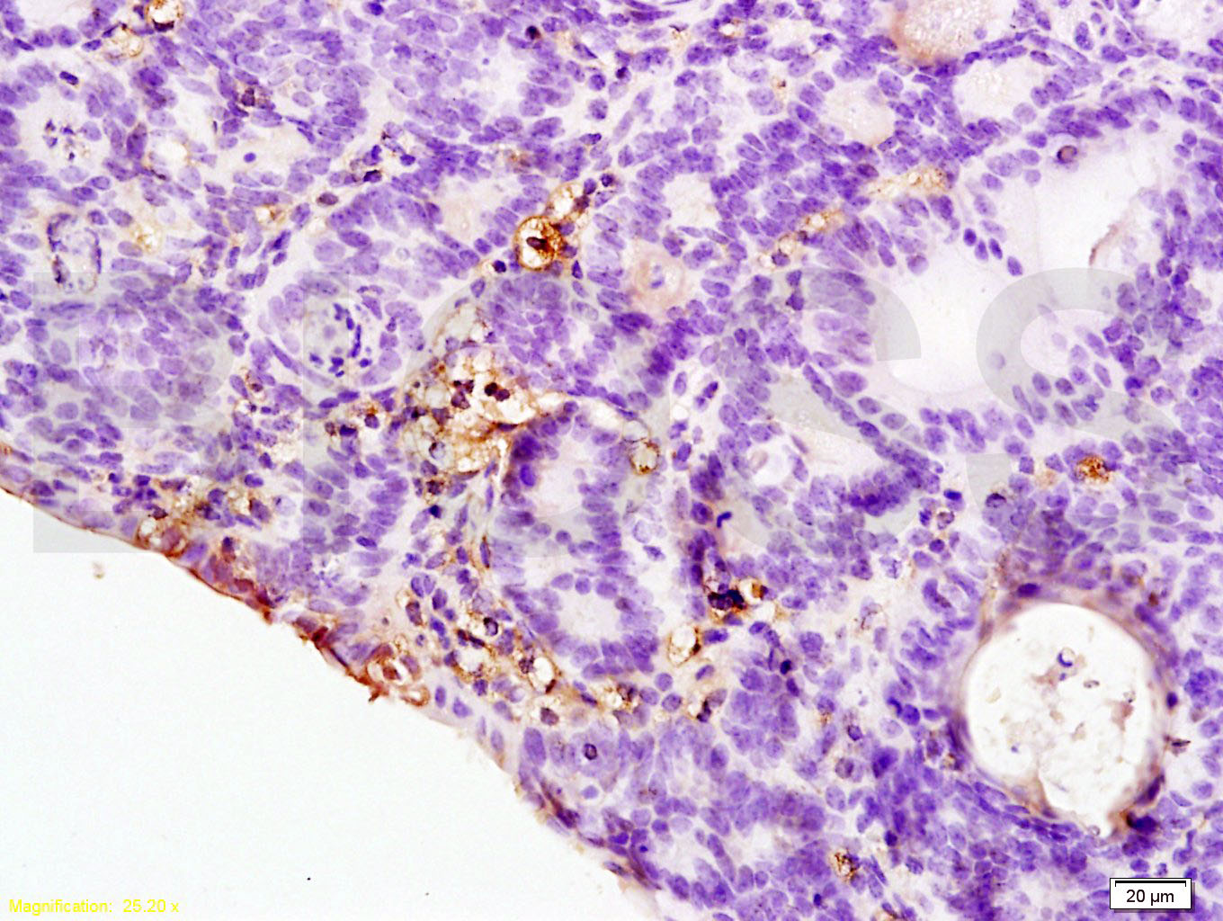 Tissue/cell: rat colitis tissue; 4% Paraformaldehyde-fixed and paraffin-embedded;
Antigen retrieval: citrate buffer ( 0.01M, pH 6.0 ), Boiling bathing for 15min; Block endogenous peroxidase by 3% Hydrogen peroxide for 30min; Blocking buffer (normal goat serum,C-0005) at 37℃ for 20 min;
Incubation: Anti-HLA-DR Polyclonal Antibody, Unconjugated(SL1198R) 1:200, overnight at 4°C, followed by conjugation to the secondary antibody(SP-0023) and DAB(C-0010) staining
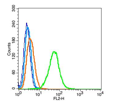 Blank control: A549(blue), the cells were fixed with 2% paraformaldehyde (10 min) and then permeabilized with ice-cold 90% methanol for 30 min on ice..
Isotype Control Antibody: Rabbit IgG(orange) ; Secondary Antibody: Goat anti-rabbit IgG-FITC(white blue), Dilution: 1:100 in 1 X PBS containing 0.5% BSA ; Primary Antibody Dilution: 1μg in 100 μL1X PBS containing 0.5% BSA(green).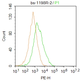 Blank control: Mouse spleen.
Primary Antibody (green line): Rabbit Anti-HLA-DR antibody (SL1198R)
Dilution: 2μg /10^6 cells;
Isotype Control Antibody (orange line): Rabbit IgG .
Secondary Antibody : Goat anti-rabbit IgG-PE
Dilution: 1μg /test.
Protocol
The cells were incubated in 5%BSA to block non-specific protein-protein interactions for 30 min at at room temperature .Cells stained with Primary Antibody for 30 min at room temperature. The secondary antibody used for 40 min at room temperature. Acquisition of 20,000 events was performed.
Cartpieces
Totalgoods,subtotals:¥Checkout
Bought notes(bought amounts latest0)
No one bought this product
User Comment(Total0User Comment Num)
- No comment


 +86 571 56623320
 +86 18668110335

