
Rabbit Anti-ITGB3 antibody
Integrin beta-3; Integrin beta chain, β3 precursor; Integrin Beta 3; CD 61; CD61; CD61 antigen; GP3A; GPIIIa; HPA 1; HPA 4; Integrin beta 3 (platelet glycoprotein IIIa antigen CD61); Integrin beta chain beta 3; ITG B3; ITGB 3; NAIT; Platelet fibrinogen re
View History [Clear]
Details
Product Name ITGB3 Chinese Name 整合素β3/CD61抗体 Alias Integrin beta-3; Integrin beta chain, β3 precursor; Integrin Beta 3; CD 61; CD61; CD61 antigen; GP3A; GPIIIa; HPA 1; HPA 4; Integrin beta 3 (platelet glycoprotein IIIa antigen CD61); Integrin beta chain beta 3; ITG B3; ITGB 3; NAIT; Platelet fibrinogen receptor beta subunit; Platelet glycoprotein IIIa; platelet glycoprotein IIIa precursor; Platelet membrane glycoprotein IIIa; PTP; ITB3_HUMAN. Integrin β3; Integrin-β3; Integrin β 3; Integrinβ3; literatures Research Area Cell biology immunology Signal transduction Stem cells Cell adhesion molecule Immunogen Species Rabbit Clonality Polyclonal React Species Human, Mouse, (predicted: Pig, Cow, Rabbit, ) Applications WB=1:500-2000 ELISA=1:5000-10000 IHC-P=1:100-500 IHC-F=1:100-500 IF=1:100-500 (Paraffin sections need antigen repair)
not yet tested in other applications.
optimal dilutions/concentrations should be determined by the end user.Theoretical molecular weight 84kDa Detection molecular weight 90 kDa Cellular localization The cell membrane Form Liquid Concentration 1mg/ml immunogen KLH conjugated synthetic peptide derived from human Integrin beta 3: 27-120/788 <Extracellular> Lsotype IgG Purification affinity purified by Protein A Buffer Solution 0.01M TBS(pH7.4) with 1% BSA, 0.03% Proclin300 and 50% Glycerol. Storage Shipped at 4℃. Store at -20 °C for one year. Avoid repeated freeze/thaw cycles. Attention This product as supplied is intended for research use only, not for use in human, therapeutic or diagnostic applications. PubMed PubMed Product Detail The ITGB3 protein product is the integrin beta chain beta 3. Integrins are integral cell-surface proteins composed of an alpha chain and a beta chain. A given chain may combine with multiple partners resulting in different integrins. Integrin beta 3 is found along with the alpha IIb chain in platelets. Integrins are known to participate in cell adhesion as well as cell-surface mediated signalling. [provided by RefSeq, Jul 2008]
Function:
Integrin alpha-V/beta-3 is a receptor for cytotactin, fibronectin, laminin, matrix metalloproteinase-2, osteopontin, osteomodulin, prothrombin, thrombospondin, vitronectin and von Willebrand factor. Integrin alpha-IIb/beta-3 is a receptor for fibronectin, fibrinogen, plasminogen, prothrombin, thrombospondin and vitronectin. Integrins alpha-IIb/beta-3 and alpha-V/beta-3 recognize the sequence R-G-D in a wide array of ligands. Integrin alpha-IIb/beta-3 recognizes the sequence H-H-L-G-G-G-A-K-Q-A-G-D-V in fibrinogen gamma chain. Following activation integrin alpha-IIb/beta-3 brings about platelet/platelet interaction through binding of soluble fibrinogen. This step leads to rapid platelet aggregation which physically plugs ruptured endothelial surface. In case of HIV-1 infection, the interaction with extracellular viral Tat protein seems to enhance angiogenesis in Kaposi's sarcoma lesions.
Subunit:
Heterodimer of an alpha and a beta subunit. Beta-3 associates with either alpha-IIb or alpha-V. Isoform Beta-3C interacts with FLNB. Interacts with COMP. Interacts with HIV-1 Tat. Interacts with PDIA6 following platelet stimulation. Interacts with SYK; upon activation by ITGB3 promotes platelet adhesion. Interacts with MYO10.
Subcellular Location:
Membrane; Single-pass type I membrane protein.
Tissue Specificity:
Isoform beta-3A and isoform beta-3C are widely expressed. Isoform beta-3A is specifically expressed in osteoblast cells; isoform beta-3C is specifically expressed in prostate and testis.
Post-translational modifications:
Phosphorylated on tyrosine residues in response to thrombin-induced platelet aggregation. Probably involved in outside-in signaling. A peptide (AA 740-762) is capable of binding GRB2 only when both Tyr-773 and Tyr-785 are phosphorylated. Phosphorylation of Thr-779 inhibits SHC binding.
DISEASE:
Defects in ITGB3 are a cause of Glanzmann thrombasthenia (GT) [MIM:273800]; also known as thrombasthenia of Glanzmann and Naegeli. GT is the most common inherited disease of platelets. It is an autosomal recessive disorder characterized by mucocutaneous bleeding of mild-to-moderate severity and the inability of this integrin to recognize macromolecular or synthetic peptide ligands. GT has been classified clinically into types I and II. In type I, platelets show absence of the glycoprotein IIb/beta-3 complexes at their surface and lack fibrinogen and clot retraction capability. In type II, the platelets express the glycoprotein IIb/beta-3 complex at reduced levels (5-20% controls), have detectable amounts of fibrinogen, and have low or moderate clot retraction capability. The platelets of GT 'variants' have normal or near normal (60-100%) expression of dysfunctional receptors.
Similarity:
Belongs to the integrin beta chain family. Contains 1 VWFA domain.
SWISS:
P05106
Gene ID:
3690
Database links:Entrez Gene: 3690 Human
Entrez Gene: 16416 Mouse
Omim: 173470 Human
SwissProt: P05106 Human
SwissProt: O54890 Mouse
Unigene: 218040 Human
Unigene: 87150 Mouse
CD61抗原又称为GP III a,是一种表达于血小板、巨核细胞、单核细胞、巨噬细胞和endothelial cells上的glycoprotein。CD61和CD41构成血小板glycoproteinII b/III b。Product Picture 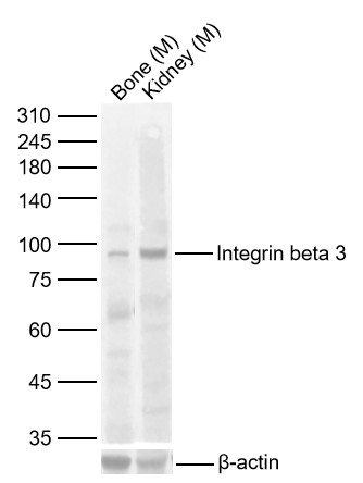 Sample:
Lane 1: Mouse Bone Lysates
Lane 2: Mouse Kidney Lysates
Primary: Anti-Integrin beta 3 (SL0342R) at 1/1000 dilution
Secondary: IRDye800CW Goat Anti-Rabbit IgG at 1/20000 dilution
Predicted band size: 84kDa
Observed band size: 84kDa
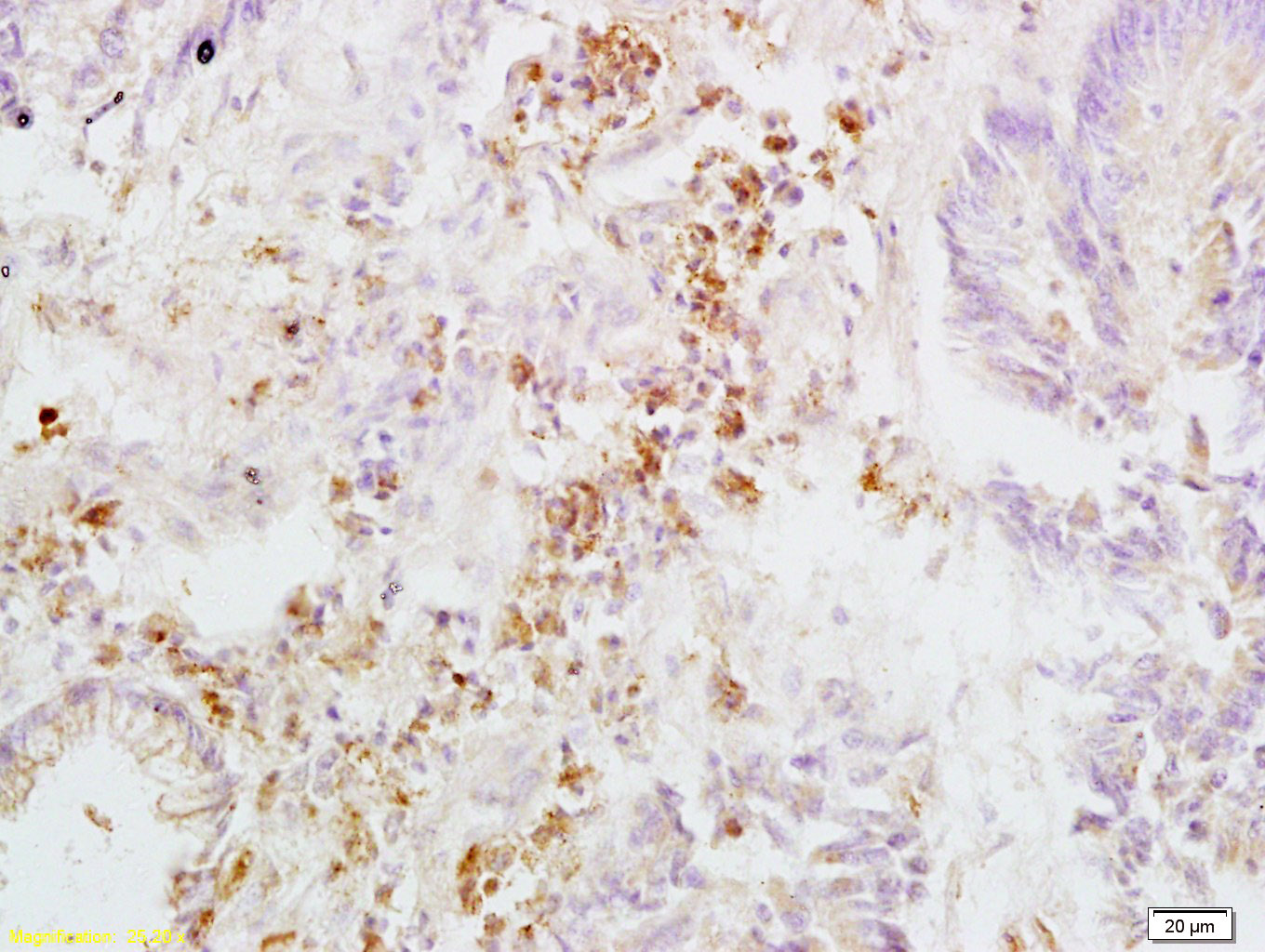 Tissue/cell: human rectal carcinoma; 4% Paraformaldehyde-fixed and paraffin-embedded;
Antigen retrieval: citrate buffer ( 0.01M, pH 6.0 ), Boiling bathing for 15min; Block endogenous peroxidase by 3% Hydrogen peroxide for 30min; Blocking buffer (normal goat serum,C-0005) at 37℃ for 20 min;
Incubation: Anti-Integrin beta 3/CD61 Polyclonal Antibody, Unconjugated(SL0342R) 1:200, overnight at 4癈, followed by conjugation to the secondary antibody(SP-0023) and DAB(C-0010) staining
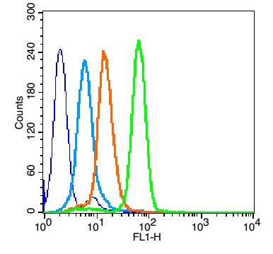 Overlay histogram showing HL 60 cells stained with SL0342R (Green line).
The cells were fixed with 90% methanol (5 min) and then permeabilized with 0.01M PBS-Tween for 20 min. The cells were then incubated in 1x PBS / 10% normal goat serum to block non-specific protein-protein interactions followed by the antibody (SL0342R,1μg/1x10^6 cells) for 30 min at 22℃. The secondary antibody used was fluorescein isothiocyanate goat anti-rabbit IgG (H+L) (SL 0295G-FITC , Brillant blue line) at 1/200 dilution for 30 min at 22℃. Isotype control antibody was rabbit IgG (polyclonal,SL0295P,Orange line) (1μg/1x10^6 cells) used under the same conditions. Unlabelled sample (blue line) was also used as a control. Acquisition of 20,000 events were collected using a 20mW Argon ion laser (488nm) and 525/30 bandpass filter.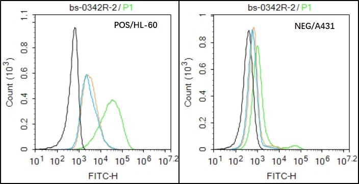 Black line : Positive blank control (HL60); Negative blank control (A431)
Green line : Primary Antibody (Rabbit Anti-CD61 antibody (SL0342R) )
Orange line:Isotype Control Antibody (Rabbit IgG) .
Blue line : Secondary Antibody (Goat anti-rabbit IgG-AF488)
HL60(Positive)and A431 Negative control)cells (black) were incubated in 5% BSA blocking buffer for 30 min at room temperature. Cells were then stained with CD61 Antibody(SL0342R)at 1:50 dilution in blocking buffer and incubated for 30 min at room temperature, washed twice with 2% BSA in PBS, followed by secondary antibody(blue) incubation for 40 min at room temperature. Acquisitions of 20,000 events were performed. Cells stained with primary antibody (green), and isotype control (orange).
Cartpieces
Totalgoods,subtotals:¥Checkout
Bought notes(bought amounts latest0)
No one bought this product
User Comment(Total0User Comment Num)
- No comment


 +86 571 56623320
 +86 18668110335

