
Rabbit Anti-AEBP1 antibody
AE binding protein 1; ACLP; Adipocyte enhancer binding protein 1; AEBP 1; AEBP1; Aortic carboxypeptidase like protein ACLP;Aortic carboxypeptidase like protein; FLJ33612; AEBP1_HUMAN; Adipocyte enhancer-binding protein 1; AE-binding protein 1; Aortic carb
View History [Clear]
Details
Product Name AEBP1 Chinese Name 脂肪细胞增强Binding protein1 Alias AE binding protein 1; ACLP; Adipocyte enhancer binding protein 1; AEBP 1; AEBP1; Aortic carboxypeptidase like protein ACLP;Aortic carboxypeptidase like protein; FLJ33612; AEBP1_HUMAN; Adipocyte enhancer-binding protein 1; AE-binding protein 1; Aortic carboxypeptidase-like protein. literatures Research Area immunology Chromatin and nuclear signals transcriptional regulatory factor Binding protein Immunogen Species Rabbit Clonality Polyclonal React Species Human, (predicted: Mouse, Rat, ) Applications ELISA=1:5000-10000 IHC-P=1:100-500 IHC-F=1:100-500 Flow-Cyt=1ug/test IF=1:100-500 (Paraffin sections need antigen repair)
not yet tested in other applications.
optimal dilutions/concentrations should be determined by the end user.Theoretical molecular weight 128/80kDa Cellular localization The nucleus cytoplasmic Secretory protein Form Liquid Concentration 1mg/ml immunogen KLH conjugated synthetic peptide derived from human AEBP1: 101-200/728 Lsotype IgG Purification affinity purified by Protein A Buffer Solution 0.01M TBS(pH7.4) with 1% BSA, 0.03% Proclin300 and 50% Glycerol. Storage Shipped at 4℃. Store at -20 °C for one year. Avoid repeated freeze/thaw cycles. Attention This product as supplied is intended for research use only, not for use in human, therapeutic or diagnostic applications. PubMed PubMed Product Detail This gene encodes a member of carboxypeptidase A protein family. The encoded protein may function as a transcriptional repressor and play a role in adipogenesis and smooth muscle cell differentiation. Studies in mice suggest that this gene functions in wound healing and abdominal wall development. Overexpression of this gene is associated with glioblastoma. [provided by RefSeq, May 2013].
Function:
May positively regulate MAP-kinase activity in adipocytes, leading to enhanced adipocyte proliferation and reduced adipocyte differentiation. May also positively regulate NF-kappa-B activity in macrophages by promoting the phosphorylation and subsequent degradation of I-kappa-B-alpha (NFKBIA), leading to enhanced macrophage inflammatory responsiveness. Can act as a transcriptional repressor.
Subunit:
Interacts with GNG5, NFKBIA, MAPK1, MAPK3 and PTEN. May interact with calmodulin. Binds to DNA in vitro.
Subcellular Location:
Isoform 1: Secreted.
Isoform 2: Cytoplasm (Probable). Nucleus (Probable).
Tissue Specificity:
Expressed in osteoblast and visceral fat.
Post-translational modifications:
Phosphorylated by MAPK1 in vitro.
Similarity:
Belongs to the peptidase M14 family.
Contains 1 F5/8 type C domain.
SWISS:
Q14113
Gene ID:
165
Database links:Entrez Gene: 165 Human
Omim: 602981 Human
SwissProt: Q14113 Human
Unigene: 439463 Human
Product Picture 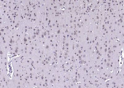 Paraformaldehyde-fixed, paraffin embedded (rat brain); Antigen retrieval by boiling in sodium citrate buffer (pH6.0) for 15min; Block endogenous peroxidase by 3% hydrogen peroxide for 20 minutes; Blocking buffer (normal goat serum) at 37°C for 30min; Antibody incubation with (AEBP1) Polyclonal Antibody, Unconjugated (SL25321R) at 1:200 overnight at 4°C, followed by operating according to SP Kit(Rabbit) (sp-0023) instructionsand DAB staining.
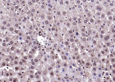 Paraformaldehyde-fixed, paraffin embedded (rat liver); Antigen retrieval by boiling in sodium citrate buffer (pH6.0) for 15min; Block endogenous peroxidase by 3% hydrogen peroxide for 20 minutes; Blocking buffer (normal goat serum) at 37°C for 30min; Antibody incubation with (AEBP1) Polyclonal Antibody, Unconjugated (SL25321R) at 1:200 overnight at 4°C, followed by operating according to SP Kit(Rabbit) (sp-0023) instructionsand DAB staining.
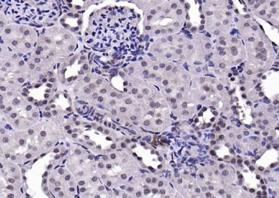 Paraformaldehyde-fixed, paraffin embedded (rat kidney); Antigen retrieval by boiling in sodium citrate buffer (pH6.0) for 15min; Block endogenous peroxidase by 3% hydrogen peroxide for 20 minutes; Blocking buffer (normal goat serum) at 37°C for 30min; Antibody incubation with (AEBP1) Polyclonal Antibody, Unconjugated (SL25321R) at 1:200 overnight at 4°C, followed by operating according to SP Kit(Rabbit) (sp-0023) instructionsand DAB staining.
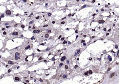 Paraformaldehyde-fixed, paraffin embedded (rat placenta); Antigen retrieval by boiling in sodium citrate buffer (pH6.0) for 15min; Block endogenous peroxidase by 3% hydrogen peroxide for 20 minutes; Blocking buffer (normal goat serum) at 37°C for 30min; Antibody incubation with (AEBP1) Polyclonal Antibody, Unconjugated (SL25321R) at 1:200 overnight at 4°C, followed by operating according to SP Kit(Rabbit) (sp-0023) instructionsand DAB staining.
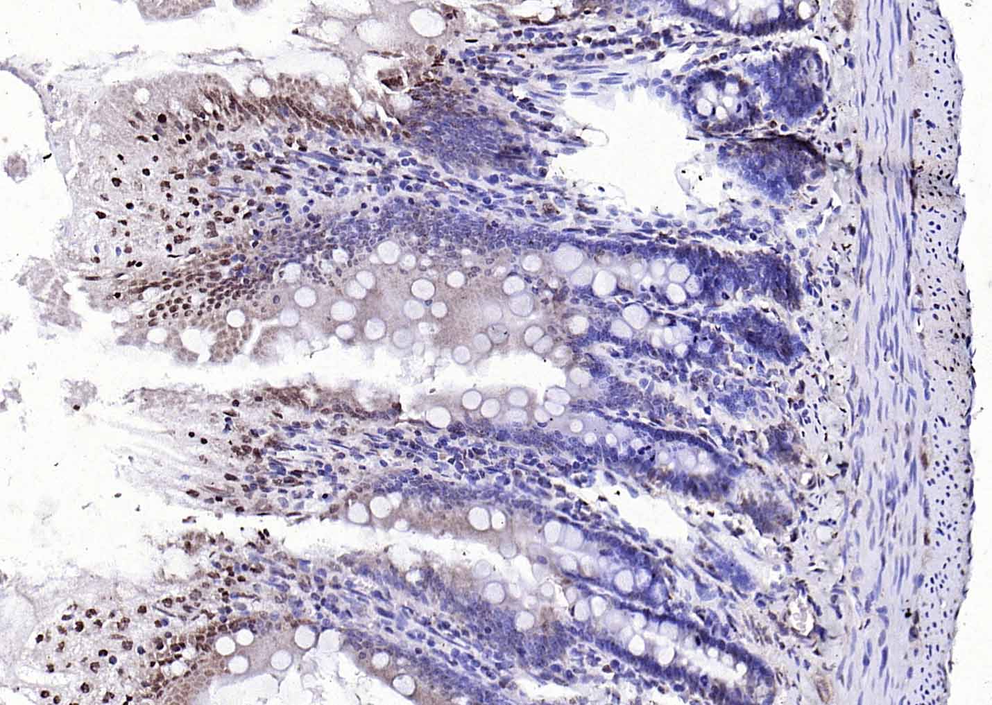 Paraformaldehyde-fixed, paraffin embedded (rat small intestine); Antigen retrieval by boiling in sodium citrate buffer (pH6.0) for 15min; Block endogenous peroxidase by 3% hydrogen peroxide for 20 minutes; Blocking buffer (normal goat serum) at 37°C for 30min; Antibody incubation with (AEBP1) Polyclonal Antibody, Unconjugated (SL25321R) at 1:200 overnight at 4°C, followed by operating according to SP Kit(Rabbit) (sp-0023) instructionsand DAB staining.
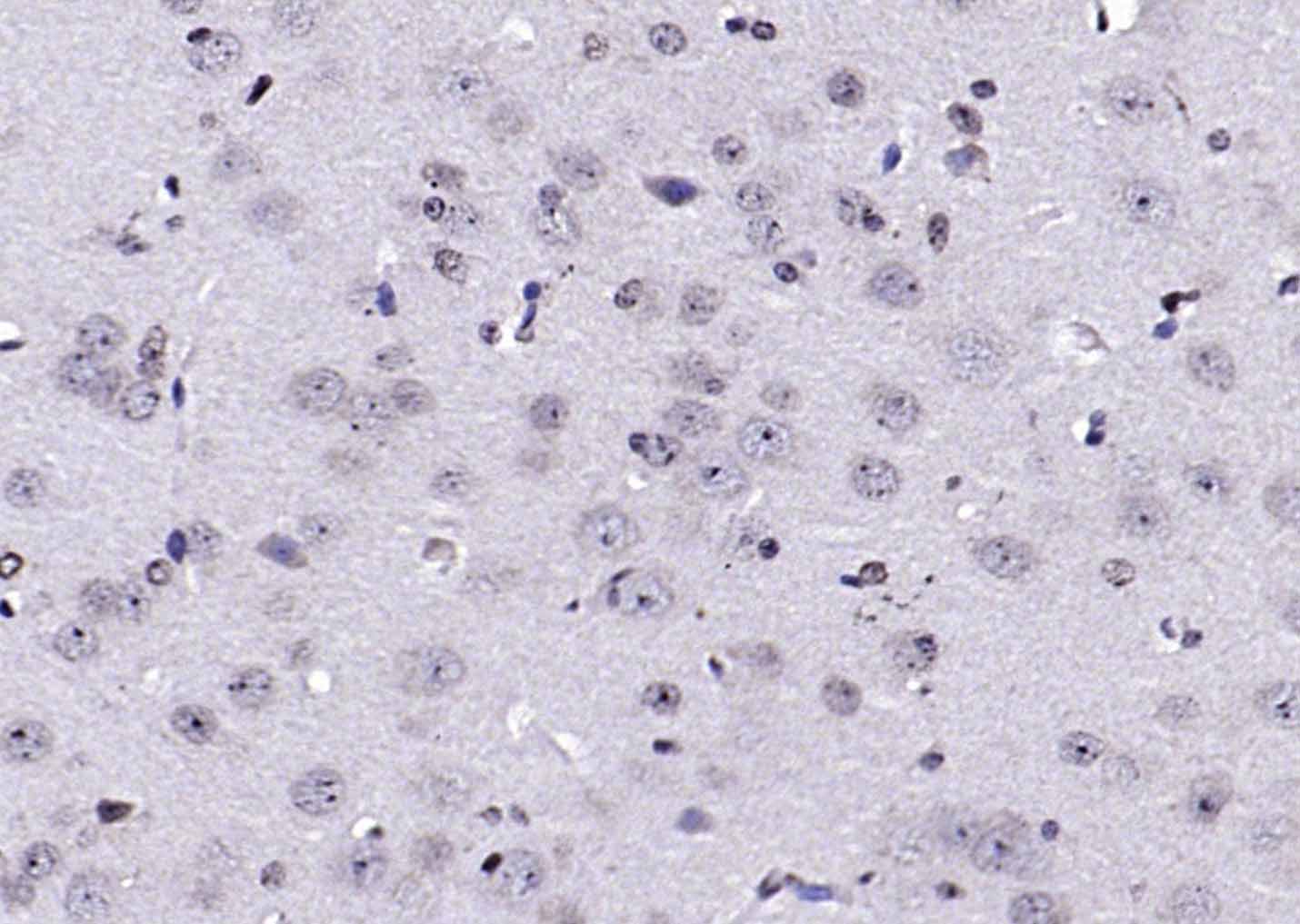 Paraformaldehyde-fixed, paraffin embedded (mouse brain); Antigen retrieval by boiling in sodium citrate buffer (pH6.0) for 15min; Block endogenous peroxidase by 3% hydrogen peroxide for 20 minutes; Blocking buffer (normal goat serum) at 37°C for 30min; Antibody incubation with (AEBP1) Polyclonal Antibody, Unconjugated (SL25321R) at 1:200 overnight at 4°C, followed by operating according to SP Kit(Rabbit) (sp-0023) instructionsand DAB staining.
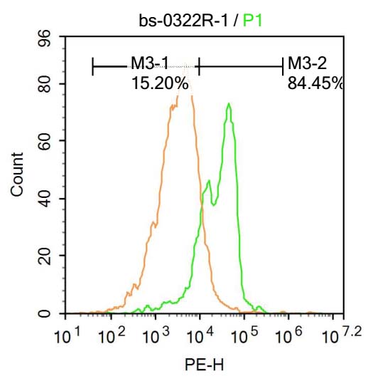 Molt-4 cells were fixed with 4% PFA for 10min at room temperature ,permeabilized with 90% ice-cold methanol for 20 min at -20℃, and incubated in 5% BSA blocking buffer for 30 min at room temperature. Cells were then stained with AEBP1 Antibody(SL0322R)at 1:500 dilution in blocking buffer and incubated for 30 min at room temperature, washed twice with 2%BSA in PBS, followed by secondary antibody incubation for 40 min at room temperature. Acquisitions of 20,000 events were performed. Cells stained with primary antibody (green), and isotype control (orange).
Cartpieces
Totalgoods,subtotals:¥Checkout
Bought notes(bought amounts latest0)
No one bought this product
User Comment(Total0User Comment Num)
- No comment


 +86 571 56623320
 +86 18668110335

