
Rabbit Anti-PRKCB antibody
PKC beta 1 + PKC beta 2; PKCB; PKC B; PKCB1; PKCB2; PRKCB; PRKCB I; PRKCB II; PRKCB1; PRKCB2; Protein kinase C beta 1; Protein kinase C beta 2; Protein kinase C beta; PKC beta 1; PKC beta 2; KPCB_HUMAN; MGC41878; PKC beta; PKC-B; PKC-beta; PRKC B; PRKC B1
View History [Clear]
Details
Product Name PRKCB Chinese Name 蛋白激酶C beta 1/2抗体 Alias PKC beta 1 + PKC beta 2; PKCB; PKC B; PKCB1; PKCB2; PRKCB; PRKCB I; PRKCB II; PRKCB1; PRKCB2; Protein kinase C beta 1; Protein kinase C beta 2; Protein kinase C beta; PKC beta 1; PKC beta 2; KPCB_HUMAN; MGC41878; PKC beta; PKC-B; PKC-beta; PRKC B; PRKC B1; Protein kinase C beta 1; Protein kinase C beta; Protein kinase C beta type. literatures Research Area Cell biology Signal transduction Kinases and Phosphatases Immunogen Species Rabbit Clonality Polyclonal React Species Human, Mouse, Rat, (predicted: Pig, Cow, Rabbit, ) Applications WB=1:500-2000 ELISA=1:5000-10000 IHC-P=1:100-500 IHC-F=1:100-500 Flow-Cyt=1:ug/Test IF=1:100-500 (Paraffin sections need antigen repair)
not yet tested in other applications.
optimal dilutions/concentrations should be determined by the end user.Theoretical molecular weight 74kDa Cellular localization The nucleus cytoplasmic The cell membrane Form Liquid Concentration 1mg/ml immunogen KLH conjugated synthetic peptide derived from human PKC beta 1: 601-673/673 Lsotype IgG Purification affinity purified by Protein A Buffer Solution 0.01M TBS(pH7.4) with 1% BSA, 0.03% Proclin300 and 50% Glycerol. Storage Shipped at 4℃. Store at -20 °C for one year. Avoid repeated freeze/thaw cycles. Attention This product as supplied is intended for research use only, not for use in human, therapeutic or diagnostic applications. PubMed PubMed Product Detail Protein kinase C (PKC) is a family of serine- and threonine-specific protein kinases that can be activated by calcium and the second messenger diacylglycerol. PKC family members phosphorylate a wide variety of protein targets and are known to be involved in diverse cellular signaling pathways. PKC family members also serve as major receptors for phorbol esters, a class of tumor promoters. Each member of the PKC family has a specific expression profile and is believed to play a distinct role. The protein encoded by this gene is one of the PKC family members. It is a calcium-independent and phospholipid-dependent protein kinase. This kinase is important for T-cell activation. It is required for the activation of the transcription factors NF-kappaB and AP-1, and may link the T cell receptor (TCR) signaling complex to the activation of the transcription factors.
Function:
Calcium-activated and phospholipid-dependent serine/threonine-protein kinase involved in various processes such as regulation of the B-cell receptor (BCR) signalosome, apoptosis and transcription regulation. Plays a key role in B-cell activation and function by regulating BCR-induced NF-kappa-B activation and B-cell suvival. Required for recruitment and activation of the IKK kinase to lipid rafts and mediates phosphorylation of CARD11/CARMA1 at 'Ser-559', 'Ser-644' and 'Ser-652', leading to activate the NF-kappa-B signaling. Involved in apoptosis following oxidative damage: in case of oxidative conditions, specifically phosphorylates 'Ser-36' of isoform p66Shc of SHC1, leading to mitochondrial accumulation of p66Shc, where p66Shc acts as a reactive oxygen species producer. Acts as a coactivator of androgen receptor (ANDR)-dependent transcription, by being recruited to ANDR target genes and specifically mediating phosphorylation of 'Thr-6' of histone H3 (H3T6ph), a specific tag for epigenetic transcriptional activation that prevents demethylation of histone H3 'Lys-4' (H3K4me) by LSD1/KDM1A. Also involved in triglyceride homeostasis. Serves as the receptor for phorbol esters, a class of tumor promoters.
Subunit:
Recruited in a circadian manner into a nuclear complex which also includes BMAL1 and GNB2L1/RACK1 (By similarity). Interacts with ADAP1/CENTA1, CSPG4 and PRKCABP. Binds to SDPR in the presence of phosphatidylserine. Interacts with PICK1 (via PDZ domain). Interacts with TRIM41.
Subcellular Location:
Cytoplasm. Cell membrane; Peripheral membrane protein. Mitochondrion membrane; Peripheral membrane protein (Probable). Nucleus.
Post-translational modifications:
Phosphorylation on Thr-500 within the activation loop renders it competent to autophosphorylate. Subsequent autophosphorylation of Thr-642 maintains catalytic competence, and autophosphorylation on Ser-661 appears to release the kinase into the cytosol. Autophosphorylation on other sites i.e. in the N-terminal and hinge regions have no effect on enzyme activity.
Similarity:
Belongs to the protein kinase superfamily. AGC Ser/Thr protein kinase family. PKC subfamily.
Contains 1 AGC-kinase C-terminal domain.
Contains 1 C2 domain.
Contains 2 phorbol-ester/DAG-type zinc fingers.
Contains 1 protein kinase domain.
SWISS:
P05771
Gene ID:
5579
Database links:Entrez Gene: 5579 Human
Entrez Gene: 18751 Mouse
Omim: 176970 Human
SwissProt: P05771 Human
SwissProt: P68404 Mouse
Unigene: 460355 Human
Unigene: 207496 Mouse
Unigene: 446371 Mouse
Unigene: 91118 Rat
Product Picture 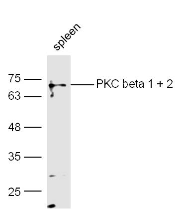 Sample:
spleen (Mouse) Lysate at 40 ug
Primary: Anti-PKC beta 1 + 2 (SL0267R) at 1/300 dilution
Secondary: IRDye800CW Goat Anti-Rabbit IgG at 1/20000 dilution
Predicted band size: 74 kD
Observed band size: 74 kD
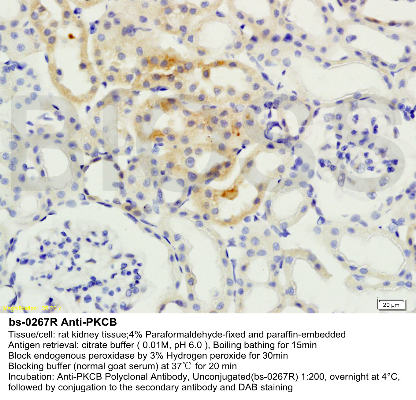
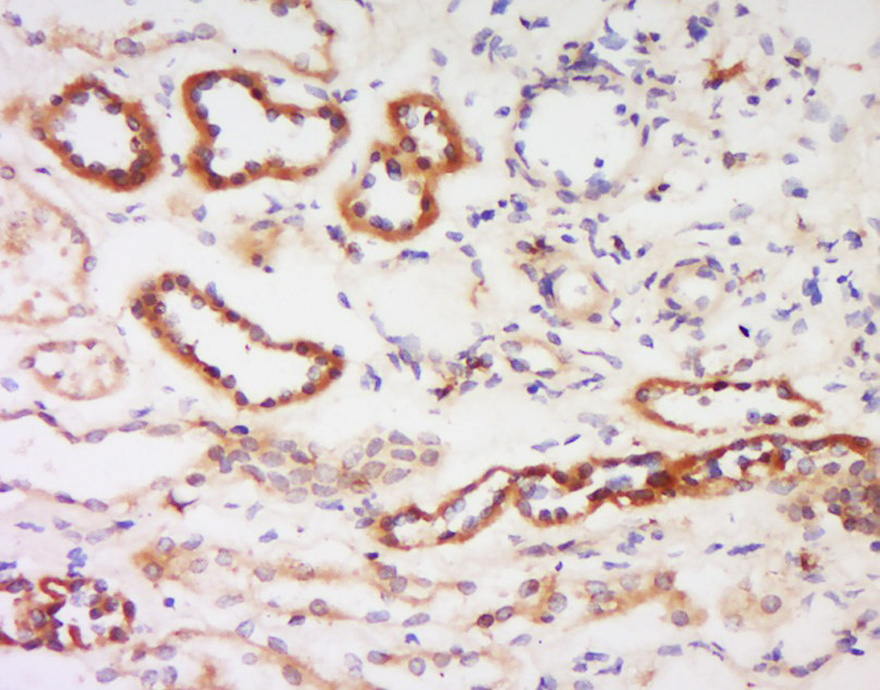 Tissue/cell: human kidney tissue; 4% Paraformaldehyde-fixed and paraffin-embedded;
Antigen retrieval: citrate buffer ( 0.01M, pH 6.0 ), Boiling bathing for 15min; Block endogenous peroxidase by 3% Hydrogen peroxide for 30min; Blocking buffer (normal goat serum,C-0005) at 37℃ for 20 min;
Incubation: Anti- PKC beta-1+2 Polyclonal Antibody, Unconjugated(SL0267R) 1:200, overnight at 4°C, followed by conjugation to the secondary antibody(SP-0023) and DAB(C-0010) staining
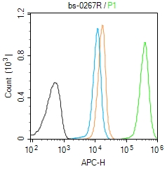 Blank control (Black line): Molt4 (Black).
Primary Antibody (green line):Rabbit Anti-PKC beta 1 + 2 antibody (SL0267R)
Dilution:1μg /10^6 cells;
Isotype Control Antibody (orange line): Rabbit IgG .
Secondary Antibody (white blue line): Goat anti-rabbit IgG-AF647
Dilution: 1μg /test.
Protocol
The cells were fixed with 4% PFA (10min at room temperature)and then permeabilized with 90% ice-cold methanol for 20 min at room temperature. The cells were then incubated in 5%BSA to block non-specific protein-protein interactions for 30 min at room temperature .Cells stained with Primary Antibody for 30 min at room temperature. The secondary antibody used for 40 min at room temperature. Acquisition of 20,000 events was performed.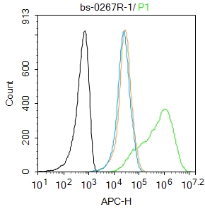 Blank control:Molt4.
Primary Antibody (green line): Rabbit Anti-PKC beta 1 + 2 antibody (SL0267R)
Dilution: 1μg /10^6 cells;
Isotype Control Antibody (orange line): Rabbit IgG .
Secondary Antibody : Goat anti-rabbit IgG-AF647
Dilution: 1μg /test.
Protocol
The cells were fixed with 4% PFA (10min at room temperature)and then permeabilized with 0.1% PBST for 20 min at room temperature. The cells were then incubated in 5%BSA to block non-specific protein-protein interactions for 30 min at room temperature .Cells stained with Primary Antibody for 30 min at room temperature. The secondary antibody used for 40 min at room temperature. Acquisition of 20,000 events was performed.
Cartpieces
Totalgoods,subtotals:¥Checkout
Bought notes(bought amounts latest0)
No one bought this product
User Comment(Total0User Comment Num)
- No comment


 +86 571 56623320
 +86 18668110335

