
Rabbit Anti-14-3-3 Alpha + Beta + Gamma + Delta + Epsilon antibody
YWHAB; YWHAZ; 14-3-3 alpha/beta/Gamma/Delta/Epsilon; 14-3-3 alpha/beta; 14 3 3 protein beta; 14 3 3 protein beta/alpha; 14 3 3 protein zeta; KCIP 1; Protein 1054; Protein kinase C inhibitor protein 1; 14 3 3 protein delta; 14 3 3 protein gamma; 14 3 3 pro
View History [Clear]
Details
Product Name 14-3-3 Alpha + Beta + Gamma + Delta + Epsilon Chinese Name 14-3-3蛋白/14-3-3 α/β/γ/δ/ε亚型抗体 Alias YWHAB; YWHAZ; 14-3-3 alpha/beta/Gamma/Delta/Epsilon; 14-3-3 alpha/beta; 14 3 3 protein beta; 14 3 3 protein beta/alpha; 14 3 3 protein zeta; KCIP 1; Protein 1054; Protein kinase C inhibitor protein 1; 14 3 3 protein delta; 14 3 3 protein gamma; 14 3 3 protein Epsilon; 1433Z_HUMAN; 14-3-3ε; 14-3-3 α; 14-3-3 β; 14-3-3 γ; 14-3-3 δ. Research Area Apoptosis Immunogen Species Rabbit Clonality Polyclonal React Species Human, Mouse, Rat, (predicted: Sheep, Fruit Fly, ) Applications WB=1:500-2000 IHC-P=1:100-500 IHC-F=1:100-500 Flow-Cyt=1μg /test. IF=1:100-500 (Paraffin sections need antigen repair)
not yet tested in other applications.
optimal dilutions/concentrations should be determined by the end user.Theoretical molecular weight 27kDa Cellular localization cytoplasmic Form Liquid Concentration 1mg/ml immunogen KLH conjugated synthetic peptide derived from human 14-3-3: 174-245/245 Lsotype IgG Purification affinity purified by Protein A Buffer Solution 0.01M TBS(pH7.4) with 1% BSA, 0.03% Proclin300 and 50% Glycerol. Storage Shipped at 4℃. Store at -20 °C for one year. Avoid repeated freeze/thaw cycles. Attention This product as supplied is intended for research use only, not for use in human, therapeutic or diagnostic applications. PubMed PubMed Product Detail 14-3-3 are activates tyrosine and tryptophan hydroxylases in the presence of Ca (2+)/calmodulin-dependent protein kinase II, and strongly activates protein kinase C. Is probably a multifunctional regulator of the cell signaling processes mediated by both kinases. Activates the ADP-ribosyltransferase (exoS) activity of bacterial origin. 14-3-3 proteins are localized in neurons, and are axonally transported to the nerve terminals. They may be also present, at lower levels, in various other eukaryotic tissues. It belongs to the 14-3-3 family.
This antibody is reactive with 14-3-3 Alpha, Beta, Gamma, Delta, Epsilon.
Function:
Adapter protein implicated in the regulation of a large spectrum of both general and specialized signaling pathways. Binds to a large number of partners, usually by recognition of a phosphoserine or phosphothreonine motif. Binding generally results in the modulation of the activity of the binding partner.
Subunit:
Interacts with CDK16 and BSPRY. Interacts with WEE1 (C-terminal). Interacts with SAMSN1. Interacts with MLF1 (phosphorylated form); the interaction retains it in the cytoplasm. Interacts with Thr-phosphorylated ITGB2. Interacts with BCL2L11. Homodimer. Heterodimerizes with YWHAE. Homo- and hetero-dimerization is inhibited by phosphorylation on Ser-58. Interacts with FOXO4, NOXA1, SSH1 and ARHGEF2. Interacts with Pseudomonas aeruginosa exoS (unphosphorylated form). Interacts with BAX; the interaction occurs in the cytoplasm. Under stress conditions, MAPK8-mediated phosphorylation releases BAX to mitochondria. Interacts with phosphorylated RAF1; the interaction is inhibited when YWHAZ is phosphorylated on Thr-232. Interacts with TP53; the interaction enhances p53 transcriptional activity. The Ser-58 phosphorylated form inhibits this interaction and p53 transcriptional activity. Interacts with ABL1 (phosphorylated form); the interaction retains ABL1 in the cytoplasm. Interacts with PKA-phosphorylated AANAT; the interaction modulates AANAT enzymatic activity by increasing affinity for arylalkylamines and acetyl-CoA and protecting the enzyme from dephosphorylation and proteasomal degradation. It may also prevent thiol-dependent inactivation. Interacts with AKT1; the interaction phosphorylates YWHAZ and modulates dimerization. Interacts with GAB2 and TLK2.
Subcellular Location:
Cytoplasm. Melanosome. Note=Located to stage I to stage IV melanosomes.
Post-translational modifications:
The delta, brain-specific form differs from the zeta form in being phosphorylated. Phosphorylation on Ser-184 by MAPK8; promotes dissociation of BAX and translocation of BAX to mitochondria. Phosphorylation on Ser-58 by PKA; disrupts homodimerization and heterodimerization with YHAE and TP53. This phosphorylation appears to be activated by sphingosine. Phosphorylation on Thr-232; inhibits binding of RAF1.
Similarity:
Belongs to the 14-3-3 family.
SWISS:
P31946
Gene ID:
7529
Database links:Entrez Gene: 7529 Human
Entrez Gene: 54401 Mouse
Omim: 601289 Human
SwissProt: P31946 Human
SwissProt: Q9CQV8 Mouse
Unigene: 643544 Human
Unigene: 34319 Mouse
Unigene: 485025 Mouse
Unigene: 8653 Rat
信号传导(Signaling Intermediates)
14-3-3蛋白是一个涉及调节Apoptosis、促细胞分裂信号传导和细胞周期关卡的蛋白质家族。它被认为是通过与丝氨酸残基磷酸化的蛋白质的结合介导的信号传导中的关键调节物。通过与Bad(相关死亡因子)的结合, 14-3-3 蛋白由于将Bad隔离于胞液而防止了Apoptosis。
蛋白是14-3-3家族成员。它广泛分布于哺乳动物、两栖类、Insect、Botany和酵母菌的真核生物高度保守性多功能蛋白质。
目前已知至少有16个成员。此抗体识别分子量为30-31kDa的14-3-3蛋白αβγδε亚型。Product Picture 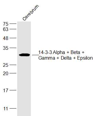 Sample:
Cerebrum (Mouse) Lysate at 40 ug
Primary: Anti-14-3-3 Alpha + Beta + Gamma + Delta + Epsilon (SL0237R) at 1/300 dilution
Secondary: IRDye800CW Goat Anti-Rabbit IgG at 1/20000 dilution
Predicted band size: 27 kD
Observed band size: 27 kD
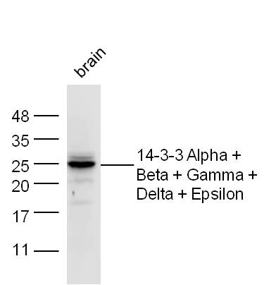 Sample:Brain Cell Lysate at 40 ug
Primary: Anti-14-3-3 Alpha + Beta + Gamma + Delta + Epsilon (SL0237R) at 1/300 dilution
Secondary: IRDye800CW Goat Anti-Rabbit IgG at 1/20000 dilution
Predicted band size: 27 kD
Observed band size: 27 kD
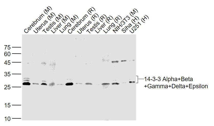 Sample:
Lane 1: Cerebrum (Mouse) Lysate at 40 ug
Lane 2: Uterus (Mouse) Lysate at 40 ug
Lane 3: Testis (Mouse) Lysate at 40 ug
Lane 4: Liver (Mouse) Lysate at 40 ug
Lane 5: Lung (Mouse) Lysate at 40 ug
Lane 6: Cerebrum (Rat) Lysate at 40 ug
Lane 7: Uterus (Rat) Lysate at 40 ug
Lane 8: Testis (Rat) Lysate at 40 ug
Lane 9: Liver (Rat) Lysate at 40 ug
Lane 10: Lung (Rat) Lysate at 40 ug
Lane 11: NIH/3T3 (Mouse) Cell Lysate at 30 ug
Lane 12: Siha (Human) Cell Lysate at 30 ug
Lane 13: U251 (Human) Cell Lysate at 30 ug
Primary: Anti-14-3-3 Alpha+Beta+Gamma+Delta+Epsilon (SL0237R) at 1/1000 dilution
Secondary: IRDye800CW Goat Anti-Rabbit IgG at 1/20000 dilution
Predicted band size: 28 kD
Observed band size: 28 kD
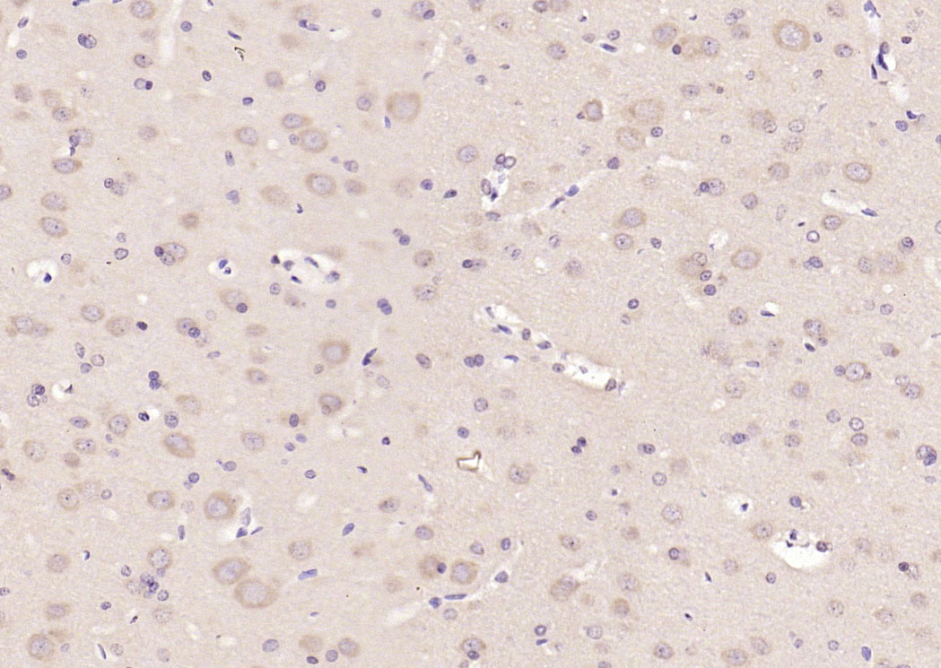 Paraformaldehyde-fixed, paraffin embedded (rat brain); Antigen retrieval by boiling in sodium citrate buffer (pH6.0) for 15min; Block endogenous peroxidase by 3% hydrogen peroxide for 20 minutes; Blocking buffer (normal goat serum) at 37°C for 30min; Antibody incubation with (14-3-3 Alpha + Beta + Gamma + Delta + Epsilon) Polyclonal Antibody, Unconjugated (SL1024R) at 1:200 overnight at 4°C, followed by operating according to SP Kit(Rabbit) (sp-0023) instructionsand DAB staining.
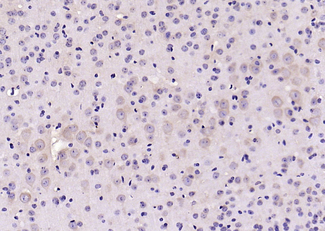 Paraformaldehyde-fixed, paraffin embedded (mouse brain); Antigen retrieval by boiling in sodium citrate buffer (pH6.0) for 15min; Block endogenous peroxidase by 3% hydrogen peroxide for 20 minutes; Blocking buffer (normal goat serum) at 37°C for 30min; Antibody incubation with (14-3-3 Alpha + Beta + Gamma + Delta + Epsilon) Polyclonal Antibody, Unconjugated (SL1024R) at 1:200 overnight at 4°C, followed by operating according to SP Kit(Rabbit) (sp-0023) instructionsand DAB staining.
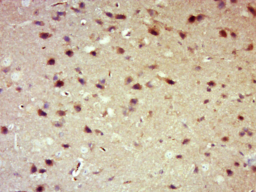 Paraformaldehyde-fixed, paraffin embedded (Mouse brain); Antigen retrieval by boiling in sodium citrate buffer (pH6.0) for 15min; Block endogenous peroxidase by 3% hydrogen peroxide for 20 minutes; Blocking buffer (normal goat serum) at 37°C for 30min; Antibody incubation with (14-3-3 Alpha + Beta + Gamma + Delta + Epsilon) Polyclonal Antibody, Unconjugated (SL0237R) at 1:500 overnight at 4°C, followed by a conjugated secondary (sp-0023) for 20 minutes and DAB staining.
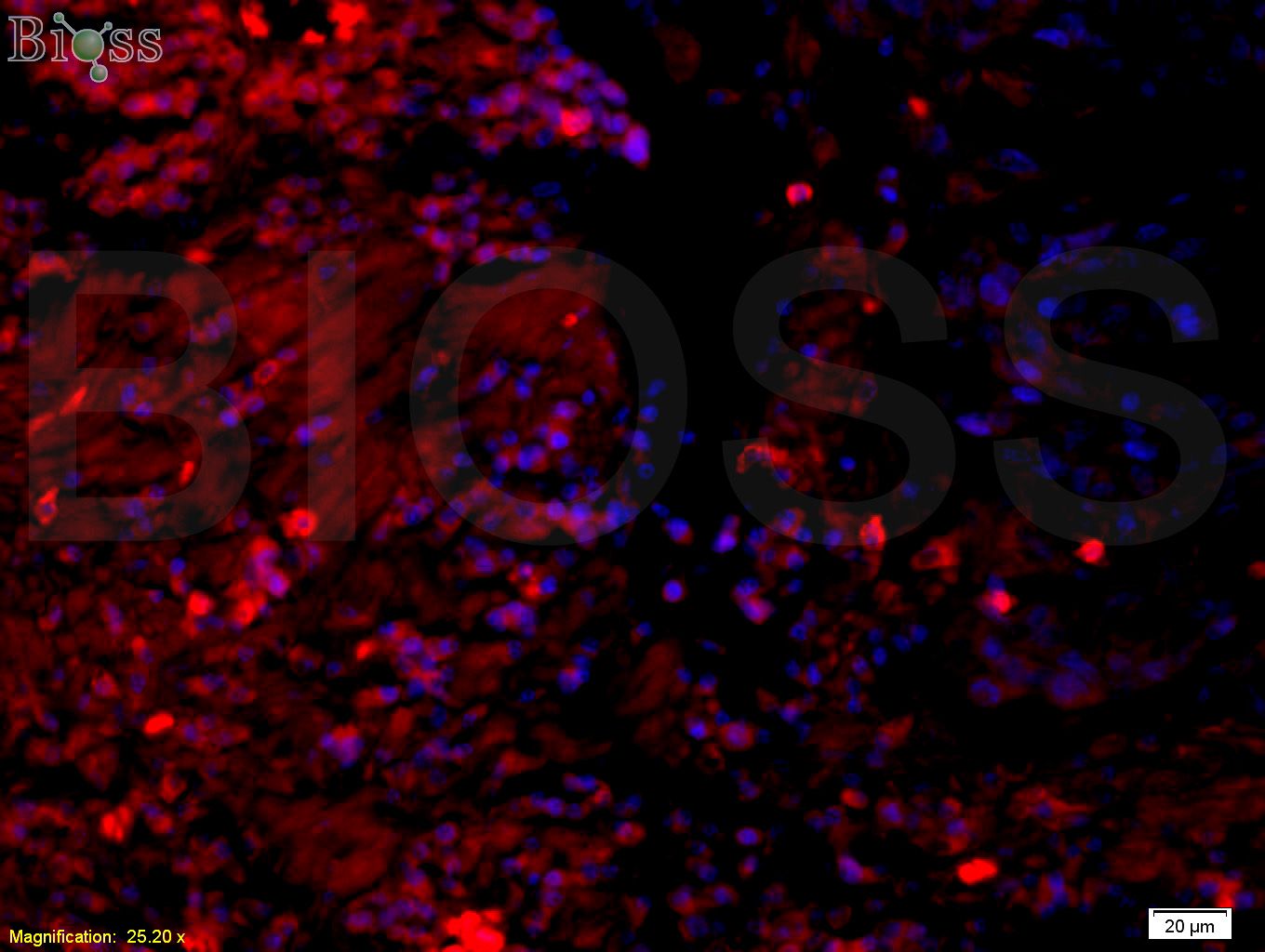 Tissue/cell: human rectal carcinoma;4% Paraformaldehyde-fixed and paraffin-embedded;
Antigen retrieval: citrate buffer ( 0.01M, pH 6.0 ), Boiling bathing for 15min; Blocking buffer (normal goat serum,C-0005) at 37℃ for 20 min;
Incubation: Anti-14-3-3 Polyclonal Antibody, Unconjugated(SL0237R) 1:200, overnight at 4°C; The secondary antibody was Goat Anti-Rabbit IgG, PE conjugated(SL0295G-PE)used at 1:200 dilution for 40 minutes at 37°C. DAPI(5ug/ml,blue,C-0033) was used to stain the cell nuclei
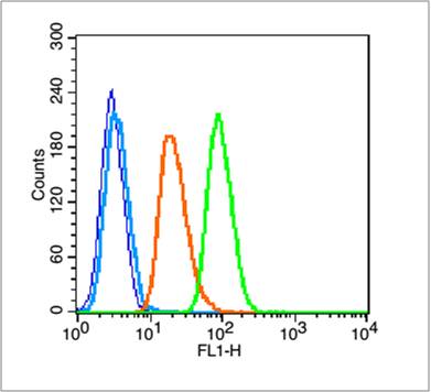 Blank control (blue line): A549 (blue).
Primary Antibody (green line): Rabbit Anti-14-3-3 Alpha + Beta + Gamma + Delta + Epsilon antibody (SL0237R)
Dilution: 1μg /10^6 cells;
Isotype Control Antibody (orange line): Rabbit IgG .
Secondary Antibody (white blue line): Goat anti-rabbit IgG-FITC
Dilution: 1μg /test.
Protocol
The cells were fixed with 70% ethanol (Overnight at 4℃) and then permeabilized with 90% ice-cold methanol for 30 min on ice. Cells stained with Primary Antibody for 30 min at room temperature. The cells were then incubated in 1 X PBS/2%BSA/10% goat serum to block non-specific protein-protein interactions followed by the antibody for 15 min at room temperature. The secondary antibody used for 40 min at room temperature. Acquisition of 20,000 events was performed.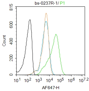 Blank control:A431.
Primary Antibody (green line): Rabbit Anti-14-3-3 Alpha + Beta + Gamma + Delta + Epsilon antibody (SL0237R)
Dilution: 1μg /10^6 cells;
Isotype Control Antibody (orange line): Rabbit IgG .
Secondary Antibody : Goat anti-rabbit IgG-AF647
Dilution: 1μg /test.
Protocol
The cells were fixed with 4% PFA (10min at room temperature)and then permeabilized with 0.1% PBST for 20 min at room temperature. The cells were then incubated in 5%BSA to block non-specific protein-protein interactions for 30 min at room temperature .Cells stained with Primary Antibody for 30 min at room temperature. The secondary antibody used for 40 min at room temperature. Acquisition of 20,000 events was performed.
Cartpieces
Totalgoods,subtotals:¥Checkout
Bought notes(bought amounts latest0)
No one bought this product
User Comment(Total0User Comment Num)
- No comment


 +86 571 56623320
 +86 18668110335

