
Rabbit Anti-TERT antibody
Telomerase catalytic subunit; EST2; hEST2; TCS1; Telomerase associated protein 2; Telomere Reverse Transcriptase; TP2; TRT; Telomerase reverse transcriptase; Telomerase Catalytic Subunit; Telomerase-associated protein 2; TERT_HUMAN.
View History [Clear]
Details
Product Name TERT Chinese Name 端粒酶反转录酶抗体 Alias Telomerase catalytic subunit; EST2; hEST2; TCS1; Telomerase associated protein 2; Telomere Reverse Transcriptase; TP2; TRT; Telomerase reverse transcriptase; Telomerase Catalytic Subunit; Telomerase-associated protein 2; TERT_HUMAN. literatures Research Area Tumour Cell biology immunology Neurobiology Signal transduction Apoptosis Cyclin Kinases and Phosphatases Immunogen Species Rabbit Clonality Polyclonal React Species Human, Mouse, Rat, Applications WB=1:500-2000 ELISA=1:5000-10000 IHC-P=1:100-500 IHC-F=1:100-500 Flow-Cyt=1μg/Test ICC=1:100 IF=1:100-500 (Paraffin sections need antigen repair)
not yet tested in other applications.
optimal dilutions/concentrations should be determined by the end user.Theoretical molecular weight 125kDa Cellular localization The nucleus cytoplasmic Form Liquid Concentration 1mg/ml immunogen KLH conjugated synthetic peptide derived from human TERT: 601-750/1132 Lsotype IgG Purification affinity purified by Protein A Buffer Solution 0.01M TBS(pH7.4) with 1% BSA, 0.03% Proclin300 and 50% Glycerol. Storage Shipped at 4℃. Store at -20 °C for one year. Avoid repeated freeze/thaw cycles. Attention This product as supplied is intended for research use only, not for use in human, therapeutic or diagnostic applications. PubMed PubMed Product Detail Telomerase is a ribonucleoprotein polymerase that maintains telomere ends by addition of the telomere repeat TTAGGG. The enzyme consists of a protein component with reverse transcriptase activity, encoded by this gene, and an RNA component which serves as a template for the telomere repeat. Telomerase expression plays a role in cellular senescence, as it is normally repressed in postnatal somatic cells resulting in progressive shortening of telomeres. Deregulation of telomerase expression in somatic cells may be involved in oncogenesis. Studies in mouse suggest that telomerase also participates in chromosomal repair, since de novo synthesis of telomere repeats may occur at double-stranded breaks. Alternatively spliced variants encoding different isoforms of telomerase reverse transcriptase have been identified; the full-length sequence of some variants has not been determined. Alternative splicing at this locus is thought to be one mechanism of regulation of telomerase activity. [provided by RefSeq, Jul 2008].
Function:
Telomerase is a ribonucleoprotein enzyme essential for the replication of chromosome termini in most eukaryotes. Active in progenitor and cancer cells. Inactive, or very low activity, in normal somatic cells. Catalytic component of the teleromerase holoenzyme complex whose main activity is the elongation of telomeres by acting as a reverse transcriptase that adds simple sequence repeats to chromosome ends by copying a template sequence within the RNA component of the enzyme. Catalyzes the RNA-dependent extension of 3'-chromosomal termini with the 6-nucleotide telomeric repeat unit, 5'-TTAGGG-3'. The catalytic cycle involves primer binding, primer extension and release of product once the template boundary has been reached or nascent product translocation followed by further extension. More active on substrates containing 2 or 3 telomeric repeats. Telomerase activity is regulated by a number of factors including telomerase complex-associated proteins, chaperones and polypeptide modifiers. Modulates Wnt signaling. Plays important roles in aging and antiapoptosis.
Subunit:
Homodimer; dimerization is required to produce a functional complex. Oligomer; can form oligomers in the absence of the telomerase RNA template component (TERC). Catalytic subunit of the telomerase holoenzyme complex composed minimally of TERT and TERC. The telomerase complex is composed of TERT, DKC1, WDR79/TCAB1, NOP10, NHP2, GAR1, TEP1, EST1A, POT1 and a telomerase RNA template component (TERC). The molecular chaperone HSP90/P23 complex is required for correct assembly and stabilization of the active telomerase. Interacts directly with HSP90A and PTGES3. Interacts with HSPA1A; the interaction occurs in the absence of TERC and dissociates once the complex has formed. Interacts with RAN; the interaction promotes nuclear export of TERT. Interacts with XPO1. Interacts with PTPN11; the interaction retains TERT in the nucleus. Interacts with NCL (via RRM1 and C-terminal RRM4/Arg/Gly-rich domains); the interaction is important for nucleolar localization of TERT. Interacts with SMARCA4 (via the bromodomain); the interaction regulates Wnt-mediated signaling. Interacts with MCRS1 (isoform MCRS2); the interaction inhibits in vitro telomerase activity. Interacts with PIF1; the interaction has no effect on the elongation activity of TERT. Interacts with PML; the interaction recruits TERT to PML bodies and inhibits telomerase activity.
Subcellular Location:
Nucleus, nucleolus. Nucleus, nucleoplasm. Nucleus. Chromosome, telomere. Cytoplasm. Nucleus, PML body. Note=Shuttling between nuclear and cytoplasm depends on cell cycle, phosphorylation states, transformation and DNA damage. Diffuse localization in the nucleoplasm. Enriched in nucleoli of certain cell types. Translocated to the cytoplasm via nuclear pores in a CRM1/RAN-dependent manner involving oxidative stress-mediated phosphorylation at Tyr-707. Dephosphorylation at this site by SHP2 retains TERT in the nucleus. Translocated to the nucleus by phosphorylation by AKT.
Tissue Specificity:
Expressed at a high level in thymocyte subpopulations, at an intermediate level in tonsil T-lymphocytes, and at a low to undetectable level in peripheral blood T-lymphocytes.
Post-translational modifications:
Ubiquitinated, leading to proteasomal degradation.
Phosphorylation at Tyr-707 under oxidative stress leads to translocation of TERT to the cytoplasm and reduces its antiapoptotic activity. Dephosphorylated by SHP2/PTPN11 leading to nuclear retention. Phosphorylation by the AKT pathway promotes nuclear location.
DISEASE:
Note=Activation of telomerase has been implicated in cell immortalization and cancer cell pathogenesis.
Defects in TERT are associated with susceptibilty to aplastic anemia (AA) [MIM:609135]. AA is a rare disease in which the reduction of the circulating blood cells results from damage to the stem cell pool in bone marrow. In most patients, the stem cell lesion is caused by an autoimmune attack. T-lymphocytes, activated by an endogenous or exogenous, and most often unknown antigenic stimulus, secrete cytokines, including IFN-gamma, which would in turn be able to suppress hematopoiesis.
Note=Genetic variations in TERT are associated with coronary artery disease (CAD).
Defects in TERT are the cause of dyskeratosis congenital autosomal dominant type 2 (DKCA2) [MIM:613989]. A rare multisystem disorder caused by defective telomere maintenance. It is characterized by progressive bone marrow failure, and the clinical triad of reticulated skin hyperpigmentation, nail dystrophy, and mucosal leukoplakia. Common but variable features include premature graying, aplastic anemia, low platelets, osteoporosis, pulmonary fibrosis, and liver fibrosis among others. Early mortality is often associated with bone marrow failure, infections, fatal pulmonary complications, or malignancy.
Defects in TERT are the cause of dyskeratosis congenital autosomal recessive type 4 (DKCB4) [MIM:613989]. A rare multisystem disorder caused by defective telomere maintenance. It is characterized by progressive bone marrow failure, and the clinical triad of reticulated skin hyperpigmentation, nail dystrophy, and mucosal leukoplakia. Common but variable features include premature graying, aplastic anemia, low platelets, osteoporosis, pulmonary fibrosis, and liver fibrosis among others. Early mortality is often associated with bone marrow failure, infections, fatal pulmonary complications, or malignancy.
Defects in TERT are a cause of susceptibility to pulmonary fibrosis idiopathic (IPF) [MIM:178500]. Pulmonary fibrosis is a lung disease characterized by shortness of breath, radiographically evident diffuse pulmonary infiltrates, and varying degrees of inflammation and fibrosis on biopsy. It results in acute lung injury with subsequent scarring and endstage lung disease.
Similarity:
Belongs to the reverse transcriptase family. Telomerase subfamily.
Contains 1 reverse transcriptase domain.
SWISS:
O14746
Gene ID:
7015
Database links:
Entrez Gene: 7015 Human
Entrez Gene: 21752 Mouse
SwissProt: O14746 Human
SwissProt: O70372 Mouse
端粒酶是一种依赖RNA的DNA聚合酶,催化合成端粒体的DNA重复序列,并引导端粒添加到染色体的尾端,对维持染色体的长度、调节细胞增殖和凋亡起重要作用。
端粒酶主要存在于恶性Tumour之中,一般在大多数正常组织中没有活性或活性极低,同时由于端粒酶在恶性Tumour的发展中起着关键作用,所以通过各种途径抑制端粒酶的活性可能有效地抑制大多数Tumour的生长,而对大多数正常细胞没有影响。这种抑制作用可以通过直接抑制端粒酶活性、抑制端粒酶RNA或端粒酶蛋白成分以及诱导Tumour细胞发生分化等方法实现。
端粒酶与恶性Tumour之间令人惊异的相关性使它在Tumour的诊断和治疗上有望成为行之有效的新的靶目标。Product Picture 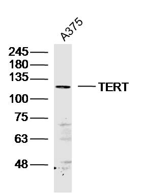 Sample: A375 (human)cell Lysate at 40 ug
Primary: Anti-TERT (SL0233R)at 1/300 dilution
Secondary: IRDye800CW Goat Anti-Rabbit IgG at 1/20000 dilution
Predicted band size: 125kD
Observed band size: 125 kD
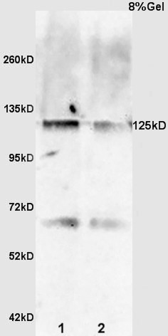 Sample:
Spleen(Rat) lysate at 30ug;
Brain(Rat) lysate at 30ug;
Primary: Anti-TERT (SL0233R) at 1:200 dilution;
Secondary: HRP conjugated Goat Anti-Rabbit IgG(SL0295G-HRP) at 1: 3000 dilution;
Predicted band size : 125kD
Observed band size : 125kD
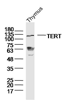 Sample: Thymus (Mouse) Lysate at 40 ug
Primary: Anti-TERT (SL0233R) at 1/300 dilution
Secondary: IRDye800CW Goat Anti-Rabbit IgG at 1/20000 dilution
Predicted band size: 125kD
Observed band size: 125kD
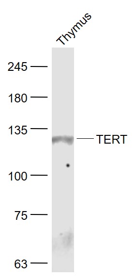 Sample:
Thymus (Mouse) Lysate at 40 ug
Primary: Anti- TERT (SL0233R) at 1/1000 dilution
Secondary: IRDye800CW Goat Anti-Rabbit IgG at 1/20000 dilution
Predicted band size: 125 kD
Observed band size: 125 kD
 Sample:
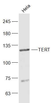
Hela(Human) Cell Lysate at 30 ug
Primary: Anti-TERT (SL0233R) at 1/1000 dilution
Secondary: IRDye800CW Goat Anti-Rabbit IgG at 1/20000 dilution
Predicted band size: 125 kD
Observed band size: 125 kD
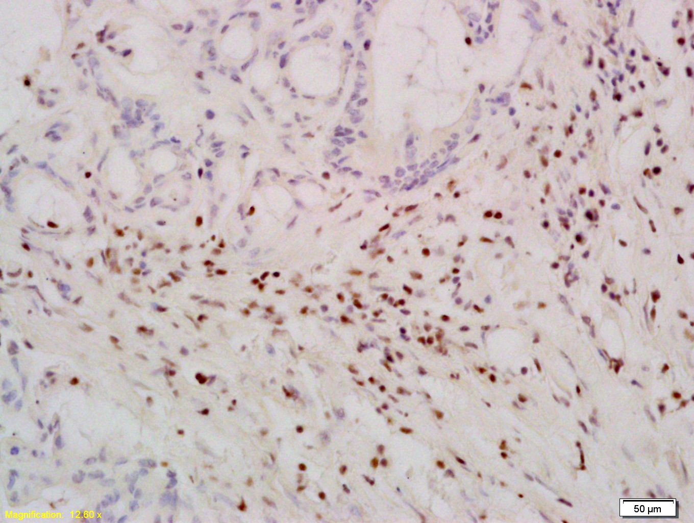
 Tissue/cell: human stomach carcinoma; 4% Paraformaldehyde-fixed and paraffin-embedded;
Antigen retrieval: citrate buffer ( 0.01M, pH 6.0 ), Boiling bathing for 15min; Block endogenous peroxidase by 3% Hydrogen peroxide for 30min; Blocking buffer (normal goat serum,C-0005) at 37℃ for 20 min;
Incubation: Anti-TERT Polyclonal Antibody, Unconjugated(SL0233R) 1:400, overnight at 4°C, followed by conjugation to the secondary antibody(SP-0023) and DAB(C-0010) staining
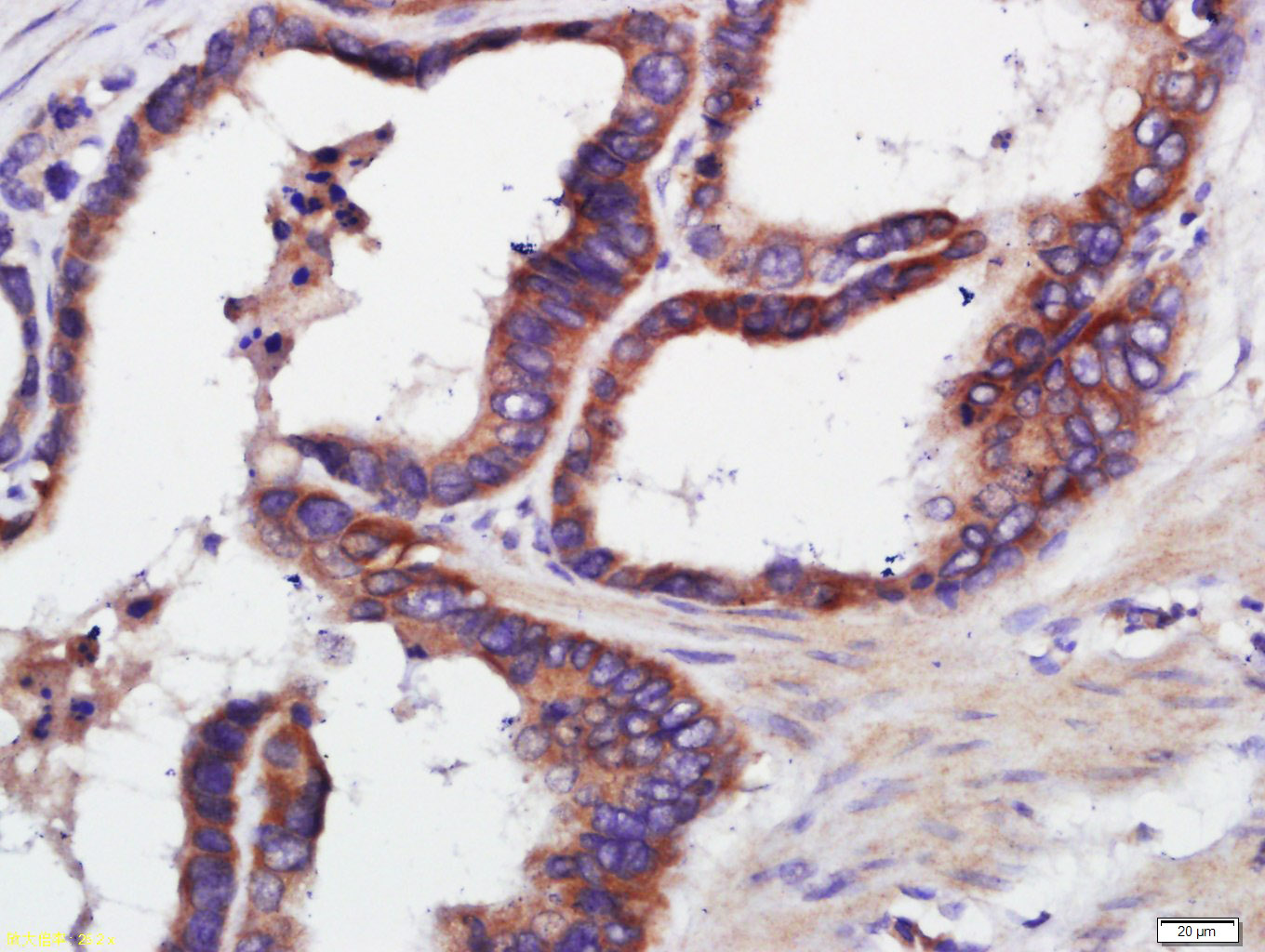
 Tissue/cell: human colon carcinoma; 4% Paraformaldehyde-fixed and paraffin-embedded;
Antigen retrieval: citrate buffer ( 0.01M, pH 6.0 ), Boiling bathing for 15min; Block endogenous peroxidase by 3% Hydrogen peroxide for 30min; Blocking buffer (normal goat serum,C-0005) at 37℃ for 20 min;
Incubation: Anti-TERT Polyclonal Antibody, Unconjugated(SL0233R) 1:200, overnight at 4°C, followed by conjugation to the secondary antibody(SP-0023) and DAB(C-0010) staining
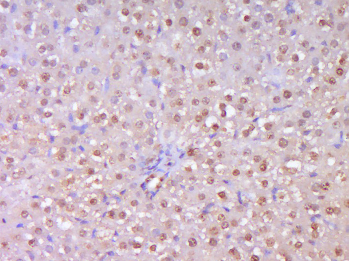 Paraformaldehyde-fixed, paraffin embedded (Rat liver); Antigen retrieval by boiling in sodium citrate buffer (pH6.0) for 15min; Block endogenous peroxidase by 3% hydrogen peroxide for 20 minutes; Blocking buffer (normal goat serum) at 37°C for 30min; Antibody incubation with (TERT) Polyclonal Antibody, Unconjugated (SL0233R) at 1:400 overnight at 4°C, followed by operating according to SP Kit(Rabbit) (sp-0023) instructions and DAB staining.
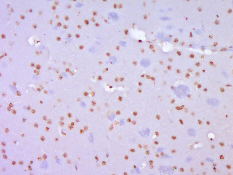 Paraformaldehyde-fixed, paraffin embedded (Mouse brain); Antigen retrieval by boiling in sodium citrate buffer (pH6.0) for 15min; Block endogenous peroxidase by 3% hydrogen peroxide for 20 minutes; Blocking buffer (normal goat serum) at 37°C for 30min; Antibody incubation with (TERT) Polyclonal Antibody, Unconjugated (SL0233R) at 1:400 overnight at 4°C, followed by operating according to SP Kit(Rabbit) (sp-0023) instructions and DAB staining.
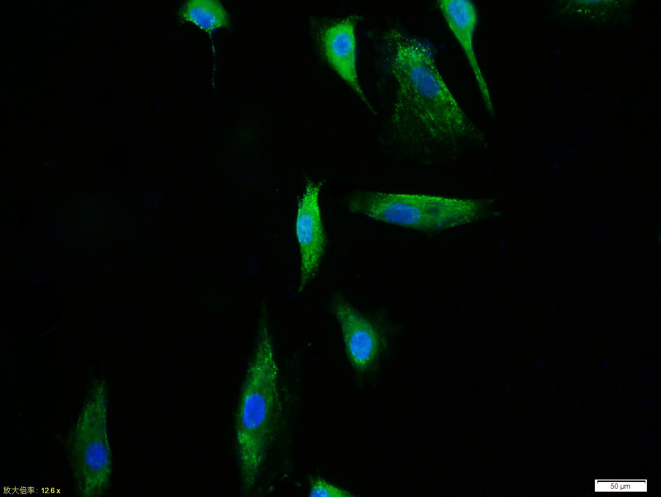 Tissue/cell:A549 cell; 4% Paraformaldehyde-fixed; Triton X-100 at room temperature for 20 min; Blocking buffer (normal goat serum,C-0005) at 37°C for 20 min; Antibody incubation with (TERT) polyclonal Antibody, Unconjugated (SL0233R) 1:100, 90 minutes at 37°C; followed by a FITC conjugated Goat Anti-Rabbit IgG antibody at 37°C for 90 minutes, DAPI (blue, C02-04002) was used to stain the cell nuclei.Tissue/cell:A549 cell; 4% Paraformaldehyde-fixed; Triton X-100 at room temperature for 20 min; Blocking buffer (normal goat serum,C-0005) at 37°C for 20 min; Antibody incubation with (TERT) polyclonal Antibody, Unconjugated (SL0233R) 1:100, 90 minutes at 37°C; followed by a FITC conjugated Goat Anti-Rabbit IgG antibody at 37°C for 90 minutes, DAPI (blue, C02-04002) was used to stain the cell nuclei.
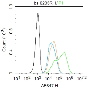 Blank control:Hela.
Primary Antibody (green line): Rabbit Anti-TERT antibody (SL0233R)
Dilution: 1μg /10^6 cells;
Isotype Control Antibody (orange line): Rabbit IgG .
Secondary Antibody : Goat anti-rabbit IgG-AF647
Dilution: 1μg /test.
Protocol
The cells were fixed with 4% PFA (10min at room temperature)and then permeabilized with 90% ice-cold methanol for 20 min at-20℃.The cells were then incubated in 5%BSA to block non-specific protein-protein interactions for 30 min at room temperature .Cells stained with Primary Antibody for 30 min at room temperature. The secondary antibody used for 40 min at room temperature. Acquisition of 20,000 events was performed.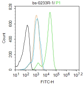 Blank control: K562.
Primary Antibody (green line): Rabbit Anti-TERT antibody (SL0233R)
Dilution: 1μg /10^6 cells;
Isotype Control Antibody (orange line): Rabbit IgG .
Secondary Antibody : Goat anti-rabbit IgG-FITC
Dilution: 1μg /test.
Protocol
The cells were fixed with 4% PFA (10min at room temperature)and then permeabilized with 90% ice-cold methanol for 20 min at -20℃.The cells were then incubated in 5%BSA to block non-specific protein-protein interactions for 30 min at room temperature .Cells stained with Primary Antibody for 30 min at room temperature. The secondary antibody used for 40 min at room temperature. Acquisition of 20,000 events was performed.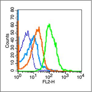 Blank control (blue line): Mouse thymus cells (blue).
Primary Antibody (green line): Rabbit Anti-TERT antibody (SL0233R)
Dilution: 1μg /10^6 cells;
Isotype Control Antibody (orange line): Rabbit IgG .
Secondary Antibody (white blue line): Goat anti-rabbit IgG-PE
Dilution: 1μg /test.
Protocol
The cells were fixed with 70% methanol (Overnight at 4℃) and then permeabilized with 90% ice-cold methanol for 20 min at -20℃. Cells stained with Primary Antibody for 30 min at room temperature. The cells were then incubated in 1 X PBS/2%BSA/10% goat serum to block non-specific protein-protein interactions followed by the antibody for 15 min at room temperature. The secondary antibody used for 40 min at room temperature. Acquisition of 20,000 events was performed.
Cartpieces
Totalgoods,subtotals:¥Checkout
Bought notes(bought amounts latest0)
No one bought this product
User Comment(Total0User Comment Num)
- No comment


 +86 571 56623320
 +86 18668110335

