
Rabbit Anti-Fas Ligand antibody
Tumor necrosis factor ligand superfamily member 6; FASLG; APO1L; Apoptosis (APO 1) antigen ligand 1; Fas-L; Apoptosis antigen ligand 1; APTL; CD178; CD-178; CD 178; CD95L; CD95 ligand; CD95-L; CD95L protein; FAS antigen ligand; Fas L; FASL; Fas ligand (TN
View History [Clear]
Details
Product Name Fas Ligand Chinese Name FasL(CD178)抗体 Alias Tumor necrosis factor ligand superfamily member 6; FASLG; APO1L; Apoptosis (APO 1) antigen ligand 1; Fas-L; Apoptosis antigen ligand 1; APTL; CD178; CD-178; CD 178; CD95L; CD95 ligand; CD95-L; CD95L protein; FAS antigen ligand; Fas L; FASL; Fas ligand (TNF superfamily member 6); Generalized lymphoproliferative disease; TNF superfamily member 6; TNFSF6; TNFL6_HUMAN; Apoptosis antigen ligand; Tumor necrosis factor ligand superfamily member 6, membrane form; Tumor necrosis factor ligand superfamily member 6, soluble form; FASLG. literatures Research Area Tumour Cell biology immunology Apoptosis Immunogen Species Rabbit Clonality Polyclonal React Species Human, Mouse, Rat, (predicted: Cow, ) Applications WB=1:500-2000 ELISA=1:5000-10000 IHC-P=1:100-500 IHC-F=1:100-500 Flow-Cyt=1μg/Test ICC=1:100-500 IF=1:100-500 (Paraffin sections need antigen repair)
not yet tested in other applications.
optimal dilutions/concentrations should be determined by the end user.Theoretical molecular weight 31kDa Detection molecular weight 38-42 kDa Cellular localization The nucleus The cell membrane Extracellular matrix Secretory protein Form Liquid Concentration 1mg/ml immunogen KLH conjugated synthetic peptide derived from human Fas Ligand: 196-281/281 Lsotype IgG Purification affinity purified by Protein A Buffer Solution 0.01M TBS(pH7.4) with 1% BSA, 0.03% Proclin300 and 50% Glycerol. Storage Shipped at 4℃. Store at -20 °C for one year. Avoid repeated freeze/thaw cycles. Attention This product as supplied is intended for research use only, not for use in human, therapeutic or diagnostic applications. PubMed PubMed Product Detail This gene is a member of the tumor necrosis factor superfamily. The primary function of the encoded transmembrane protein is the induction of apoptosis triggered by binding to FAS. The FAS/FASLG signaling pathway is essential for immune system regulation, including activation-induced cell death (AICD) of T cells and cytotoxic T lymphocyte induced cell death. It has also been implicated in the progression of several cancers. Defects in this gene may be related to some cases of systemic lupus erythematosus (SLE). Alternatively spliced transcript variants have been described. [provided by RefSeq, Nov 2014]
Function:
Cytokine that binds to TNFRSF6/FAS, a receptor that transduces the apoptotic signal into cells. May be involved in cytotoxic T-cell mediated apoptosis and in T-cell development. TNFRSF6/FAS-mediated apoptosis may have a role in the induction of peripheral tolerance, in the antigen-stimulated suicide of mature T-cells, or both. Binding to the decoy receptor TNFRSF6B/DcR3 modulates its effects.
Subunit:
Homotrimer (Probable). Interacts with ARHGAP9, BAIAP2L1, BTK, CACNB3, CACNB4, CRK, DLG2, DNMBP, DOCK4, EPS8L3, FGR, FYB, FYN, HCK, ITK, ITSN2, KALRN, LYN, MACC1, MIA, MPP4, MYO15A, NCF1, NCK1, NCK2, NCKIPSD, OSTF1, PIK3R1, PSTPIP1, RIMBP3C, SAMSN1, SH3GL3, SH3PXD2B, SH3PXD2A, SH3RF2, SKAP2, SNX33, SNX9, SORBS3, SPTA1, SRC, SRGAP1, SRGAP2, SRGAP3, TEC, TJP3 and YES1.
Subcellular Location:
Cell membrane; Single-pass type II membrane protein. Secreted. Cytoplasmic vesicle lumen. Lysosome lumen. Note=May be released into the extracellular fluid, probably by cleavage form the cell surface. Is internalized into multivesicular bodies of secretory lysosomes after phosphorylation by FGR and monoubiquitination.
Post-translational modifications:
N-glycosylated.
The soluble form derives from the membrane form by proteolytic processing.
Phosphorylated by FGR on tyrosine residues; this is required for ubiquitination and subsequent internalization.
Monoubiquitinated.
DISEASE:
Defects in FASLG are the cause of autoimmune lymphoproliferative syndrome type 1B (ALPS1B) [MIM:601859]; also known as Canale-Smith syndrome (CSS). ALPS is a childhood syndrome involving hemolytic anemia and thrombocytopenia with massive lymphadenopathy and splenomegaly.
Similarity:
Belongs to the tumor necrosis factor family.
SWISS:
P48023
Gene ID:
356
Database links:Entrez Gene: 356 Human
Entrez Gene: 14103 Mouse
Omim: 134638 Human
SwissProt: P48023 Human
SwissProt: P41047 Mouse
Unigene: 2007 Human
Unigene: 3355 Mouse
Unigene: 9725 Rat
Product Picture 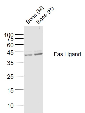 Sample:
Lane 1: Bone (Mouse) Lysate at 40 ug
Lane 2: Bone (Rat) Lysate at 40 ug
Primary: Anti-Fas Ligand (SL0216R) at 1/1000 dilution
Secondary: IRDye800CW Goat Anti-Rabbit IgG at 1/20000 dilution
Predicted band size: 40 kD
Observed band size: 40 kD
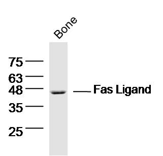 Sample: Bone (mouse) Lysate at 40 ug
Primary: Anti- Fas ligand (SL0216R) at 1/300 dilution
Secondary: IRDye800CW Goat Anti-Rabbit IgG at 1/20000 dilution
Predicted band size: 31 kD
Observed band size: 46 kD
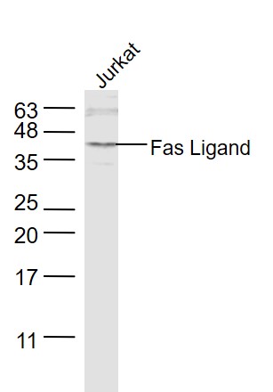 Sample:
Jurkat(Human) Cell Lysate at 30 ug
Primary: Anti-Fas Ligand (SL0216R) at 1/1000 dilution
Secondary: IRDye800CW Goat Anti-Rabbit IgG at 1/20000 dilution
Predicted band size: 31 kD
Observed band size: 40 kD
 Paraformaldehyde-fixed, paraffin embedded (mouse placenta); Antigen retrieval by boiling in sodium citrate buffer (pH6.0) for 15min; Block endogenous peroxidase by 3% hydrogen peroxide for 20 minutes; Blocking buffer (normal goat serum) at 37°C for 30min; Antibody incubation with (Fas Ligand) Polyclonal Antibody, Unconjugated (SL0216R) at 1:200 overnight at 4°C, followed by operating according to SP Kit(Rabbit) (sp-0023) instructionsand DAB staining.
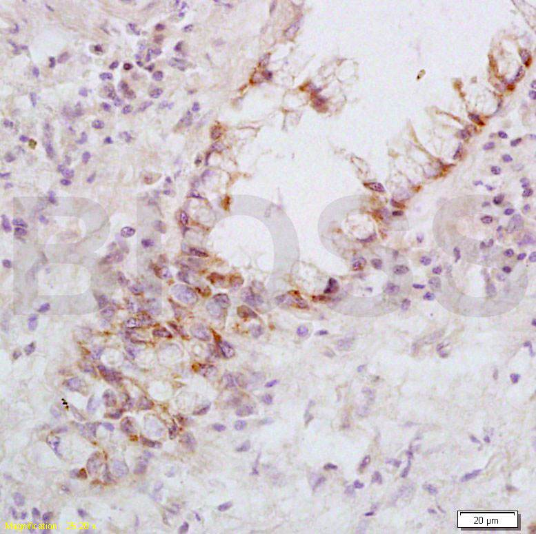 Tissue/cell: human rectal carcinoma; 4% Paraformaldehyde-fixed and paraffin-embedded;
Antigen retrieval: citrate buffer ( 0.01M, pH 6.0 ), Boiling bathing for 15min; Block endogenous peroxidase by 3% Hydrogen peroxide for 30min; Blocking buffer (normal goat serum,C-0005) at 37℃ for 20 min;
Incubation: Anti-FasL Polyclonal Antibody, Unconjugated(SL0216R) 1:200, overnight at 4°C, followed by conjugation to the secondary antibody(SP-0023) and DAB(C-0010) staining
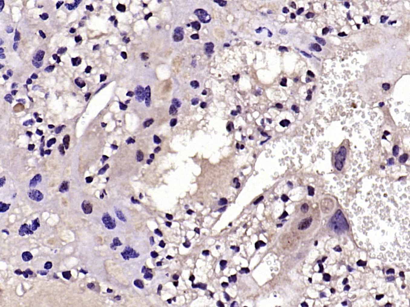
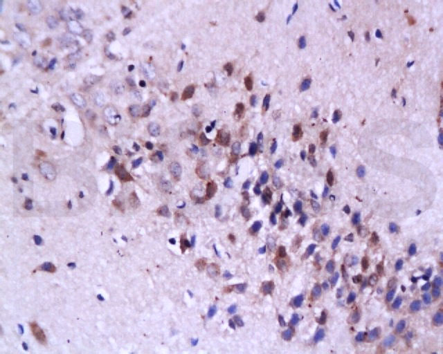 Tissue/cell: rat brain tissue; 4% Paraformaldehyde-fixed and paraffin-embedded;
Antigen retrieval: citrate buffer ( 0.01M, pH 6.0 ), Boiling bathing for 15min; Block endogenous peroxidase by 3% Hydrogen peroxide for 30min; Blocking buffer (normal goat serum,C-0005) at 37℃ for 20 min;
Incubation: Anti-FasL Polyclonal Antibody, Unconjugated(SL0216R) 1:200, overnight at 4°C, followed by conjugation to the secondary antibody(SP-0023) and DAB(C-0010) staining
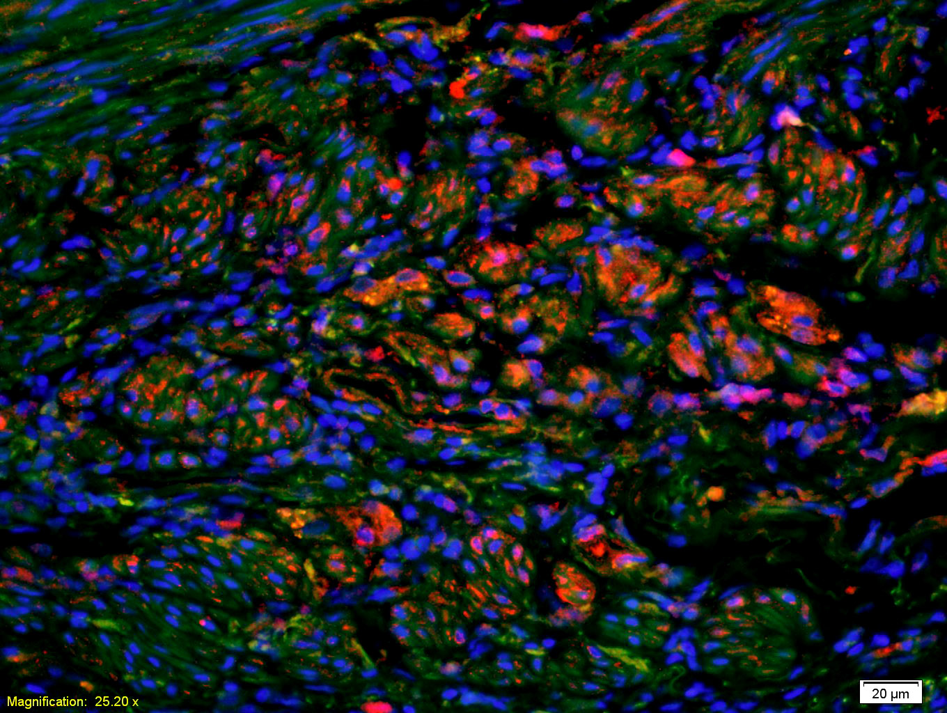 Tissue/cell: human rectal carcinoma;4% Paraformaldehyde-fixed and paraffin-embedded;
Antigen retrieval: citrate buffer ( 0.01M, pH 6.0 ), Boiling bathing for 15min; Blocking buffer (normal goat serum,C-0005) at 37℃ for 20 min;
Incubation: Anti-FasL Polyclonal Antibody, Unconjugated(SL0216R) 1:200, overnight at 4°C; The secondary antibody was Goat Anti-Rabbit IgG, Cy3 conjugated(SL0295G-Cy3)used at 1:200 dilution for 40 minutes at 37°C. DAPI(5ug/ml,blue,C-0033) was used to stain the cell nuclei
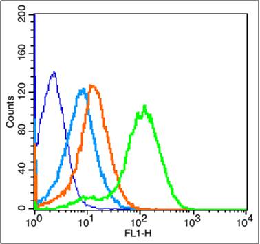 Blank control: Mouse Kidney (blue).
Primary Antibody:Rabbit Anti-phospho-Fas Ligand antibody (SL0216R,Green); Dilution: 1μg in 100 μL 1X PBS containing 0.5% BSA;
Isotype Control Antibody: Rabbit IgG(orange) ,used under the same conditions;
Secondary Antibody: Goat anti-rabbit IgG-FITC(white blue), Dilution: 1:200 in 1 X PBS containing 0.5% BSA.
Protocol
The cells were fixed with 2% paraformaldehyde for 10 min at 37℃. Primary antibody (SL0216R, 1μg /1x10^6 cells) were incubated for 30 min at room temperature, followed by 1 X PBS containing 0.5% BSA + 1 0% goat serum (15 min) to block non-specific protein-protein interactions. Then the Goat Anti-rabbit IgG/FITC antibody was added into the blocking buffer mentioned above to react with the primary antibody at 1/200 dilution for 40 min on ice. Acquisition of 20,000 events was performed.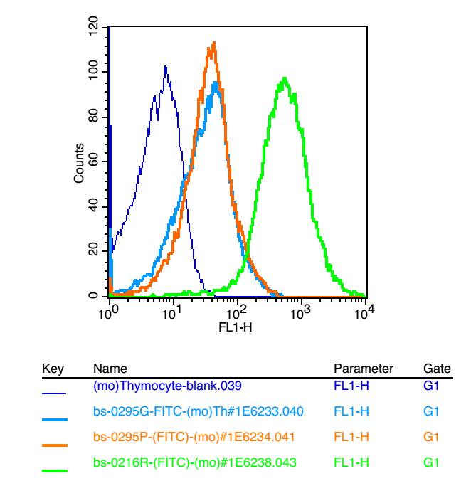 Blank control: mouse thymouses(blue)
Isotype Control Antibody: Rabbit IgG(orange) ; Secondary Antibody: Goat anti-rabbit IgG-FITC(white blue), Dilution: 1:100 in 1 X PBS containing 0.5% BSA ; Primary Antibody Dilution: 1μl in 100 μL1X PBS containing 0.5% BSA(green).
Cartpieces
Totalgoods,subtotals:¥Checkout
Bought notes(bought amounts latest0)
No one bought this product
User Comment(Total0User Comment Num)
- No comment


 +86 571 56623320
 +86 18668110335

