
Rabbit Anti-RAGE antibody
Advanced glycosylation end product specific receptor; Advanced glycosylation end product-specific receptor; AGER; EC 2.7.11.22; LE 9211 A antigen;LE-9211-A antigen; MGC22357; MOK; RAGE 1; RAGE1; MOK protein kinase; Receptor for advanced glycation endprodu
View History [Clear]
Details
Product Name RAGE Chinese Name 晚期糖基化终末产物特异性受体抗体 Alias Advanced glycosylation end product specific receptor; Advanced glycosylation end product-specific receptor; AGER; EC 2.7.11.22; LE 9211 A antigen;LE-9211-A antigen; MGC22357; MOK; RAGE 1; RAGE1; MOK protein kinase; Receptor for advanced glycation endproducts;Renal tumor antigen 1; Renal tumor antigen; Renal cell carcinoma antigen (MOK protein kinase); Renal tumor antigen 1; RAGE_HUMAN. literatures Research Area Tumour Cardiovascular immunology Growth factors and hormones Diabetes Endocrinopathy Immunogen Species Rabbit Clonality Polyclonal React Species Human, Mouse, Rat, Applications WB=1:500-2000 ELISA=1:5000-10000 IHC-P=1:100-500 IHC-F=1:100-500 Flow-Cyt=1μg /test IF=1:100-500 (Paraffin sections need antigen repair)
not yet tested in other applications.
optimal dilutions/concentrations should be determined by the end user.Theoretical molecular weight 42kDa Cellular localization The cell membrane Secretory protein Form Liquid Concentration 1mg/ml immunogen KLH conjugated synthetic peptide derived from rat AGER: 151-250/403 <Extracellular> Lsotype IgG Purification affinity purified by Protein A Buffer Solution 0.01M TBS(pH7.4) with 1% BSA, 0.03% Proclin300 and 50% Glycerol. Storage Shipped at 4℃. Store at -20 °C for one year. Avoid repeated freeze/thaw cycles. Attention This product as supplied is intended for research use only, not for use in human, therapeutic or diagnostic applications. PubMed PubMed Product Detail Advanced glycosylation end product-specific receptor (AGER; RAGE) is a member of the immunoglobulin superfamily of cell surface molecules that binds molecules that have been irreversibly modified by non-enzymatic glycation and oxidation, and are know as advanced glycation end products (AGEs). It is expressed by endothelium, mononuclear phagocytes, neurons and smooth muscle cells. Whereas RAGE is present at high levels during development, especially in the central nervous system, its levels decline during maturity.The increased expression of RAGE is associated with several pathological states, such as diabetic vasculopathy, neuropathy, retinopathy and other disorders, including Alzheimer's disease and immune/inflammatory reactions of the vessel walls. In diabetic tissues, the production of RAGE is due to the overproduction of AGEs that eventually overwhelm the protective properties of RAGE. This results in oxidative stress and endothelial cell dysfunction that leads to vascular disease in diabetics. In the brain, RAGE also binds amyloid beta (Ab). Because Ab is overproduced in neurons and vessels in the brains of Alzheimer disease, this leads to the hyperstimulation of RAGE. The RAGE-Ab interaction is thought to result in oxidative stress leading to neuronal degeneration.
Function:
Mediates interactions of advanced glycosylation end products (AGE). These are nonenzymatically glycosylated proteins which accumulate in vascular tissue in aging and at an accelerated rate in diabetes. Acts as a mediator of both acute and chronic vascular inflammation in conditions such as atherosclerosis and in particular as a complication of diabetes. AGE/RAGE signaling plays an important role in regulating the production/expression of TNF-alpha, oxidative stress, and endothelial dysfunction in type 2 diabetes. Interaction with S100A12 on endothelium, mononuclear phagocytes, and lymphocytes triggers cellular activation, with generation of key proinflammatory mediators. Receptor for amyloid beta peptide. Contributes to the translocation of amyloid-beta peptide (ABPP) across the cell membrane from the extracellular to the intracellular space in cortical neurons. ABPP-initiated RAGE signaling, especially stimulation of p38 mitogen-activated protein kinase (MAPK), has the capacity to drive a transport system delivering ABPP as a complex with RAGE to the intraneuronal space. Interaction with S100B after myocardial infarction may play a role in myocyte apoptosis by activating ERK1/2 and p53/TP53 signaling.
Subunit:
Interacts with S100B, S100A1 and APP. Interacts with S100A12.
Subcellular Location:
Isoform 1: Cell membrane; Single-pass type I membrane protein.
Isoform 2: Secreted.
Tissue Specificity:
Endothelial cells and cardiomyocytes.
Similarity:
Contains 2 Ig-like C2-type (immunoglobulin-like) domains.
Contains 1 Ig-like V-type (immunoglobulin-like) domain.
SWISS:
Q63495
Gene ID:
81722
Database links:Entrez Gene: 177 Human
Entrez Gene: 11596 Mouse
Omim: 600214 Human
SwissProt: Q15109 Human
SwissProt: Q62151 Mouse
Unigene: 534342 Human
Unigene: 3383 Mouse
Unigene: 9829 Rat
晚期糖基化终末产物受体(AGER)与其配体AGEs形成的AGEs-AGER 系统在Diabetes血管病变的发生、发展过程中起着重要作用.
年龄及晚期糖基化终末产物(AGEs)等多种因素均能调节AGER基因的表达. Diabetes患者体内晚期糖基化终末产物受体(AGER)的高表达加速了病人血管病变的发展过程,并增加了病变的复杂性.阻断AGER通路可缓解Diabetes血管的病变过程。
因此,AGER可以作为治疗Diabetes血管病变的药物靶点,并为临床治疗Diabetes血管病变提供了新的思路.Product Picture 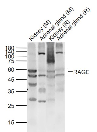 Sample:
Lane 1: Kidney (Mouse) Lysate at 40 ug
Lane 2: Adrenal gland (Mouse) Lysate at 40 ug
Lane 3: Kidney (Rat) Lysate at 40 ug
Lane 4: Adrenal gland (Rat) Lysate at 40 ug
Primary: Anti-RAGE (SL0177R) at 1/1000 dilution
Secondary: IRDye800CW Goat Anti-Rabbit IgG at 1/20000 dilution
Predicted band size: 50/43/55 kD
Observed band size: 58/50 kD
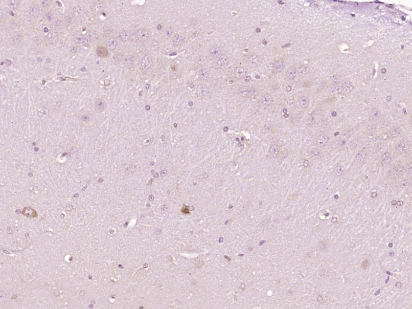 Paraformaldehyde-fixed, paraffin embedded (Rat brain); Antigen retrieval by boiling in sodium citrate buffer (pH6.0) for 15min; Block endogenous peroxidase by 3% hydrogen peroxide for 20 minutes; Blocking buffer (normal goat serum) at 37°C for 30min; Antibody incubation with (RAGE) Polyclonal Antibody, Unconjugated (SL0177R) at 1:400 overnight at 4°C, followed by operating according to SP Kit(Rabbit) (sp-0023) instructionsand DAB staining.
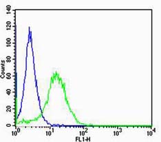 Cell: NIH/3T3
Concentration:1:100
Host/Isotype:Rabbit/IgG
Flow cytometric analysis of Rabbit IgG isotype control (Cat#: SL0177R) on NIH/3T3(green) compared with control in the absence of primary antibody (blue) followed by Alexa Fluor 488-conjugated goat anti-rabbit IgG(H+L) secondary antibody .
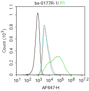 Blank control: MCF7.
Primary Antibody (green line): Rabbit Anti-RAGE antibody (SL0177R)
Dilution: 1μg /10^6 cells;
Isotype Control Antibody (orange line): Rabbit IgG .
Secondary Antibody : Goat anti-rabbit IgG-AF647
Dilution: 1μg /test.
Protocol
The cells were incubated in 5%BSA to block non-specific protein-protein interactions for 30 min at room temperature .Cells stained with Primary Antibody for 30 min at room temperature. The secondary antibody used for 40 min at room temperature. Acquisition of 20,000 events was performed.
Cartpieces
Totalgoods,subtotals:¥Checkout
References (0)
No References
Bought notes(bought amounts latest0)
No one bought this product
User Comment(Total0User Comment Num)
- No comment


 +86 571 56623320
 +86 18668110335

