
Rabbit Anti-VEGFR1 antibody
Vascular endothelial growth factor receptor 1; VEGF-R1; VEGFR-1; VEGF Receptor 1; FLT-1; vascular permeability factor receptor; vascular endothelial growth factor receptor; vascular endothelial growth factor receptor-1; fms-related tyrosine kinase 1; vasc
View History [Clear]
Details
Product Name VEGFR1 Chinese Name 血管内皮生长因子受体1抗体 Alias Vascular endothelial growth factor receptor 1; VEGF-R1; VEGFR-1; VEGF Receptor 1; FLT-1; vascular permeability factor receptor; vascular endothelial growth factor receptor; vascular endothelial growth factor receptor-1; fms-related tyrosine kinase 1; vascular endothelial growth factor/vascular permeability factor receptor; AI323757; FLT; FLT1; sFlt1; VGFR1_HUMAN; VGFR1_MOUSE. literatures Research Area Tumour Cell biology immunology Developmental biology Neurobiology Growth factors and hormones Kinases and Phosphatases The cell membrane受体 Cell Surface Molecule Immunogen Species Rabbit Clonality Polyclonal React Species Human, Mouse, Rat, (predicted: Pig, Horse, ) Applications ELISA=1:5000-10000 IHC-P=1:100-500 IHC-F=1:100-500 ICC=1:100 IF=1:100-500 (Paraffin sections need antigen repair)
not yet tested in other applications.
optimal dilutions/concentrations should be determined by the end user.Theoretical molecular weight 147kDa Cellular localization cytoplasmic The cell membrane Secretory protein Form Liquid Concentration 1mg/ml immunogen KLH conjugated synthetic peptide derived from mouse VEGFR1: 1162-1260/1333 <Cytoplasmic> Lsotype IgG Purification affinity purified by Protein A Buffer Solution 0.01M TBS(pH7.4) with 1% BSA, 0.03% Proclin300 and 50% Glycerol. Storage Shipped at 4℃. Store at -20 °C for one year. Avoid repeated freeze/thaw cycles. Attention This product as supplied is intended for research use only, not for use in human, therapeutic or diagnostic applications. PubMed PubMed Product Detail VEGF Receptor 1 (also known as FLT) belongs to the src gene family and shows tyrosine protein kinase activity that is important for the control of cell proliferation and differentiation. The protein acts as a receptor for VEGF, VEGFB and PGF. An alternatively spliced form of the gene produces a soluble protein (sFlt1) which binds vascular endothelial growth factor (VEGF) with high affinity. sFlt1 has a higher affinity for VEGF indicating that it may function as an inhibitor in the VEGF response. VEGF Receptor 1 is specifically expressed in most vascular endothelial cells and peripheral blood monocytes.
Function:
Tyrosine-protein kinase that acts as a cell-surface receptor for VEGFA, VEGFB and PGF, and plays an essential role in the development of embryonic vasculature, the regulation of angiogenesis, cell survival, cell migration, macrophage function, chemotaxis, and cancer cell invasion. May play an essential role as a negative regulator of embryonic angiogenesis by inhibiting excessive proliferation of endothelial cells. Can promote endothelial cell proliferation, survival and angiogenesis in adulthood. Its function in promoting cell proliferation seems to be cell-type specific. Promotes PGF-mediated proliferation of endothelial cells, and proliferation of some types of cancer cells, but does not promote proliferation of normal fibroblasts. Has very high affinity for VEGFA and relatively low protein kinase activity; may function as a negative regulator of VEGFA signaling by limiting the amount of free VEGFA and preventing its binding to KDR. Modulates KDR signaling by forming heterodimers with KDR. Ligand binding leads to the activation of several signaling cascades. Activation of PLCG1 leads to the production of the cellular signaling molecules diacylglycerol and inositol 1,4,5-trisphosphate and the activation of protein kinase C. Mediates phosphorylation of PIK3R1, the regulatory subunit of phosphatidylinositol 3-kinase, leading to the activation of phosphatidylinositol kinase and the downstream signaling pathway. Mediates activation of MAPK1/ERK2, MAPK3/ERK1 and the MAP kinase signaling pathway, as well as of the AKT1 signaling pathway. Phosphorylates PLCG1. Promotes phosphorylation of AKT1, PTK2/FAK1; YES1 and CBL.
Subunit:
Interacts with VEGFA, VEGFB and PGF. Monomer in the absence of bound VEGFA, VEGFB or PGF. Homodimer in the presence of bound VEGFA, VEGFB and PGF. Can also form a heterodimer with KDR.Interacts (when tyrosine phosphorylated) with CBL, CRK, GRB2, NCK1,PIK3R1, PLCG1 and PTPN11. Interacts with GNB2L1/RACK1. Identified in a complex with CBL and CD2AP.
Subcellular Location:
Isoform 1: Cell membrane; Single-pass type I membrane protein. Endosome. Note=Autophosphorylation promotes ubiquitination and endocytosis;Isoform 2: Secreted;Isoform 3: Secreted;Isoform 4: Secreted;Isoform 5: Cytoplasm (Potential);Isoform 6: Cytoplasm (Potential);Isoform 7: Cytoplasm (Potential).
Tissue Specificity:
Detected in normal lung, but also in placenta,liver, kidney, heart and brain tissues. Specifically expressed in most of the vascular endothelial cells, and also expressed in peripheral blood monocytes. Isoform 2 is strongly expressed in placenta. Isoform 3 is expressed in corneal epithelial cells (at protein level). Isoform 3 is expressed in vascular smooth muscle cells (VSMC).
Post-translational modifications:
N-glycosylated.
Ubiquitinated after VEGFA-mediated autophosphorylation, leading to proteolytic degradation.
Autophosphorylated on tyrosine residues upon ligand binding. Autophosphorylation occurs in trans, i.e. one subunit of the dimeric receptor phosphorylates tyrosine residues on the other subunit. Phosphorylation at Tyr-1169 is important for interaction with PLCG1. Phosphorylation at Tyr-1213 is important for interaction with PIK3R1, PTPN11, GRB2, and PLCG1. Phosphorylation at Tyr-1331 is important for endocytosis and for interaction with CBL, NCK1 and CRK.
Similarity:
Belongs to the protein kinase superfamily. Tyr protein kinase family. CSF-1/PDGF receptor subfamily.Contains 7 Ig-like C2-type (immunoglobulin-like)domains.Contains 1 protein kinase domain.
SWISS:
P53767
Gene ID:
14254
Database links:Entrez Gene: 2321 Human
Entrez Gene: 14254 Mouse
Omim: 165070 Human
SwissProt: P17948 Human
SwissProt: P35969 Mouse
Unigene: 594454 Human
Unigene: 389712 Mouse
Unigene: 10239 Rat
VEGFR1/Flt1是一种The cell membrane受体激酶,对血管内皮生长因子有高度的亲和性,主要功能是参与vascular endothelial cell生长和血管生成的调控。用于各种恶性Tumour的研究。Product Picture  Tissue/cell: rat brain tissue; 4% Paraformaldehyde-fixed and paraffin-embedded;
Antigen retrieval: citrate buffer ( 0.01M, pH 6.0 ), Boiling bathing for 15min; Block endogenous peroxidase by 3% Hydrogen peroxide for 30min; Blocking buffer (normal goat serum,C-0005) at 37℃ for 20 min;
Incubation: Anti-VEGFR1/FLT1 Polyclonal Antibody, Unconjugated(SL0170R) 1:400, overnight at 4°C, followed by conjugation to the secondary antibody(SP-0023) and DAB(C-0010) staining
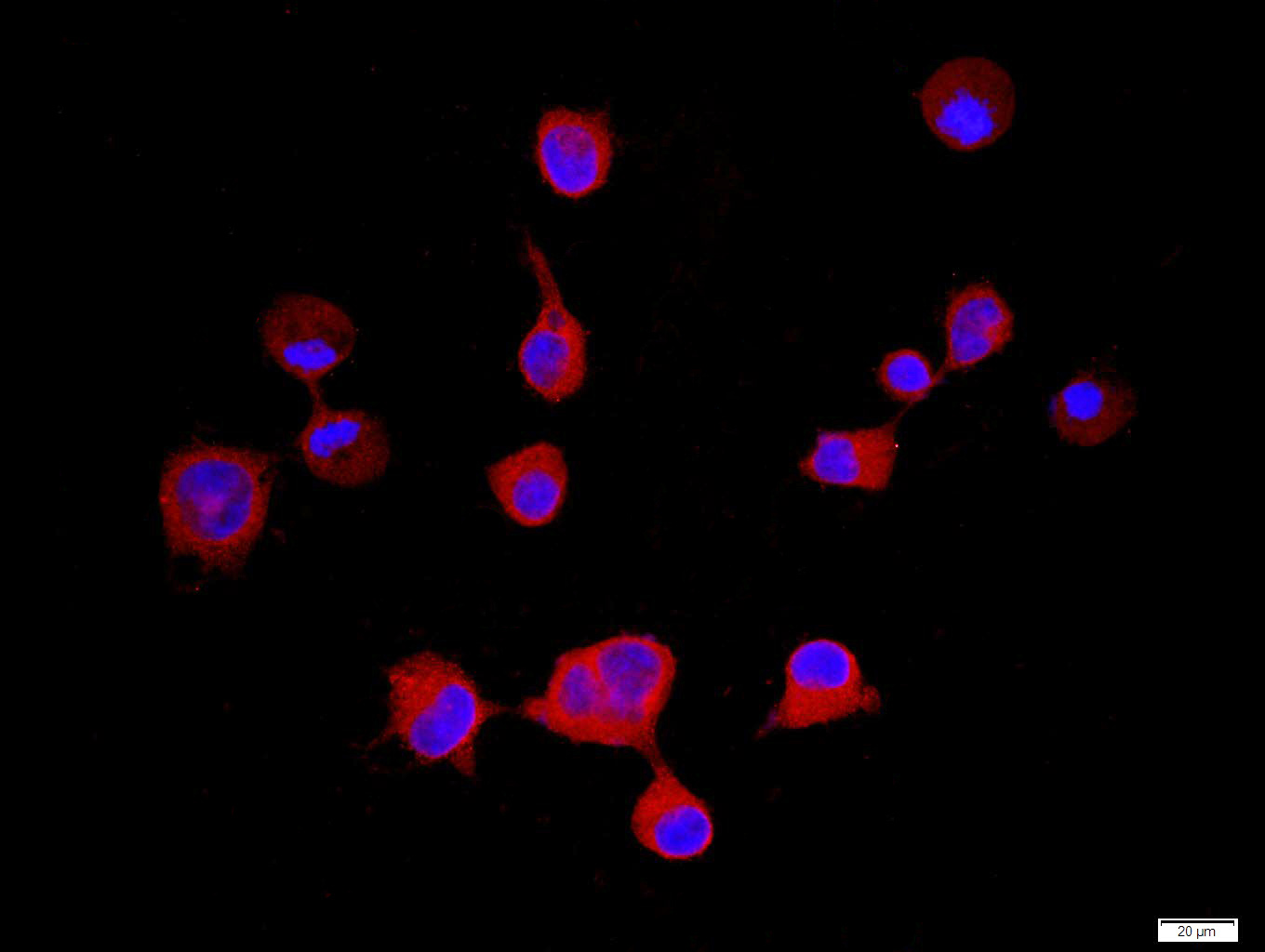 Tissue/cell: 293T cell; 4% Paraformaldehyde-fixed; Triton X-100 at room temperature for 20 min; Blocking buffer (normal goat serum, C-0005) at 37°C for 20 min; Antibody incubation with (VEGFR1) polyclonal Antibody, Unconjugated (SL0170R) 1:100, 90 minutes at 37°C; followed by a FITC conjugated Goat Anti-Rabbit IgG antibody at 37°C for 90 minutes, DAPI (blue, C02-04002) was used to stain the cell nuclei.
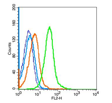 Blank control: mouse spleen cells(blue), the cells were fixed with 2% paraformaldehyde (10 min) and then permeabilized with ice-cold 90% methanol for 30 min on ice.
Isotype Control Antibody: Rabbit IgG(orange) ; Secondary Antibody: Goat anti-rabbit IgG-PE(white blue), Dilution: 1:200 in 1 X PBS containing 0.5% BSA ; Primary Antibody Dilution: 1μg in 100 μL1X PBS containing 0.5% BSA(green).
Cartpieces
Totalgoods,subtotals:¥Checkout
Bought notes(bought amounts latest0)
No one bought this product
User Comment(Total0User Comment Num)
- No comment


 +86 571 56623320
 +86 18668110335

