
Rabbit Anti-Neurokin B receptor antibody
MGC148060; MGC148061; Neurokinin B receptor; Neurokinin beta receptor; Neuromedin K Receptor; Neuromedin-K receptor; NK 3 receptor; NK 3R; NK-3 receptor; NK-3R; NK3 receptor; NK3R; NK3R_HUMAN; NKR; TAC 3R; TAC3R; TAC3RL; Tachykinin receptor 3; TACR 3; Tac
View History [Clear]
Details
Product Name Neurokin B receptor Chinese Name 神经激肽B受体抗体 Alias MGC148060; MGC148061; Neurokinin B receptor; Neurokinin beta receptor; Neuromedin K Receptor; Neuromedin-K receptor; NK 3 receptor; NK 3R; NK-3 receptor; NK-3R; NK3 receptor; NK3R; NK3R_HUMAN; NKR; TAC 3R; TAC3R; TAC3RL; Tachykinin receptor 3; TACR 3; Tacr3. literatures Research Area Cell biology Neurobiology The cell membrane受体 G protein signal Immunogen Species Rabbit Clonality Polyclonal React Species Mouse, Rat, (predicted: Human, Chicken, Dog, Pig, Cow, Horse, Rabbit, Guinea Pig, ) Applications ELISA=1:5000-10000 IHC-P=1:100-500 IHC-F=1:100-500 Flow-Cyt=1μg/Test IF=1:100-500 (Paraffin sections need antigen repair)
not yet tested in other applications.
optimal dilutions/concentrations should be determined by the end user.Theoretical molecular weight 52kDa Cellular localization The cell membrane Form Liquid Concentration 1mg/ml immunogen KLH conjugated synthetic peptide derived from human NKR: 151-250/440 <Extracellular> Lsotype IgG Purification affinity purified by Protein A Buffer Solution 0.01M TBS(pH7.4) with 1% BSA, 0.03% Proclin300 and 50% Glycerol. Storage Shipped at 4℃. Store at -20 °C for one year. Avoid repeated freeze/thaw cycles. Attention This product as supplied is intended for research use only, not for use in human, therapeutic or diagnostic applications. PubMed PubMed Product Detail The tachykinins belong to an evolutionary conserved family of peptide neurotransmitters that share the C-terminal sequence Phe-X-Gly-Leu-Met-NH2 and have an established role in neurotransmission. The mammalian tachykinins include substance P, neurokinin A (NKA) and neurokinin B (NKB) which exert their effects by binding to specific receptors. Tachykinin peptides are important in the mediation of many physiological and pathological processes including inflammation, pain, migraine headache and allergy induced asthma.
Three tachykinin receptor types have been characterized, NK-1, NK-2 and NK-3 which have preferential affinities for SP, NKA and NKB respectively. All three receptors share a high degree of sequence homology, have seven transmembrane spanning domains and similar signal transduction mechanisms (e.g. G-protein coupled activation of phospholipase C).
Function:
This is a receptor for the tachykinin neuropeptide neuromedin-K (neurokinin B). It is associated with G proteins that activate a phosphatidylinositol-calcium second messenger system. The rank order of affinity of this receptor to tachykinins is: neuromedin-K > substance K > substance P.
Subcellular Location:
Cell membrane; Multi-pass membrane protein.
Post-translational modifications:
The anchoring of this receptor to the plasma membrane is probably mediated by the palmitoylation of a cysteine residue.
DISEASE:
Hypogonadotropic hypogonadism 11 with or without anosmia (HH11) [MIM:614840]: A disorder characterized by absent or incomplete sexual maturation by the age of 18 years, in conjunction with low levels of circulating gonadotropins and testosterone and no other abnormalities of the hypothalamic-pituitary axis. In some cases, it is associated with non-reproductive phenotypes, such as anosmia, cleft palate, and sensorineural hearing loss. Anosmia or hyposmia is related to the absence or hypoplasia of the olfactory bulbs and tracts. Hypogonadism is due to deficiency in gonadotropin-releasing hormone and probably results from a failure of embryonic migration of gonadotropin-releasing hormone-synthesizing neurons. In the presence of anosmia, idiopathic hypogonadotropic hypogonadism is referred to as Kallmann syndrome, whereas in the presence of a normal sense of smell, it has been termed normosmic idiopathic hypogonadotropic hypogonadism (nIHH). Note=The disease is caused by mutations affecting the gene represented in this entry.
Similarity:
Belongs to the G-protein coupled receptor 1 family.
SWISS:
P29371
Gene ID:
6870
Database links:Entrez Gene: 6870 Human
Entrez Gene: 21338 Mouse
Entrez Gene: 100008721 Rabbit
Omim: 162332 Human
SwissProt: P29371 Human
SwissProt: P47937 Mouse
SwissProt: O97512 Rabbit
Unigene: 942 Human
Unigene: 103810 Mouse
Unigene: 9702 Rat
Product Picture 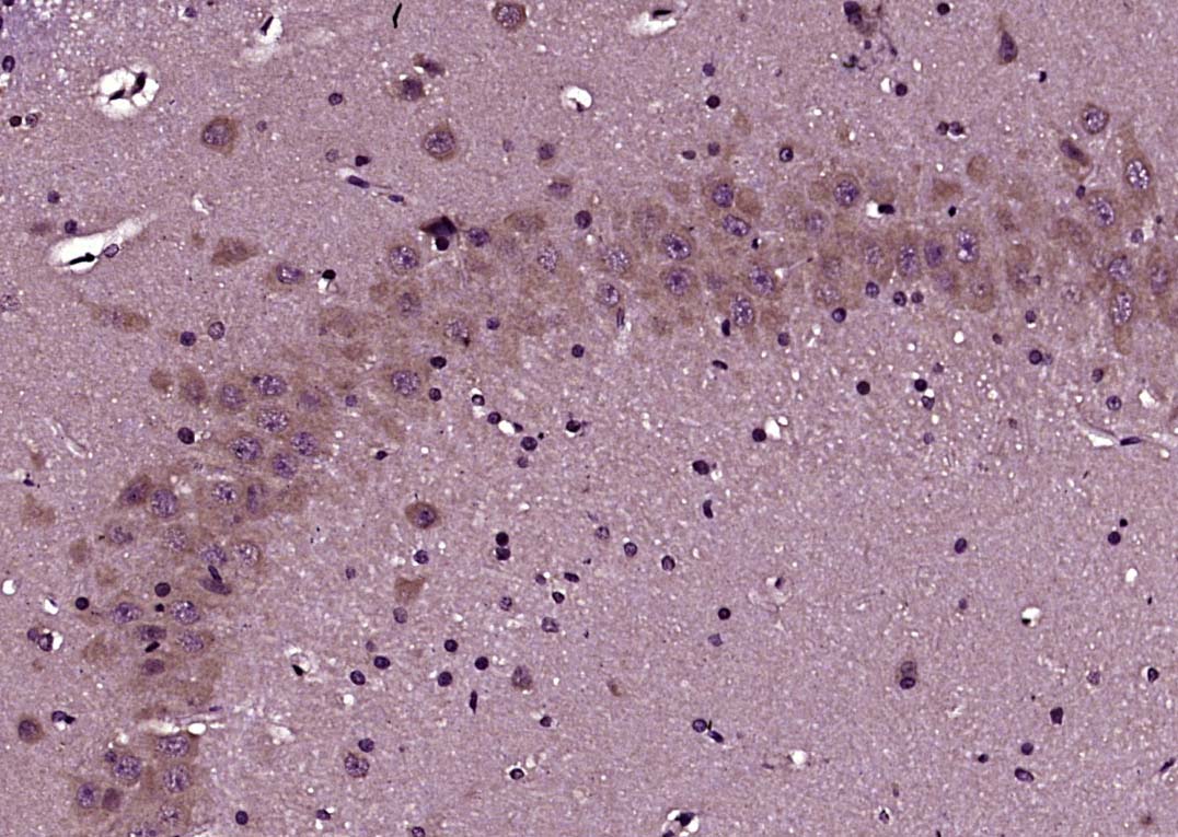 Paraformaldehyde-fixed, paraffin embedded (Rat brain); Antigen retrieval by boiling in sodium citrate buffer (pH6.0) for 15min; Block endogenous peroxidase by 3% hydrogen peroxide for 20 minutes; Blocking buffer (normal goat serum) at 37°C for 30min; Antibody incubation with (Neurokin B receptor) Polyclonal Antibody, Unconjugated (SL0166R) at 1:400 overnight at 4°C, followed by operating according to SP Kit(Rabbit) (sp-0023) instructionsand DAB staining.
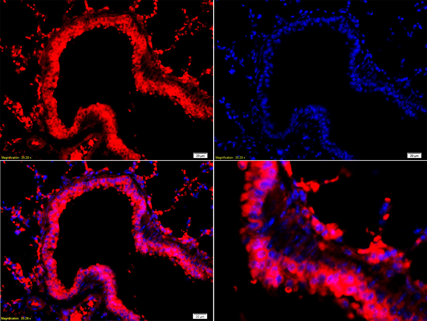 Tissue/cell: Mouse lung tissue;4% Paraformaldehyde-fixed and paraffin-embedded;
Antigen retrieval: citrate buffer ( 0.01M, pH 6.0 ), Boiling bathing for 15min; Blocking buffer (normal goat serum,C-0005) at 37℃ for 20 min;
Incubation: Anti-NK3R Polyclonal Antibody, Unconjugated(SL0166R) 1:200, overnight at 4°C; The secondary antibody was Goat Anti-Rabbit IgG, Cy3 conjugated(SL0295G-Cy3)used at 1:200 dilution for 40 minutes at 37°C. DAPI(5ug/ml,blue,C-0033) was used to stain the cell nuclei
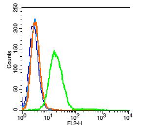 Blank control: RSC96(blue).
Primary Antibody: Rabbit Anti- Neurokin B receptor antibody(SL0166R), Dilution: 1μg in 100 μL 1X PBS containing 0.5% BSA;
Isotype Control Antibody: Rabbit IgG (orange) ,used under the same conditions.
Secondary Antibody: Goat anti-rabbit IgG-PE(white blue), Dilution: 1:200 in 1 X PBS containing 0.5% BSA.
Protocol
Primary antibody (SL0166R, 1μg /1x10^6 cells) were incubated for 30 min on the ice, followed by 1 X PBS containing 0.5% BSA + 1 0% goat serum (15 min) to block non-specific protein-protein interactions. Then the Goat Anti-rabbit IgG/PE antibody was added into the blocking buffer mentioned above to react with the primary antibody at 1/200 dilution for 30 min on ice. Acquisition of 20,000 events was performed.
Cartpieces
Totalgoods,subtotals:¥Checkout
Bought notes(bought amounts latest0)
No one bought this product
User Comment(Total0User Comment Num)
- No comment


 +86 571 56623320
 +86 18668110335

