
Rabbit Anti-PIK3R1 antibody
P85A_HUMAN; Phosphatidylinositol 3-kinase regulatory subunit alpha; GRB1; PI3-kinase regulatory subunit alpha; PI3K regulatory subunit alpha; PtdIns-3-kinase regulatory subunit alpha; Phosphatidylinositol 3-kinase 85 kDa regulatory subunit alpha (PI3-kina
View History [Clear]
Details
Product Name PIK3R1 Chinese Name 磷脂酰肌醇激酶抗体 Alias P85A_HUMAN; Phosphatidylinositol 3-kinase regulatory subunit alpha; GRB1; PI3-kinase regulatory subunit alpha; PI3K regulatory subunit alpha; PtdIns-3-kinase regulatory subunit alpha; Phosphatidylinositol 3-kinase 85 kDa regulatory subunit alpha (PI3-kinase subunit p85-alpha; PtdIns-3-kinase regulatory subunit p85-alpha); PI 3-kinase p85α; PI 3-kinase p85 α; PI 3-kinase p85-α; SH3_PI3K_p85alpha; PI3-kinase p85 subunit alpha; phosphoinositide-3-kinase regulatory subunit 1; p85; AGM7; IMD36; p85-ALPHA; PI 3 Kinase p85 alpha; literatures Research Area Tumour Cell biology immunology Neurobiology Signal transduction Apoptosis Kinases and Phosphatases Immunogen Species Rabbit Clonality Polyclonal React Species Human, Mouse, Rat, (predicted: Chicken, Dog, Cow, Horse, ) Applications WB=1:500-2000 ELISA=1:5000-10000 IHC-P=1:100-500 IHC-F=1:100-500 Flow-Cyt=1μg/Test ICC=1:100 IF=1:100-500 (Paraffin sections need antigen repair)
not yet tested in other applications.
optimal dilutions/concentrations should be determined by the end user.Theoretical molecular weight 80kDa Cellular localization cytoplasmic Form Liquid Concentration 1mg/ml immunogen KLH conjugated synthetic peptide derived from human PI3 kinase p85 subunit alpha: 501-600/724 Lsotype IgG Purification affinity purified by Protein A Buffer Solution 0.01M TBS(pH7.4) with 1% BSA, 0.03% Proclin300 and 50% Glycerol. Storage Shipped at 4℃. Store at -20 °C for one year. Avoid repeated freeze/thaw cycles. Attention This product as supplied is intended for research use only, not for use in human, therapeutic or diagnostic applications. PubMed PubMed Product Detail The enzyme phosphatidylinositol 3 kinase (PI3 kinase) is a lipid kinase that generates phosphatidylinositol 3, 4, 5-triphosphate in response to receptor activation in many signal transduction pathways. Class IA PI3Ks exist as a heterodimer of a catalytic 110 kDa (p110) and a regulatory p85 subunit (e.g. p85 alpha). p85 alpha is an adaptor molecule that regulates the activity of the catalytic p110 subunit by binding to phosphorylated receptor tyrosine kinases (RTKs) through its SH2 domain and mediating the interaction between p110 and the plasma membrane. p85 alpha is necessary for insulin-stimulated increase in glucose uptake and glycogen synthesis in insulin-sensitive tissues.
Function:
Binds to activated (phosphorylated) protein-Tyr kinases, through its SH2 domain, and acts as an adapter, mediating the association of the p110 catalytic unit to the plasma membrane. Necessary for the insulin-stimulated increase in glucose uptake and glycogen synthesis in insulin-sensitive tissues.
Subunit:
Heterodimer of a regulatory subunit PIK3R1 and a p110 catalytic subunit (PIK3CA, PIK3CB or PIK3CD). Interacts with FER. Interacts (via SH2 domain) with TEK/TIE2 (tyrosine phosphorylated). Interacts with PTK2/FAK1 (By similarity). Interacts with phosphorylated TOM1L1. Interacts with phosphorylated LIME1 upon TCR and/or BCR activation. Interacts with SOCS7. Interacts with RUFY3. Interacts (via SH2 domain) with CSF1R (tyrosine phosphorylated). Interacts with LYN (via SH3 domain); this enhances enzyme activity. Interacts with phosphorylated LAT, LAX1 and TRAT1 upon TCR activation. Interacts with CBLB. Interacts with HIV-1 Nef to activate the Nef associated p21-activated kinase (PAK). This interaction depends on the C-terminus of both proteins and leads to increased production of HIV. Interacts with HCV NS5A. The SH2 domains interact with the YTHM motif of phosphorylated INSR in vitro. Also interacts with tyrosine-phosphorylated IGF1R in vitro. Interacts with CD28 and CD3Z upon T-cell activation. Interacts with IRS1 and phosphorylated IRS4, as well as with NISCH and HCST. Interacts with FASLG, KIT and BCR. Interacts with AXL, FGFR1, FGFR2, FGFR3 and FGFR4 (phosphorylated). Interacts with FGR and HCK. Interacts with PDGFRA (tyrosine phosphorylated) and PDGFRB (tyrosine phosphorylated). Interacts with ERBB4 (phosphorylated). Interacts with NTRK1 (phosphorylated upon ligand-binding).
Tissue Specificity:
Isoform 2 is expressed in skeletal muscle and brain, and at lower levels in kidney and cardiac muscle. Isoform 2 and isoform 4 are present in skeletal muscle (at protein level).
Post-translational modifications:
Polyubiquitinated in T-cells by CBLB; which does not promote proteasomal degradation but impairs association with CD28 and CD3Z upon T-cell activation.
Phosphorylated. Tyrosine phosphorylated in response to signaling by FGFR1, FGFR2, FGFR3 and FGFR4. Phosphorylated by CSF1R. Phosphorylated by ERBB4. Phosphorylated on tyrosine residues by TEK/TIE2. Dephosphorylated by PTPRJ. Phosphorylated by PIK3CA at Ser-608; phosphorylation is stimulated by insulin and PDGF. The relevance of phosphorylation by PIK3CA is however unclear. Phosphorylated in response to KIT and KITLG/SCF. Phosphorylated by FGR.
Similarity:
Belongs to the PI3K p85 subunit family.
Contains 1 Rho-GAP domain.
Contains 2 SH2 domains.
Contains 1 SH3 domain.
SWISS:
P27986
Gene ID:
5295
Database links:Entrez Gene: 5295 Human
Entrez Gene: 18708 Mouse
Omim: 171833 Human
SwissProt: P27986 Human
SwissProt: P26450 Mouse
Unigene: 132225 Human
Kinases and Phosphatases(Kinases and Phosphatases)
Product Picture 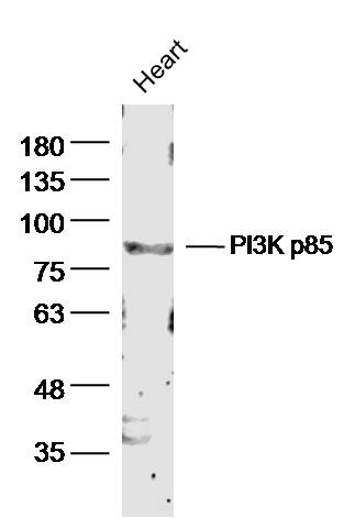 Sample: Heart (mouse) Lysate at 40 ug
Primary: Anti- PI3K p85 (SL0128R)at 1/300 dilution
Secondary: IRDye800CW Goat Anti-Rabbit IgG at 1/20000 dilution
Predicted band size: 80kD
Observed band size: 85 kD
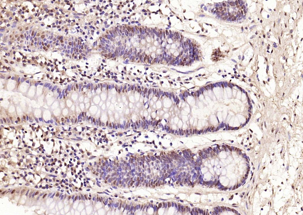 Paraformaldehyde-fixed, paraffin embedded (human rectal carcinoma); Antigen retrieval by boiling in sodium citrate buffer (pH6.0) for 15min; Block endogenous peroxidase by 3% hydrogen peroxide for 20 minutes; Blocking buffer (normal goat serum) at 37°C for 30min; Antibody incubation with (PI 3 Kinase p85 alpha) Polyclonal Antibody, Unconjugated (SL0128R) at 1:2000 overnight at 4°C, followed by operating according to SP Kit(Rabbit) (sp-0023) instructionsand DAB staining.
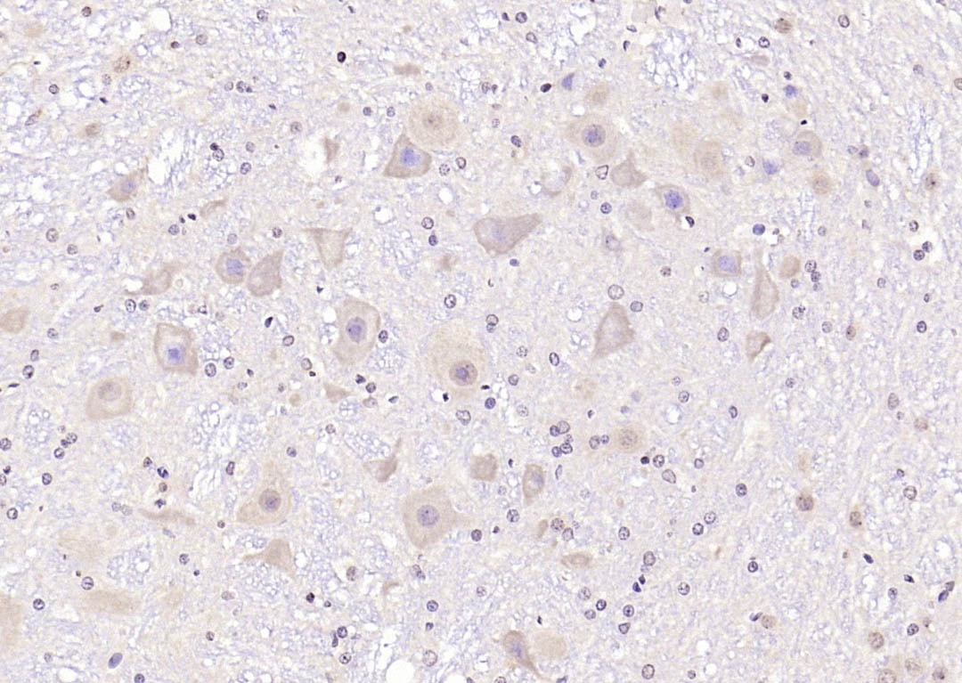 Paraformaldehyde-fixed, paraffin embedded (rat brain); Antigen retrieval by boiling in sodium citrate buffer (pH6.0) for 15min; Block endogenous peroxidase by 3% hydrogen peroxide for 20 minutes; Blocking buffer (normal goat serum) at 37°C for 30min; Antibody incubation with (PI 3 Kinase p85 alpha) Polyclonal Antibody, Unconjugated (SL0128R) at 1:2000 overnight at 4°C, followed by operating according to SP Kit(Rabbit) (sp-0023) instructionsand DAB staining.
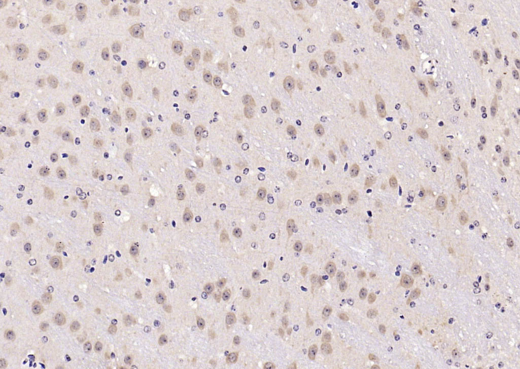 Paraformaldehyde-fixed, paraffin embedded (mouse brain); Antigen retrieval by boiling in sodium citrate buffer (pH6.0) for 15min; Block endogenous peroxidase by 3% hydrogen peroxide for 20 minutes; Blocking buffer (normal goat serum) at 37°C for 30min; Antibody incubation with (PI 3 Kinase p85 alpha) Polyclonal Antibody, Unconjugated (SL0128R) at 1:2000 overnight at 4°C, followed by operating according to SP Kit(Rabbit) (sp-0023) instructionsand DAB staining.
 Paraformaldehyde-fixed, paraffin embedded (mouse brain); Antigen retrieval by boiling in sodium citrate buffer (pH6.0) for 15min; Block endogenous peroxidase by 3% hydrogen peroxide for 20 minutes; Blocking buffer (normal goat serum) at 37°C for 30min; Antibody incubation with (PI 3 Kinase p85 alpha) Polyclonal Antibody, Unconjugated (SL0128R) at 1:2000 overnight at 4°C, followed by operating according to SP Kit(Rabbit) (sp-0023) instructionsand DAB staining.
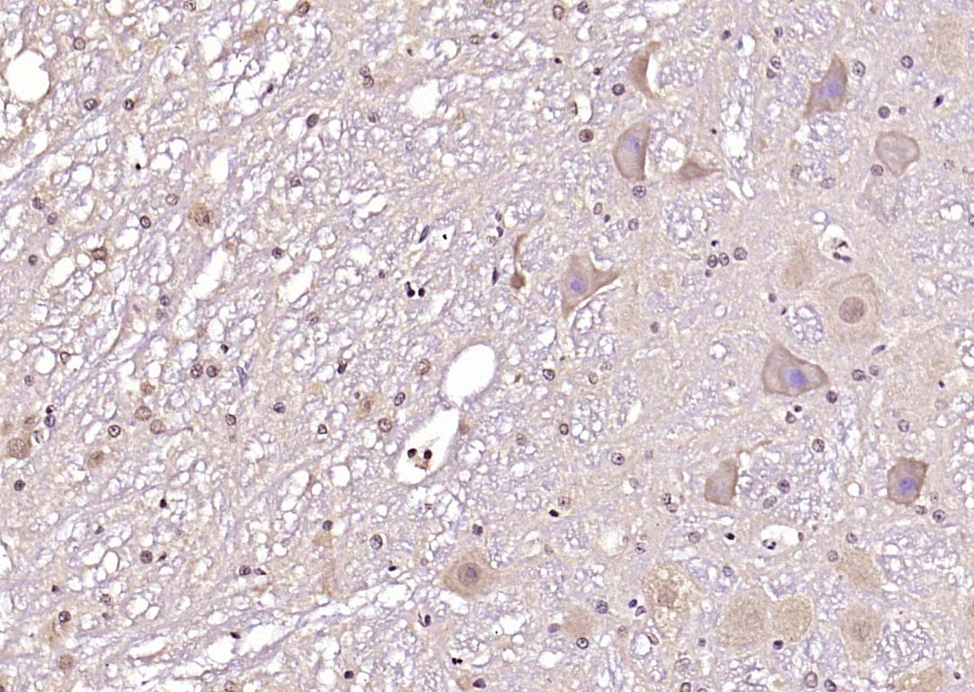 Paraformaldehyde-fixed, paraffin embedded (rat brain); Antigen retrieval by boiling in sodium citrate buffer (pH6.0) for 15min; Block endogenous peroxidase by 3% hydrogen peroxide for 20 minutes; Blocking buffer (normal goat serum) at 37°C for 30min; Antibody incubation with (PI 3 Kinase p85 alpha) Polyclonal Antibody, Unconjugated (SL0128R) at 1:2000 overnight at 4°C, followed by operating according to SP Kit(Rabbit) (sp-0023) instructionsand DAB staining.
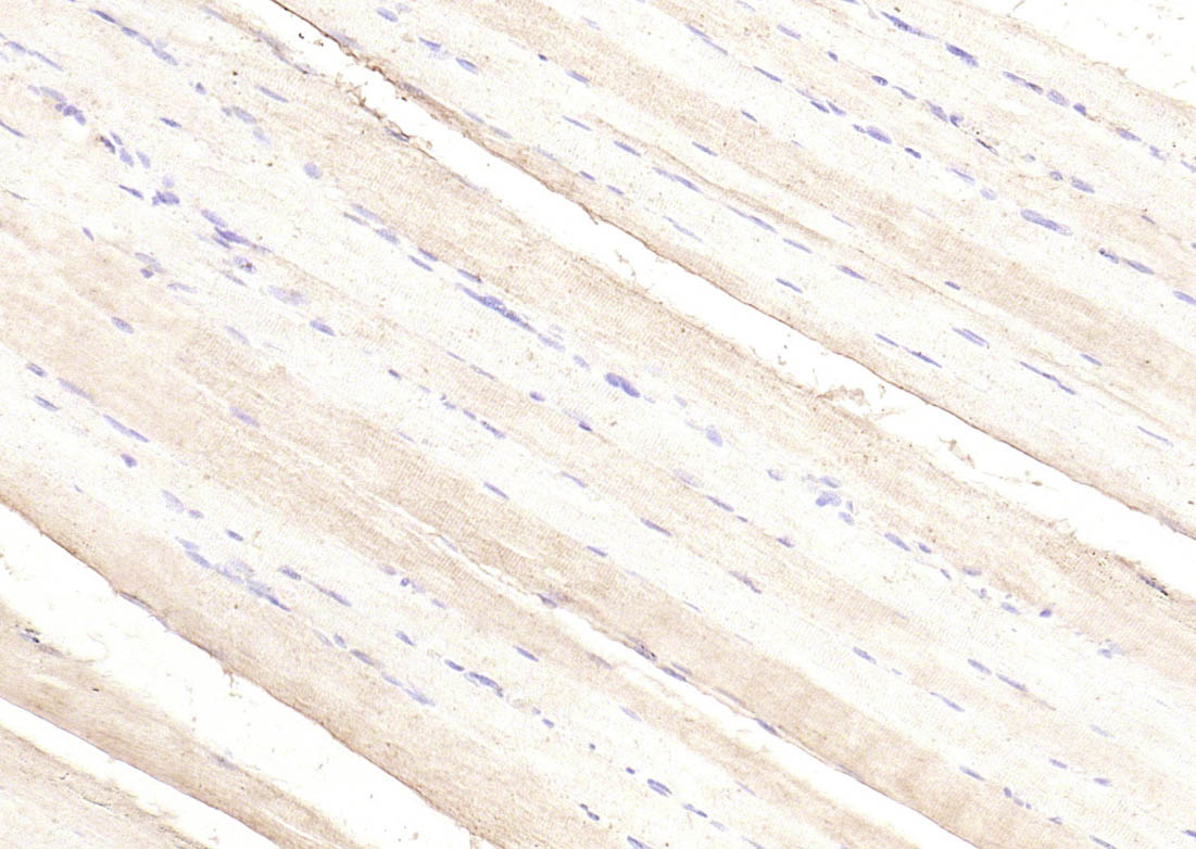 Paraformaldehyde-fixed, paraffin embedded (rat skeletal muscle); Antigen retrieval by boiling in sodium citrate buffer (pH6.0) for 15min; Block endogenous peroxidase by 3% hydrogen peroxide for 20 minutes; Blocking buffer (normal goat serum) at 37°C for 30min; Antibody incubation with (PI 3 Kinase p85 alpha) Polyclonal Antibody, Unconjugated (SL0128R) at 1:200 overnight at 4°C, followed by operating according to SP Kit(Rabbit) (sp-0023) instructionsand DAB staining.
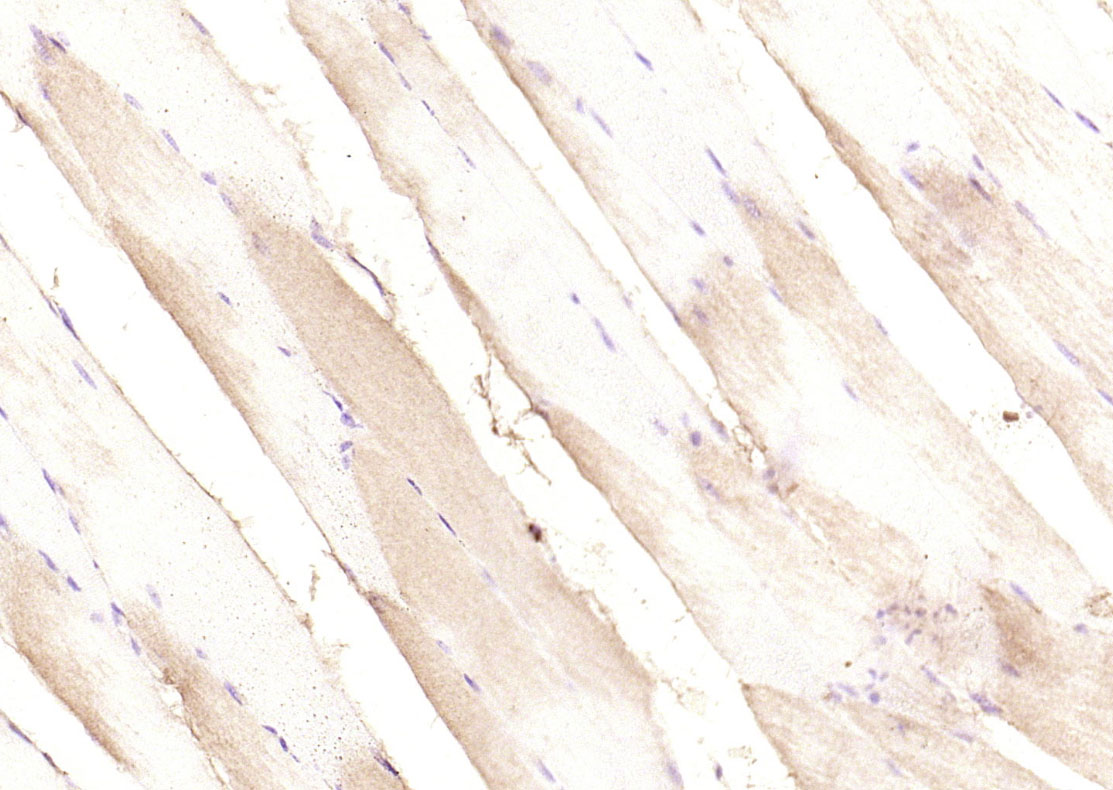 Paraformaldehyde-fixed, paraffin embedded (mouse skeletal muscle); Antigen retrieval by boiling in sodium citrate buffer (pH6.0) for 15min; Block endogenous peroxidase by 3% hydrogen peroxide for 20 minutes; Blocking buffer (normal goat serum) at 37°C for 30min; Antibody incubation with (PI 3 Kinase p85 alpha) Polyclonal Antibody, Unconjugated (SL0128R) at 1:200 overnight at 4°C, followed by operating according to SP Kit(Rabbit) (sp-0023) instructionsand DAB staining.
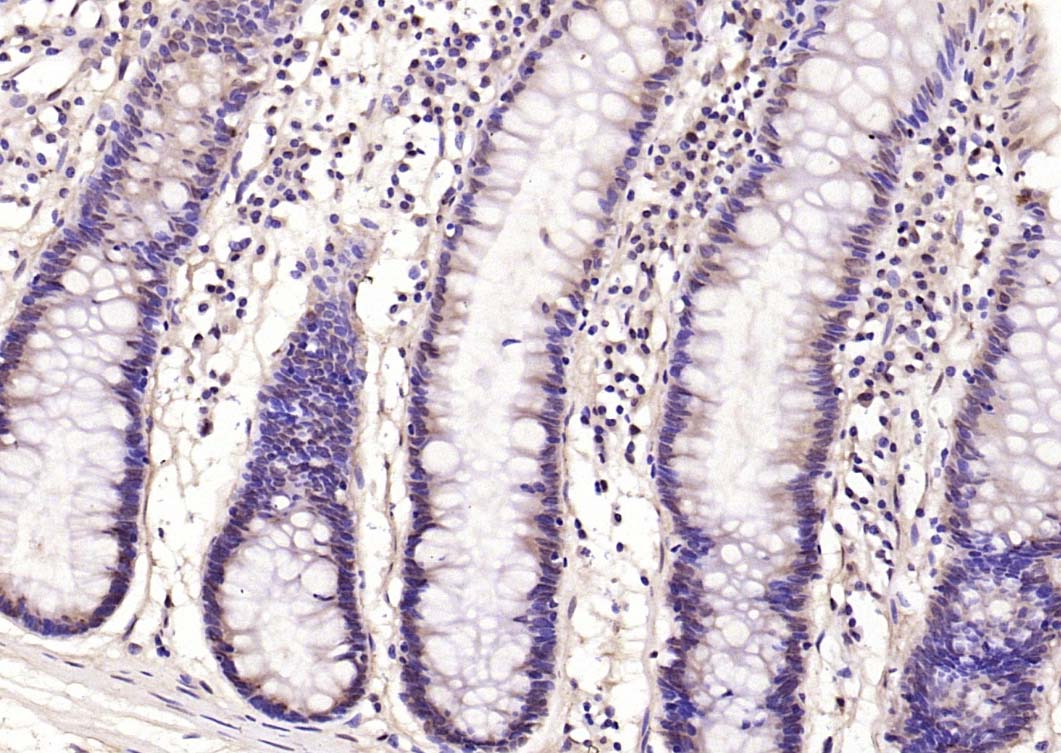 Paraformaldehyde-fixed, paraffin embedded (human rectal carcinoma); Antigen retrieval by boiling in sodium citrate buffer (pH6.0) for 15min; Block endogenous peroxidase by 3% hydrogen peroxide for 20 minutes; Blocking buffer (normal goat serum) at 37°C for 30min; Antibody incubation with (PI 3 Kinase p85 alpha) Polyclonal Antibody, Unconjugated (SL0128R) at 1:2000 overnight at 4°C, followed by operating according to SP Kit(Rabbit) (sp-0023) instructionsand DAB staining.
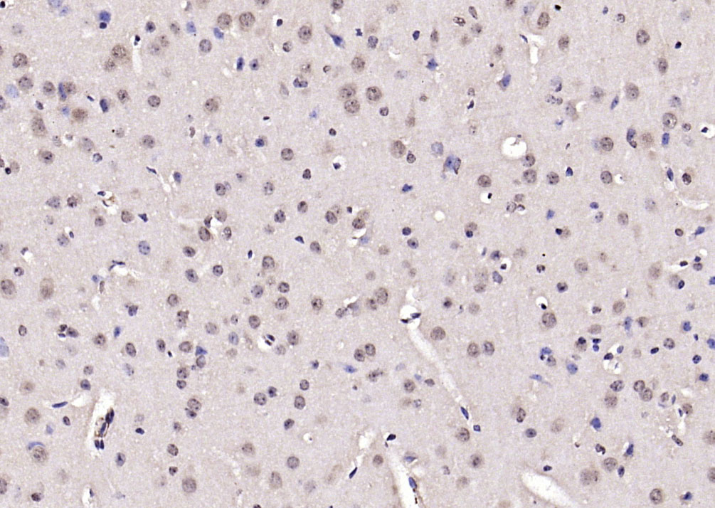 Paraformaldehyde-fixed, paraffin embedded (mouse brain); Antigen retrieval by boiling in sodium citrate buffer (pH6.0) for 15min; Block endogenous peroxidase by 3% hydrogen peroxide for 20 minutes; Blocking buffer (normal goat serum) at 37°C for 30min; Antibody incubation with (PI 3 Kinase p85 alpha) Polyclonal Antibody, Unconjugated (SL0128R) at 1:2000 overnight at 4°C, followed by operating according to SP Kit(Rabbit) (sp-0023) instructionsand DAB staining.
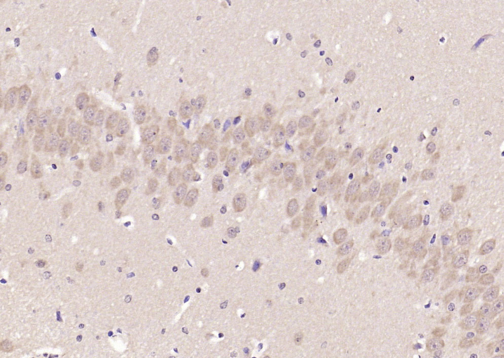 Paraformaldehyde-fixed, paraffin embedded (rat brain); Antigen retrieval by boiling in sodium citrate buffer (pH6.0) for 15min; Block endogenous peroxidase by 3% hydrogen peroxide for 20 minutes; Blocking buffer (normal goat serum) at 37°C for 30min; Antibody incubation with (PI 3 Kinase p85 alpha) Polyclonal Antibody, Unconjugated (SL0128R) at 1:2000 overnight at 4°C, followed by operating according to SP Kit(Rabbit) (sp-0023) instructionsand DAB staining.
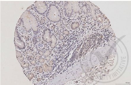 Images provided the Independent Validation Program (badge number 029650)Formalin-fixed and paraffin embedded human stomach labeled with Rabbit Anti-PI3 kinase p85 alpha subunit Polyclonal Antibody (SL0128R) at 1:250 overnight at room temperature followed by conjugation to secondary antibody.
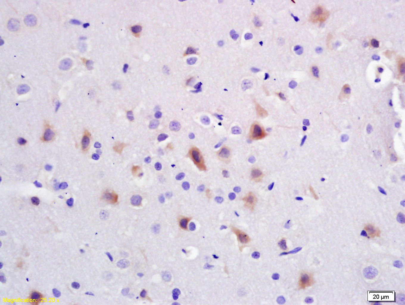 Tissue/cell: rat brain tissue; 4% Paraformaldehyde-fixed and paraffin-embedded;
Antigen retrieval: citrate buffer ( 0.01M, pH 6.0 ), Boiling bathing for 15min; Block endogenous peroxidase by 3% Hydrogen peroxide for 30min; Blocking buffer (normal goat serum,C-0005) at 37℃ for 20 min;
Incubation: Anti-PI3K/PI3 kinase p85 alpha subunit Polyclonal Antibody, Unconjugated(SL0128R) 1:200, overnight at 4°C, followed by conjugation to the secondary antibody(SP-0023) and DAB(C-0010) staining
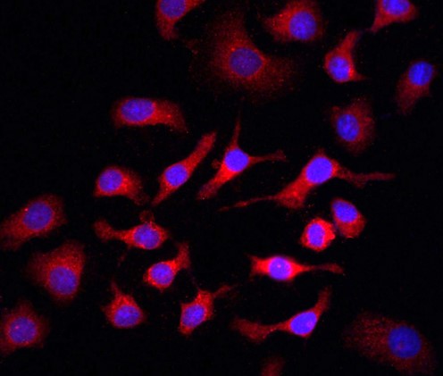 Tissue/cell: HepG2 cell; 4% Paraformaldehyde-fixed; Triton X-100 at room temperature for 20 min; Blocking buffer (normal goat serum, C-0005) at 37°C for 20 min; Antibody incubation with (PI3K p85) polyclonal Antibody, Unconjugated (SL0128R) 1:100, 90 minutes at 37°C; followed by a FITC conjugated Goat Anti-Rabbit IgG antibody at 37°C for 90 minutes, DAPI (blue, C02-04002) was used to stain the cell nuclei.
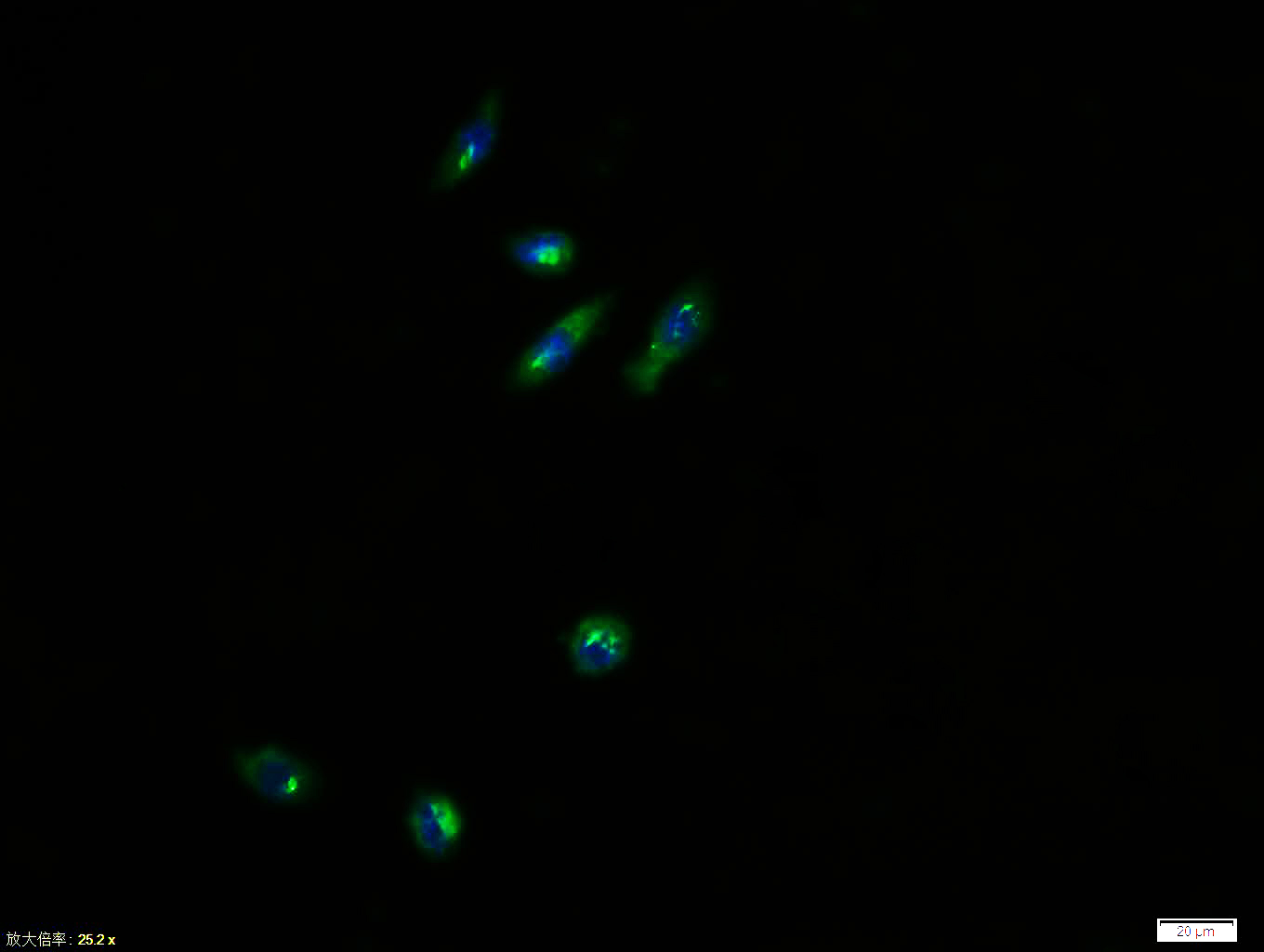 Tissue/cell: NIH/3T3 cell; 4% Paraformaldehyde-fixed; Triton X-100 at room temperature for 20 min; Blocking buffer (normal goat serum, C-0005) at 37°C for 20 min; Antibody incubation with (PI 3 Kinase p85 alpha) polyclonal Antibody, Unconjugated (SL0128R) 1:100, 90 minutes at 37°C; followed by a FITC conjugated Goat Anti-Rabbit IgG antibody at 37°C for 90 minutes, DAPI (blue, C02-04002) was used to stain the cell nuclei.
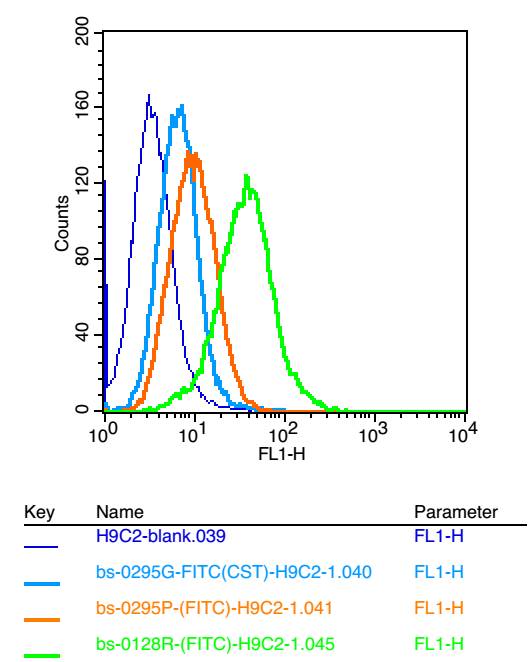 Positive control: H9C2(2% Paraformaldehyde-fixed )
Isotype Control Antibody
Antibody: Rabbit IgG; Dilution: 1μg in 100 μl 1 X PBS containing 0.5% BSA
Secondary Antibody
Antibody: Goat anti-rabbit IgG-FITC; Dilution: 1:200 in 1 X PBS containing 0.5% BSA
Primary Antibody
Supplier catalog number: SL1297R; Dilution: 1μg in 100 μl 1X PBS containing 0.5% BSA
Cartpieces
Totalgoods,subtotals:¥Checkout
Bought notes(bought amounts latest0)
No one bought this product
User Comment(Total0User Comment Num)
- No comment


 +86 571 56623320
 +86 18668110335

