
Rabbit Anti-Estrogen Receptor alpha antibody
Estradiol receptor; Estrogen receptor alpha; Estradiol Receptor-alpha; Estrogen Receptor 1; Atherosclerosis, susceptibility to, included; DKFZp686N23123; ER Alpha; ER; ER-alpha; ERalpha; ER[a]; Era; ESR; ESR1; ESR1_HUMAN; ESR2; ESRA; Estr; Estrogen recept
View History [Clear]
Details
Product Name Estrogen Receptor alpha Chinese Name 雌激素受体α抗体 Alias Estradiol receptor; Estrogen receptor alpha; Estradiol Receptor-alpha; Estrogen Receptor 1; Atherosclerosis, susceptibility to, included; DKFZp686N23123; ER Alpha; ER; ER-alpha; ERalpha; ER[a]; Era; ESR; ESR1; ESR1_HUMAN; ESR2; ESRA; Estr; Estrogen receptor 1 (alpha); Estrogen resistance, included; HDL cholesterol, augmented response of, to hormone replacement, included; Myocardial infarction, susceptibility to, included; NR3A1; Nuclear receptor subfamily 3 group A member 1; OTTHUMP00000017718; OTTHUMP00000017719; RNESTROR. literatures Research Area Tumour immunology Growth factors and hormones Immunogen Species Rabbit Clonality Polyclonal React Species Human, Rat, (predicted: Mouse, Dog, Cow, Rabbit, ) Applications WB=1:500-2000 ELISA=1:5000-10000 ICC=1:100
not yet tested in other applications.
optimal dilutions/concentrations should be determined by the end user.Theoretical molecular weight 67kDa Cellular localization The nucleus Form Liquid Concentration 1mg/ml immunogen KLH conjugated synthetic peptide derived from the middle of human Estrogen Receptor alpha: 331-360/595 Lsotype IgG Purification affinity purified by Protein A Buffer Solution 0.01M TBS(pH7.4) with 1% BSA, 0.03% Proclin300 and 50% Glycerol. Storage Shipped at 4℃. Store at -20 °C for one year. Avoid repeated freeze/thaw cycles. Attention This product as supplied is intended for research use only, not for use in human, therapeutic or diagnostic applications. PubMed PubMed Product Detail Estrogen and progesterone receptor are members of a family of transcription factors that are regulated by the binding of their cognate ligands. The interaction of hormone-bound estrogen receptors with estrogen responsive elements(EREs) alters transcription of ERE-containing genes. The carboxy terminal region of the estrgen receptor contains the ligand binding domain, the amino terminus serves as the transactivation domain, and the DNA binding domain is centrally located. Two forms of estrogen receptor have been identified, ER Alpha and ER Beta. ER Alpha and ER Beta have been shown to be differentially activated by various ligands. The biological response to progesterone is mediated by two distinct forms of the human progesterone receptor (hPR-A and hPR-B), which arise from alternative splicing. In most cells, hPR-B functions as a transcriptional activator of progesterone-responsive gene, whereas hPR-A function as a transcriptional inhibitor of all steroid hormone receptors.
Function:
Nuclear hormone receptor. The steroid hormones and their receptors are involved in the regulation of eukaryotic gene expression and affect cellular proliferation and differentiation in target tissues.
Subunit:
Interacts with SLC30A9 (By similarity). Binds DNA as a homodimer. Can form a heterodimer with ESR2. Interacts with NCOA3, NCOA5 and NCOA6 coactivators, leading to a strong increase of transcription of target genes. Interacts with NCOA7 in a ligand-inducible manner. Interacts with PHB2, PELP1 and UBE1C. Interacts with AKAP13. Interacts with CUEDC2. Interacts with KDM5A. Interacts with SMARD1. Interacts with HEXIM1 and MAP1S. Interacts with PBXIP1. Interaction with MUC1 is stimulated by 7 beta-estradiol (E2) and enhances ERS1-mediated transcription. Interacts with DNTTIP2, FAM120B and UIMC1. Interacts with isoform 4 of TXNRD1. Interacts with MLL2. Interacts with ATAD2 and this interaction is enhanced by estradiol.
Subcellular Location:
Nucleus.
Post-translational modifications:
Phosphorylated by cyclin A/CDK2. Phosphorylation probably enhances transcriptional activity.
Glycosylated; contains N-acetylglucosamine, probably O-linked.
Similarity:
Belongs to the nuclear hormone receptor family. NR3 subfamily.
Contains 1 nuclear receptor DNA-binding domain.
SWISS:
P03372
Gene ID:
2099
Database links:
Entrez Gene: 2099 Human
Entrez Gene: 13982 Mouse
Omim: 133430 Human
SwissProt: P03372 Human
类固醇受体(Steroid Receptors)
雌激素受体ER-α(Estrogen Receptor-α, ERα)是一类位于细胞表面或细胞内的特异性蛋白质。
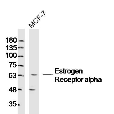
某些特异的雌激素、雌激素类似物、衍生物、拮抗剂或阻断剂与它结合后可引发或阻断受体蛋白质的转化作用。并将雌激素-受体复合物从细胞质再定位进入The nucleus以启动蛋白质的生物合成。Product Picture 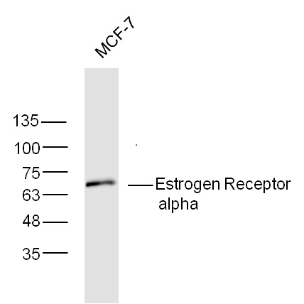 Sample:MCF-7 Cell Lysate at 30ug;
Primary: Anti-Estrogen Receptor alpha (SL0122R) at 1:300;
Secondary: HRP conjugated Goat-Anti-rabbit IgG(SL0295G-HRP) at 1: 5000;
Predicted band size: 67 kD
Observed band size: 67 kD
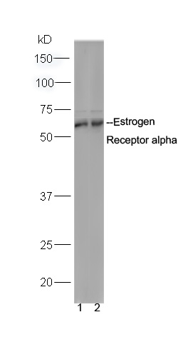 Sample:
MCF-7 Cell Lysate at 30ug;
DU145 Cell Lysate at 30 ug;
Primary: Anti-Estrogen Receptor alpha (SL0122R) at 1:300;
Secondary: HRP conjugated Goat-Anti-rabbit IgG(SL0295G-HRP) at 1: 5000;
Predicted band size: 67 kD
Observed band size: 67 kD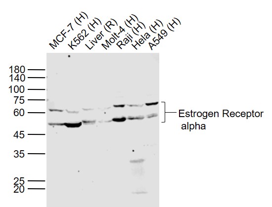 Sample:
Lane 1: MCF-7 (Human) Cell Lysate at 30 ug
Lane 2: K562 (Human) Cell Lysate at 30 ug
Lane 3: Liver (Rat) Lysate at 40 ug
Lane 4: Molt-4 (Human) Cell Lysate at 30 ug
Lane 5: Raji (Human) Cell Lysate at 30 ug
Lane 6: Hela (Human) Cell Lysate at 30 ug
Lane 7: A549 (Human) Cell Lysate at 30 ug
Primary: Anti- Estrogen Receptor alpha (SL0122R) at 1/300 dilution
Secondary: IRDye800CW Goat Anti-Rabbit IgG at 1/20000 dilution
Predicted band size: 66/46 kD
Observed band size: 62/50 kD
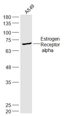 Sample:
A549(Human) Cell Lysate at 30 ug
Primary: Anti-Estrogen Receptor alpha (SL0122R) at 1/300 dilution
Secondary: IRDye800CW Goat Anti-Rabbit IgG at 1/20000 dilution
Predicted band size: 67 kD
Observed band size: 67 kD
 Sample: MCF-7 Cell (Human) Lysate at 30 ug
Primary: Anti- Estrogen Receptor alpha (SL0122R)at 1/300 dilution
Secondary: IRDye800CW Goat Anti-Rabbit IgG at 1/20000 dilution
Predicted band size: 67kD
Observed band size: 65kD
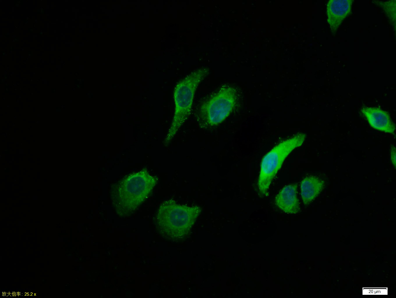 Tissue/cell:MCF7 cell; 4% Paraformaldehyde-fixed; Triton X-100 at room temperature for 20 min; Blocking buffer (normal goat serum, C-0005) at 37°C for 20 min; Antibody incubation with (Estrogen Receptor alpha) polyclonal Antibody, Unconjugated (SL0122R) 1:100, 90 minutes at 37°C; followed by a FITC conjugated Goat Anti-Rabbit IgG antibody at 37°C for 90 minutes, DAPI (blue, C02-04002) was used to stain the cell nuclei.
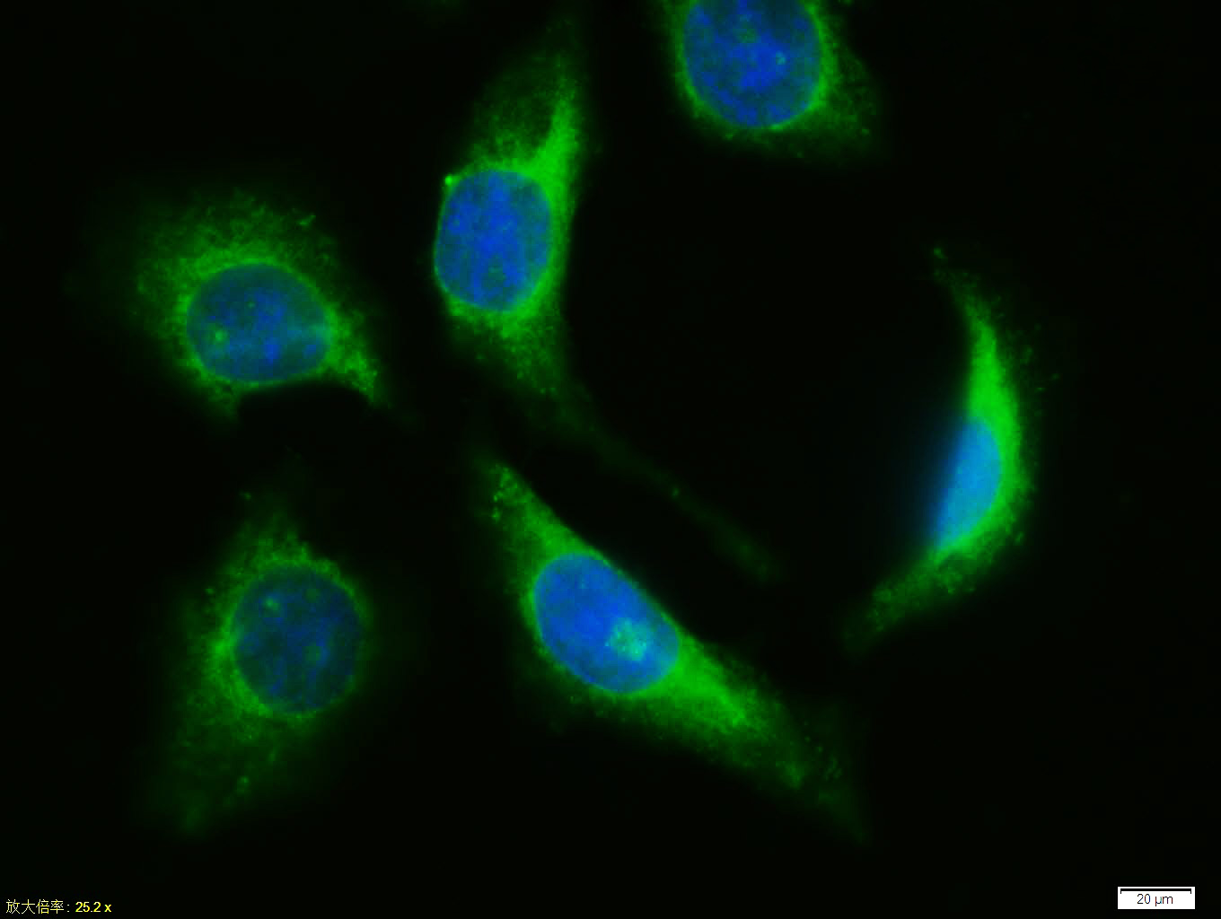 Tissue/cell:MCF7 cell; 4% Paraformaldehyde-fixed; Triton X-100 at room temperature for 20 min; Blocking buffer (normal goat serum, C-0005) at 37°C for 20 min; Antibody incubation with (Estrogen Receptor alpha) polyclonal Antibody, Unconjugated (SL0122R) 1:100, 90 minutes at 37°C; followed by a FITC conjugated Goat Anti-Rabbit IgG antibody at 37°C for 90 minutes, DAPI (blue, C02-04002) was used to stain the cell nuclei.Tissue/cell:MCF7 cell; 4% Paraformaldehyde-fixed; Triton X-100 at room temperature for 20 min; Blocking buffer (normal goat serum, C-0005) at 37°C for 20 min; Antibody incubation with (Estrogen Receptor alpha) polyclonal Antibody, Unconjugated (SL0122R) 1:100, 90 minutes at 37°C; followed by a FITC conjugated Goat Anti-Rabbit IgG antibody at 37°C for 90 minutes, DAPI (blue, C02-04002) was used to stain the cell nuclei.Tissue/cell:MCF7 cell; 4% Paraformaldehyde-fixed; Triton X-100 at room temperature for 20 min; Blocking buffer (normal goat serum, C-0005) at 37°C for 20 min; Antibody incubation with (Estrogen Receptor alpha) polyclonal Antibody, Unconjugated (SL0122R) 1:100, 90 minutes at 37°C; followed by a FITC conjugated Goat Anti-Rabbit IgG antibody at 37°C for 90 minutes, DAPI (blue, C02-04002) was used to stain the cell nuclei.
Cartpieces
Totalgoods,subtotals:¥Checkout
Bought notes(bought amounts latest0)
No one bought this product
User Comment(Total0User Comment Num)
- No comment


 +86 571 56623320
 +86 18668110335

