
Rabbit Anti-Caspase-3 antibody
Caspase-3 subunit p17; cleaved Caspase 3; cleaved Caspase-3; APOPAIN; CASP3; Caspase 3 apoptosis related cysteine protease; Caspase3; CPP32; CPP32B; Cysteine protease CPP32; Human cysteine protease CPP32 isoform alpha mRNA complete cds; PARP cleavage prot
View History [Clear]
Details
Product Name Caspase-3 Chinese Name 活化半胱胺酸蛋白酶蛋白-3抗体 Alias Caspase-3 subunit p17; cleaved Caspase 3; cleaved Caspase-3; APOPAIN; CASP3; Caspase 3 apoptosis related cysteine protease; Caspase3; CPP32; CPP32B; Cysteine protease CPP32; Human cysteine protease CPP32 isoform alpha mRNA complete cds; PARP cleavage protease; SCA 1; SCA1; SREBP cleavage activity 1; Yama; CASP3_HUMAN; Caspase-3; CASP-3; Apopain; Protein Yama; SREBP cleavage activity 1; SCA-1. literatures Research Area Tumour Cell biology Neurobiology Signal transduction Apoptosis Alzheimer's Immunogen Species Rabbit Clonality Polyclonal React Species Human, Mouse, Rat, Applications WB=1:500-2000 ELISA=1:5000-10000 IHC-P=1:100-500 ICC=1:100 IF=1:100-500 (Paraffin sections need antigen repair)
not yet tested in other applications.
optimal dilutions/concentrations should be determined by the end user.Theoretical molecular weight 17/32kDa Cellular localization cytoplasmic Form Liquid Concentration 1mg/ml immunogen KLH conjugated synthetic peptide derived from human caspase-3 p17 subunit: 80-175/277 Lsotype IgG Purification affinity purified by Protein A Buffer Solution 0.01M TBS(pH7.4) with 1% BSA, 0.03% Proclin300 and 50% Glycerol. Storage Shipped at 4℃. Store at -20 °C for one year. Avoid repeated freeze/thaw cycles. Attention This product as supplied is intended for research use only, not for use in human, therapeutic or diagnostic applications. PubMed PubMed Product Detail The caspase family of cysteine proteases play a key role in apoptosis. Caspase 3 is the most extensively studied apoptotic protein among caspase family members. Caspase 3 is synthesized as inactive pro enzyme that is processed in cells undergoing apoptosis by self proteolysis and/or cleavage by other upstream proteases (e.g. Caspases 8, 9 and 10). The processed form of Caspase 3 consists of large (17kDa) and small (12kDa) subunits which associate to form an active enzyme. Caspase 3 is cleaved at Asp28 Ser29 and Asp175 Ser176. The active Caspase 3 proteolytically cleaves and activates other caspases (e.g. Caspases 6, 7 and 9), as well as relevant targets in the cells (e.g. PARP and DFF). Alternative splicing of this gene results in two transcript variants which encode the same protein. In immunohistochemical studies Caspase 3 expression has been shown to be widespread but not present in all cell types (e.g. commonly reported in epithelial cells of skin, renal proximal tubules and collecting ducts). Differences in the level of Caspase 3 have been reported in cells of short lived nature (eg germinal centre B cells) and those that are long lived (eg mantle zone B cells). Caspase 3 is the predominant caspase involved in the cleavage of amyloid beta 4A precursor protein, which is associated with neuronal death in Alzheimer's disease.
Reacts with Caspase-3 subunit p17 and precursor.
Function:
Involved in the activation cascade of caspases responsible for apoptosis execution. At the onset of apoptosis it proteolytically cleaves poly(ADP-ribose) polymerase (PARP) at a '216-Asp-|-Gly-217' bond. Cleaves and activates sterol regulatory element binding proteins (SREBPs) between the basic helix-loop-helix leucine zipper domain and the membrane attachment domain. Cleaves and activates caspase-6, -7 and -9. Involved in the cleavage of huntingtin. Triggers cell adhesion in sympathetic neurons through RET cleavage.
Subunit:
Heterotetramer that consists of two anti-parallel arranged heterodimers, each one formed by a 17 kDa (p17) and a 12 kDa (p12) subunit. Interacts with BIRC6/bruce.
Subcellular Location:
Cytoplasm.
Tissue Specificity:
Highly expressed in lung, spleen, heart, liver and kidney. Moderate levels in brain and skeletal muscle, and low in testis. Also found in many cell lines, highest expression in cells of the immune system.
Post-translational modifications:
Cleavage by granzyme B, caspase-6, caspase-8 and caspase-10 generates the two active subunits. Additional processing of the propeptides is likely due to the autocatalytic activity of the activated protease. Active heterodimers between the small subunit of caspase-7 protease and the large subunit of caspase-3 also occur and vice versa.
S-nitrosylated on its catalytic site cysteine in unstimulated human cell lines and denitrosylated upon activation of the Fas apoptotic pathway, associated with an increase in intracellular caspase activity. Fas therefore activates caspase-3 not only by inducing the cleavage of the caspase zymogen to its active subunits, but also by stimulating the denitrosylation of its active site thiol.
Similarity:
Belongs to the peptidase C14A family.
SWISS:
P42574
Gene ID:
836
Database links:
Entrez Gene: 836 Human
Entrez Gene: 12367 Mouse
Entrez Gene: 100008840 Rabbit
Omim: 600636 Human
SwissProt: P42574 Human
SwissProt: P70677 Mouse
SwissProt: Q8MJC3 Rabbit
Unigene: 141125 Human
Unigene: 34405 Mouse
Unigene: 10562 Rat
Caspase3广泛分布于各种不同类型的细胞中,是Caspase家族中最重要的凋亡执行者之一,激活的Caspase-3能使许多与细胞结构、细胞周期及DNA修复等相关蛋白或激酶失活,从而使Apoptosis.Product Picture 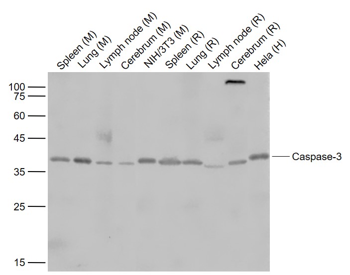 Sample:
Lane 1: Spleen (Mouse) Lysate at 40 ug
Lane 2: Lung (Mouse) Lysate at 40 ug
Lane 3: Lymph node (Mouse) Lysate at 40 ug
Lane 4: Cerebrum (Mouse) Lysate at 40 ug
Lane 5: NIH/3T3 (Mouse) Cell Lysate at 30 ug
Lane 6: Spleen (Rat) Lysate at 40 ug
Lane 7: Lung (Rat) Lysate at 40 ug
Lane 8: Lymph node (Rat) Lysate at 40 ug
Lane 9: Cerebrum (Rat) Lysate at 40 ug
Lane 10: Hela (Human) Cell Lysate at 30 ug
Primary: Anti-Caspase-3 (SL0081R) at 1/1000 dilution
Secondary: IRDye800CW Goat Anti-Rabbit IgG at 1/20000 dilution
Predicted band size: 35 kD
Observed band size: 37 kD
 Sample:
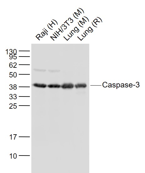
Lane 1: Raji (Human) Cell Lysate at 30 ug
Lane 2: NIH/3T3 (Mouse) Cell Lysate at 30 ug
Lane 3: Lung (Mouse) Lysate at 40 ug
Lane 4: Lung (Rat) Lysate at 40 ug
Primary: Anti-Caspase-3 (SL0081R) at 1/1000 dilution
Secondary: IRDye800CW Goat Anti-Rabbit IgG at 1/20000 dilution
Predicted band size: 35 kD
Observed band size: 37 kD
 Sample:
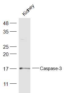
Kidney (Mouse) Lysate at 40 ug
Primary: Anti-Caspase-3 (SL0081R) at 1/300 dilution
Secondary: IRDye800CW Goat Anti-Rabbit IgG at 1/20000 dilution
Predicted band size: 28 kD
Observed band size: 17 kD
 Sample:
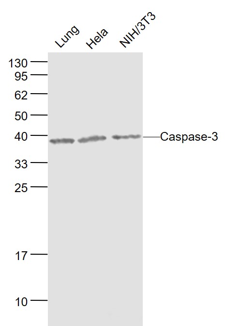
Lung(Mouse) Lysate at 40 ug
Hela(Human) Cell Lysate at 30 ug
NIH/3T3(Mouse) Cell Lysate at 30 ug
Primary: Anti-Caspase-3 (SL0081R) at 1/1000 dilution
Secondary: IRDye800CW Goat Anti-Rabbit IgG at 1/20000 dilution
Predicted band size: 35/29/19/17 kD
Observed band size: 38 kD
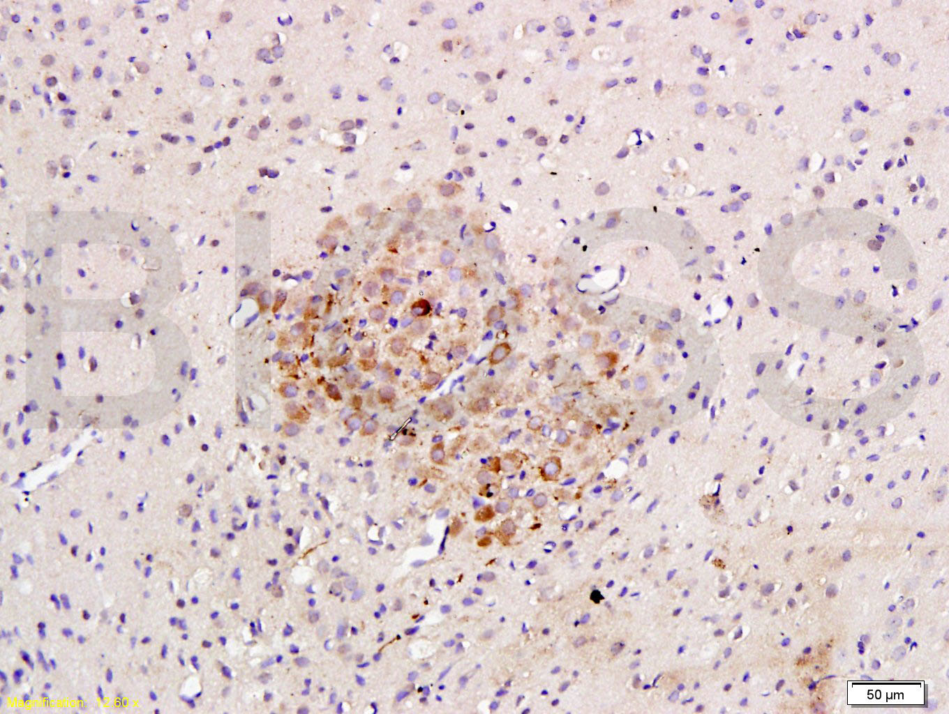 Tissue/cell: rat brain tissue; 4% Paraformaldehyde-fixed and paraffin-embedded;
Antigen retrieval: citrate buffer ( 0.01M, pH 6.0 ), Boiling bathing for 15min; Block endogenous peroxidase by 3% Hydrogen peroxide for 30min; Blocking buffer (normal goat serum,C-0005) at 37℃ for 20 min;
Incubation: Anti-Caspase-3 Polyclonal Antibody, Unconjugated(SL0081R) 1:200, overnight at 4癈, followed by conjugation to the secondary antibody(SP-0023) and DAB(C-0010) staining
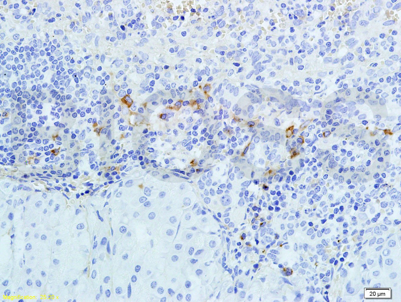 Tissue/cell: rabbit pancreas tissue; 4% Paraformaldehyde-fixed and paraffin-embedded;
Antigen retrieval: citrate buffer ( 0.01M, pH 6.0 ), Boiling bathing for 15min; Block endogenous peroxidase by 3% Hydrogen peroxide for 30min; Blocking buffer (normal goat serum,C-0005) at 37℃ for 20 min;
Incubation: Anti-Caspase-3 Polyclonal Antibody, Unconjugated(SL0081R) 1:300, overnight at 4癈, followed by conjugation to the secondary antibody(SP-0023) and DAB(C-0010) staining
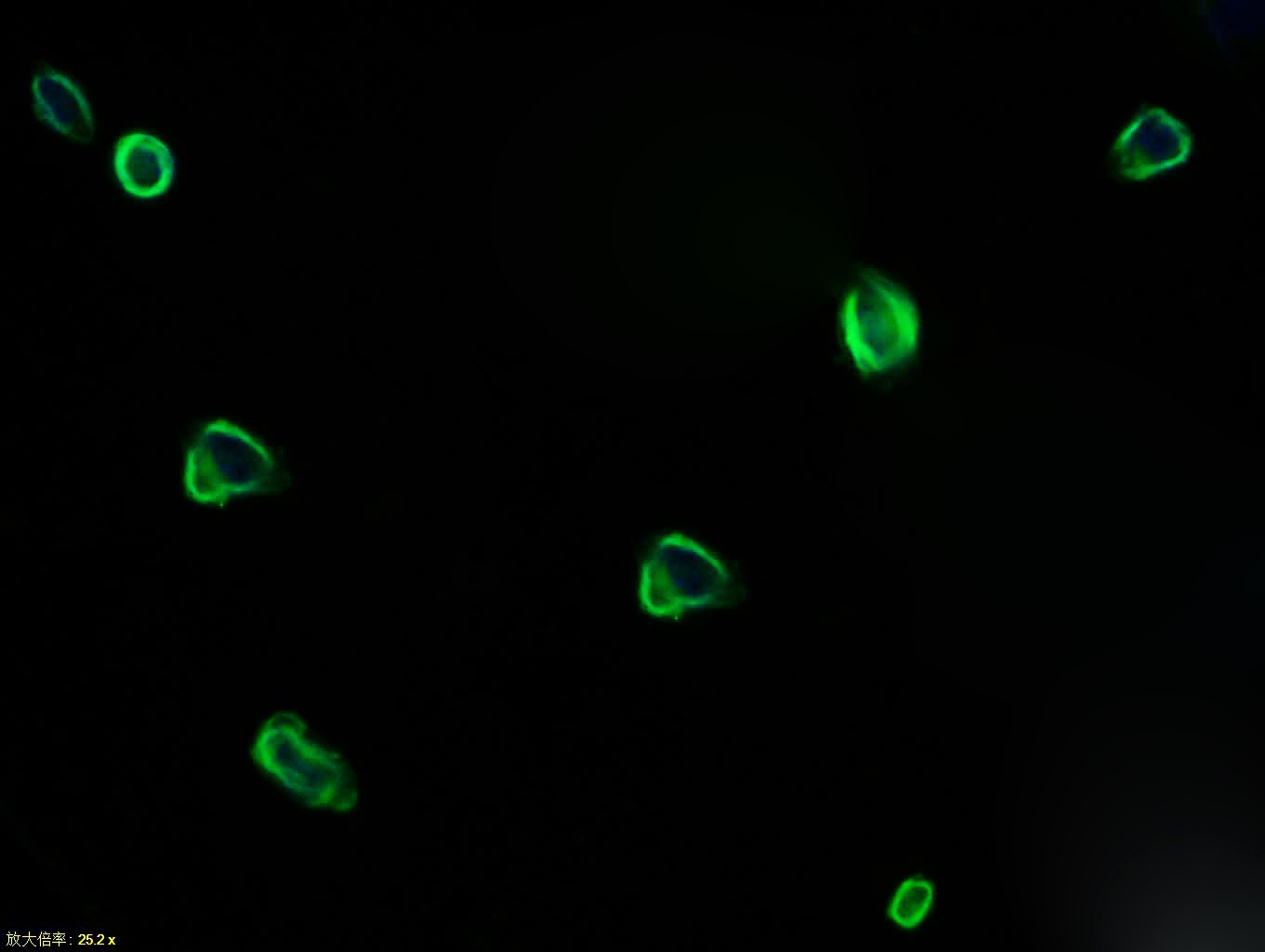 Tissue/cell: NIH/3T3 cell; 4% Paraformaldehyde-fixed; Triton X-100 at room temperature for 20 min; Blocking buffer (normal goat serum, C-0005) at 37°C for 20 min; Antibody incubation with (Caspase-3) polyclonal Antibody, Unconjugated (SL0081R) 1:100, 90 minutes at 37°C; followed by a FITC conjugated Goat Anti-Rabbit IgG antibody at 37°C for 90 minutes, DAPI (blue, C02-04002) was used to stain the cell nuclei.
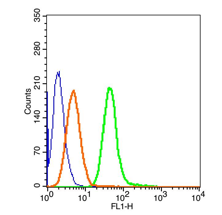 The figure annotation:
The blue histogram is unstained cells
. The Orange histogram is cells stained with Rabbit IgG/FITC (SL0295P-FITC).
The green histogram is cells stained with Rabbit Anti-Caspase-3/FITC Conjugated antibody (SL0081R-FITC).
Controls
Positive control: HepG 2 cells
Isotype control: Cell lines treated with Rabbit IgG/FITC (SL0295P-FITC) instead of the primary antibody to confirm that primary antibody binding is specific. 2μg in 1 00μL1 X PBS containing 0.5% BSA.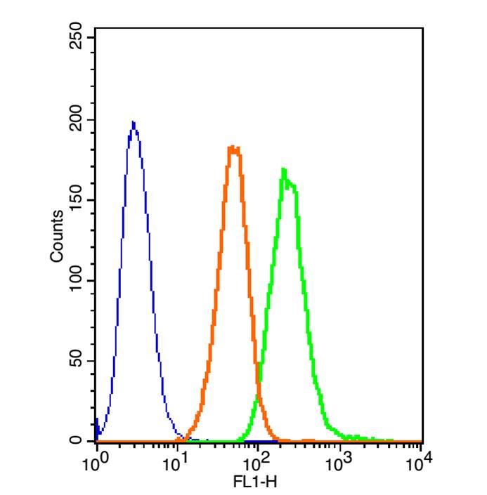 The figure annotation:
The blue histogram is unstained cells.
The Orange histogram is cells stained with Rabbit IgG/FITC (SL0295P-FITC).
The green histogram is cells stained with Rabbit Anti-Caspase-3/FITC Conjugated antibody (SL0081R-FITC).
Controls
Positive control: A549 cells
Isotype control: Cell lines treated with Rabbit IgG/FITC(SL0295P-FITC) instead of the primary antibody to confirm that primary antibody binding is specific. 3μg in 1 00 μL 1 X PBS containing 0.5% BSA.
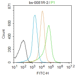 Blank control:Mouse spleen.
Primary Antibody (green line): Rabbit Anti-Caspase-3 antibody (SL0081R)
Dilution: 2μg /10^6 cells;
Isotype Control Antibody (orange line): Rabbit IgG .
Secondary Antibody : Goat anti-rabbit IgG-AF488
Dilution: 1μg /test.
Protocol
The cells were fixed with 4% PFA (10min at room temperature)and then permeabilized with 0.1% PBST for 20 min at room temperature. The cells were then incubated in 5%BSA to block non-specific protein-protein interactions for 30 min at room temperature .Cells stained with Primary Antibody for 30 min at room temperature. The secondary antibody used for 40 min at room temperature. Acquisition of 20,000 events was performed.
Cartpieces
Totalgoods,subtotals:¥Checkout
Partial purchase records(bought amounts latest0)
No one bought this product
User Comment(Total0User Comment Num)
- No comment


 +86 571 56623320


