
Rabbit Anti-GSK-3 Beta antibody
Glycogen synthase kinase-3 beta; glycogen synthase kinase 3 beta; GSK 3 beta; GSK 3B; GSK3B; GSK3B protein; GSK3beta isoform; GSK3 beta; Serine/threonine-protein kinase GSK3B; GSK3β; GSK3B_HUMAN.GSK 3β; GSK 3 β; GSK-3β; GSK3β;
View History [Clear]
Details
Product Name GSK-3 Beta Chinese Name 糖原合酶激酶-3β抗体 Alias Glycogen synthase kinase-3 beta; glycogen synthase kinase 3 beta; GSK 3 beta; GSK 3B; GSK3B; GSK3B protein; GSK3beta isoform; GSK3 beta; Serine/threonine-protein kinase GSK3B; GSK3β; GSK3B_HUMAN. GSK 3β; GSK 3 β; GSK-3β; GSK3β; literatures Research Area Cardiovascular Neurobiology Signal transduction Stem cells Kinases and Phosphatases Diabetes The new supersedes the old Immunogen Species Rabbit Clonality Polyclonal React Species Human, Mouse, Rat, (predicted: Chicken, Dog, Pig, Cow, Horse, Rabbit, Sheep, ) Applications ELISA=1:5000-10000 IHC-P=1:100-500 IHC-F=1:100-500 Flow-Cyt=1μg /test ICC=1:100 IF=1:100-500 (Paraffin sections need antigen repair)
not yet tested in other applications.
optimal dilutions/concentrations should be determined by the end user.Theoretical molecular weight 47kDa Cellular localization The nucleus cytoplasmic The cell membrane Form Liquid Concentration 1mg/ml immunogen KLH conjugated synthetic peptide derived from human GSK-3 Beta: 341-420/420 Lsotype IgG Purification affinity purified by Protein A Buffer Solution 0.01M TBS(pH7.4) with 1% BSA, 0.03% Proclin300 and 50% Glycerol. Storage Shipped at 4℃. Store at -20 °C for one year. Avoid repeated freeze/thaw cycles. Attention This product as supplied is intended for research use only, not for use in human, therapeutic or diagnostic applications. PubMed PubMed Product Detail The protein encoded by this gene is a serine-threonine kinase, belonging to the glycogen synthase kinase subfamily. It is involved in energy metabolism, neuronal cell development, and body pattern formation. Polymorphisms in this gene have been implicated in modifying risk of Parkinson disease, and studies in mice show that overexpression of this gene may be relevant to the pathogenesis of Alzheimer disease. Alternatively spliced transcript variants encoding different isoforms have been found for this gene.[provided by RefSeq, Sep 2009]
Function:
Constitutively active protein kinase that acts as a negative regulator in the hormonal control of glucose homeostasis, Wnt signaling and regulation of transcription factors and microtubules, by phosphorylating and inactivating glycogen synthase (GYS1 or GYS2), EIF2B, CTNNB1/beta-catenin, APC, AXIN1, JUN, NFATC1/NFATC, MAPT/TAU and MACF1. Requires primed phosphorylation of the majority of its substrates. In skeletal muscle, contributes to insulin regulation of glycogen synthesis by phosphorylating and inhibiting GYS1 activity and hence glycogen synthesis. May also mediate the development of insulin resistance by regulating activation of transcription factors. Regulates protein synthesis by controlling the activity of initiation factor 2B (EIF2BE/EIF2B5) in the same manner as glycogen synthase. In Wnt signaling, GSK3B forms a multimeric complex with APC, AXIN1 and CTNNB1/beta-catenin and phosphorylates the N-terminus of CTNNB1 leading to its degradation mediated by ubiquitin/proteasomes. Phosphorylates JUN at sites proximal to its DNA-binding domain, thereby reducing its affinity for DNA. Phosphorylates NFATC1/NFATC on conserved serine residues promoting NFATC1/NFATC nuclear export, shutting off NFATC1/NFATC gene regulation, and thereby opposing the action of calcineurin. Phosphorylates MAPT/TAU on 'Thr-548', decreasing significantly MAPT/TAU ability to bind and stabilize microtubules. MAPT/TAU is the principal component of neurofibrillary tangles in Alzheimer disease. Plays an important role in ERBB2-dependent stabilization of microtubules at the cell cortex. Phosphorylates MACF1, inhibiting its binding to microtubules which is critical for its role in bulge stem cell migration and skin wound repair. Probably regulates NF-kappa-B (NFKB1) at the transcriptional level and is required for the NF-kappa-B-mediated anti-apoptotic response to TNF-alpha (TNF/TNFA). Negatively regulates replication in pancreatic beta-cells, resulting in apoptosis, loss of beta-cells and diabetes. Phosphorylates MUC1 in breast cancer cells, decreasing the interaction of MUC1 with CTNNB1/beta-catenin. Is necessary for the establishment of neuronal polarity and axon outgrowth. Phosphorylates MARK2, leading to inhibit its activity. Phosphorylates SIK1 at 'Thr-182', leading to sustain its activity.
Subunit:
Monomer (By similarity). Interacts with ARRB2 and DISC1 (By similarity). Interacts with CABYR, MMP2, MUC1, NIN and PRUNE Interacts with AXIN1; the interaction mediates hyperphosphorylation of CTNNB1 leading to its ubiquitination and destruction. Interacts with and phosphorylates SNAI1. Interacts with DNM1L (via a C-terminal domain). Found in a complex composed of MACF1, APC, AXIN1, CTNNB1 and GSK3B (By similarity). Interacts with SGK3.
Subcellular Location:
Cytoplasm. Nucleus. Cell membrane. Note=The phosphorylated form shows localization to cytoplasm and cell membrane. The MEMO1-RHOA-DIAPH1 signaling pathway controls localization of the phosophorylated form to the cell membrane.
Tissue Specificity:
Expressed in testis, thymus, prostate and ovary and weakly expressed in lung, brain and kidney.
Post-translational modifications:
Phosphorylated by AKT1 and ILK1. Upon insulin-mediated signaling, the activated PKB/AKT1 protein kinase phosphorylates and desactivates GSK3B, resulting in the dephosphorylation and activation of GYS1. Activated by phosphorylation at Tyr-216.
Similarity:
Belongs to the protein kinase superfamily. CMGC Ser/Thr protein kinase family. GSK-3 subfamily.
Contains 1 protein kinase domain.
SWISS:
P49841
Gene ID:
2932
Database links:Entrez Gene: 2932 Human
Entrez Gene: 56637 Mouse
Omim: 605004 Human
SwissProt: P49841 Human
SwissProt: Q9WV60 Mouse
Unigene: 445733 Human
Unigene: 394930 Mouse
Kinases and Phosphatases(Kinases and Phosphatases)
GSK3β是一个serine/threonine,proline 直接激酶,参与多个信号通路的排列,包括糖原合成和细胞黏附,GSK3β与AD病有关。此抗体识别分子量为47kDa 的GSK-3β蛋白。
Product Picture 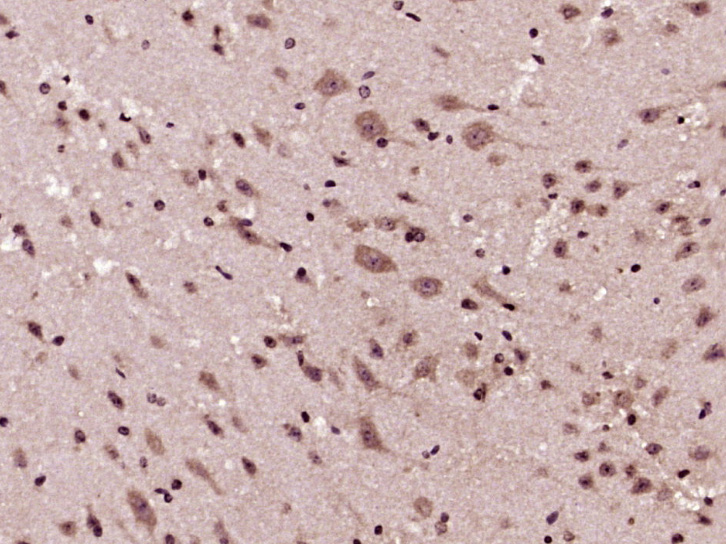 Paraformaldehyde-fixed, paraffin embedded (Mouse brain); Antigen retrieval by boiling in sodium citrate buffer (pH6.0) for 15min; Block endogenous peroxidase by 3% hydrogen peroxide for 20 minutes; Blocking buffer (normal goat serum) at 37°C for 30min; Antibody incubation with (GSK-3 Beta) Polyclonal Antibody, Unconjugated (SL0028R) at 1:400 overnight at 4°C, followed by operating according to SP Kit(Rabbit) (sp-0023) instructions and DAB staining.
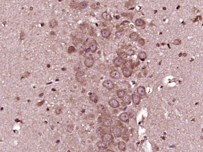 Paraformaldehyde-fixed, paraffin embedded (Rat brain); Antigen retrieval by boiling in sodium citrate buffer (pH6.0) for 15min; Block endogenous peroxidase by 3% hydrogen peroxide for 20 minutes; Blocking buffer (normal goat serum) at 37°C for 30min; Antibody incubation with (GSK-3 Beta) Polyclonal Antibody, Unconjugated (SL0028R) at 1:400 overnight at 4°C, followed by operating according to SP Kit(Rabbit) (sp-0023) instructions and DAB staining.
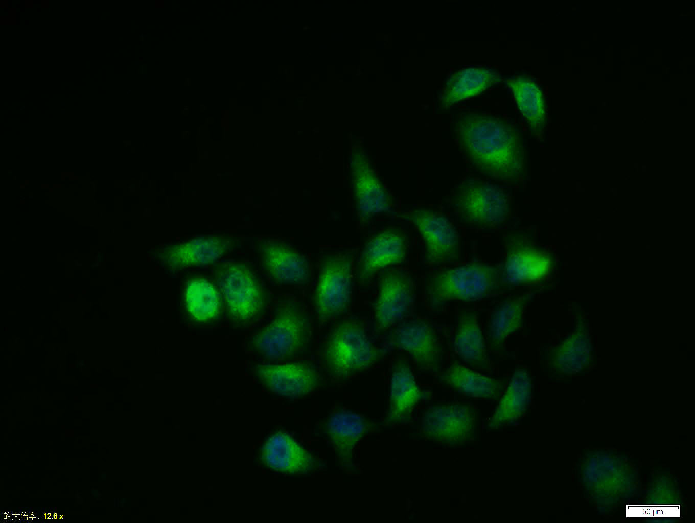 Tissue/cell:Hela cell; 4% Paraformaldehyde-fixed; Triton X-100 at room temperature for 20 min; Blocking buffer (normal goat serum, C-0005) at 37°C for 20 min; Antibody incubation with (GSK-3 Beta) polyclonal Antibody, Unconjugated (SL0028R) 1:100, 90 minutes at 37°C; followed by a FITC conjugated Goat Anti-Rabbit IgG antibody at 37°C for 90 minutes, DAPI (blue, C02-04002) was used to stain the cell nuclei.
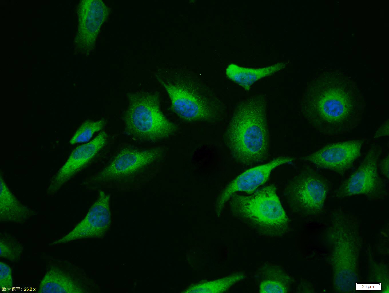 Tissue/cell:Hela cell; 4% Paraformaldehyde-fixed; Triton X-100 at room temperature for 20 min; Blocking buffer (normal goat serum,C-0005) at 37°C for 20 min; Antibody incubation with (GSK-3 Beta) polyclonal Antibody, Unconjugated (SL0028R) 1:100, 90 minutes at 37°C; followed by a FITC conjugated Goat Anti-Rabbit IgG antibody at 37°C for 90 minutes, DAPI (blue, C02-04002) was used to stain the cell nuclei.
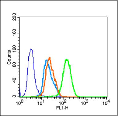 Blank control (blue line): MCF 7 (blue).
Primary Antibody (green line): Rabbit Anti- GSK-3 Beta (CT) antibody (SL0028R)
Dilution: 1μg /10^5 cells;
Isotype Control Antibody (orange line): Rabbit IgG .
Secondary Antibody (white blue line): Goat anti-rabbit IgG-FITC
Dilution: 1μg /test.
Protocol
The cells were fixed with 70% methanol (Overnight at 4℃) and then permeabilized with 90% ice-cold methanol for 30 min on ice. Cells stained with Primary Antibody for 30 min at room temperature. The cells were then incubated in 1 X PBS/2%BSA/10% goat serum to block non-specific protein-protein interactions followed by the antibody for 15 min at room temperature. The secondary antibody used for 40 min at room temperature. Acquisition of 20,000 events was performed.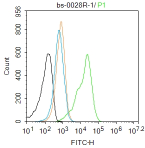 Blank control: A431.
Primary Antibody (green line): Rabbit Anti-GSK-3 Beta antibody (SL0028R)
Dilution: 1μg /10^6 cells;
Isotype Control Antibody (orange line): Rabbit IgG .
Secondary Antibody : Goat anti-rabbit IgG-AF488
Dilution: 1μg /test.
Protocol
The cells were fixed with 4% PFA (10min at room temperature)and then permeabilized with 90% ice-cold methanol for 20 min at-20℃. The cells were then incubated in 5%BSA to block non-specific protein-protein interactions for 30 min at room temperature .Cells stained with Primary Antibody for 30 min at room temperature. The secondary antibody used for 40 min at room temperature. Acquisition of 20,000 events was performed.
Cartpieces
Totalgoods,subtotals:¥Checkout
Partial purchase records(bought amounts latest0)
No one bought this product
User Comment(Total0User Comment Num)
- No comment


 +86 571 56623320


