![[KD-Validated] Anti-Aconitase 2 Rabbit Monoclonal Antibody](images/default_images/52.jpg)
[KD-Validated] Anti-Aconitase 2 Rabbit Monoclonal Antibody
ACO2; Aconitase 2; Aconitate Hydratase, Mitochondrial; ACONM; Aconitase 2, Mitochondrial; Mitochondrial Aconitase; Citrate Hydro-Lyase; EC 4.2.1.3 ; Epididymis Secretory Sperm Binding Protein Li 284; HEL-S-284; Aconitase ; EC 4.2.1; ICRD; OCA8; OPA9
View History [Clear]
Details
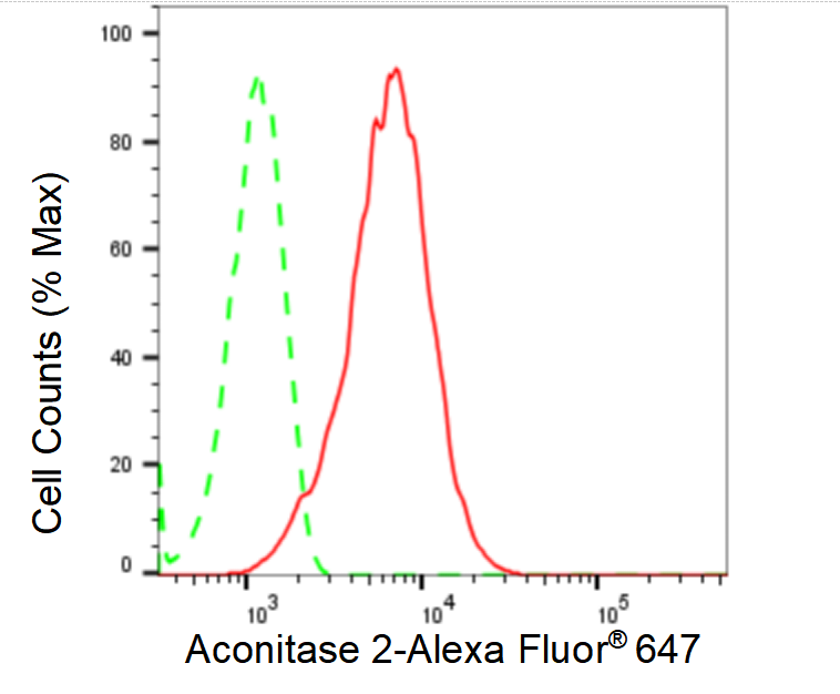
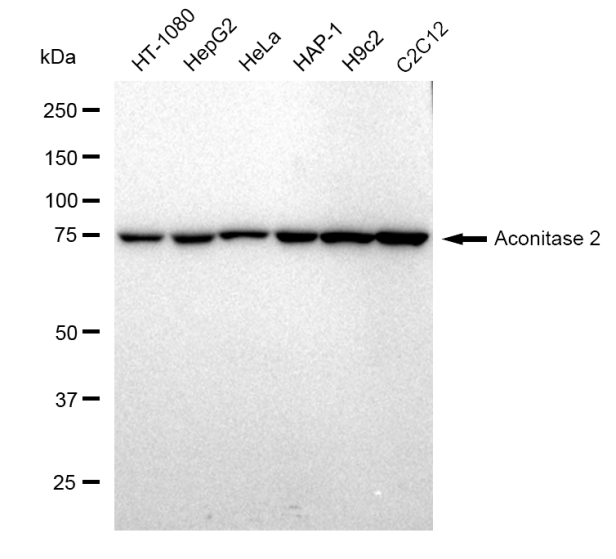
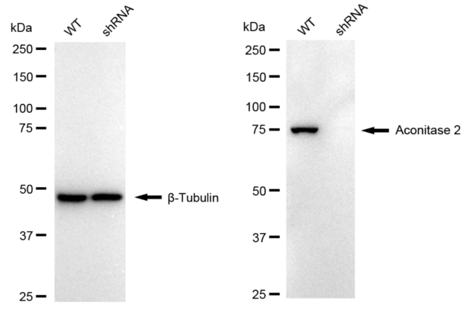
Aliases: ACO2; Aconitase 2; Aconitate Hydratase, Mitochondrial; ACONM; Aconitase 2, Mitochondrial; Mitochondrial Aconitase; Citrate Hydro-Lyase; EC 4.2.1.3 ; Epididymis Secretory Sperm Binding Protein Li 284; HEL-S-284; Aconitase ; EC 4.2.1; ICRD; OCA8; OPA9 Background: UniProt Entry: Q99798;NCBI Gene Entry: 50 Application Information Molecular Weight: Predicted, 85 kDa, observed, 85 kDa Clonality: Rabbit monoclonal antibody Clone ID: 23GB1585 Species Reactivity: Human, Mouse, Rat Applications Tested: Western Blotting (WB), Flow Cytometry (FCM), Immunocytochemistry (IC) Immunogen A synthesized peptide derived from human Aconitase 2 Isotype Rabbit IgG Storage Buffer Supplied in PBS (pH 7.4) containing 50% glycerol, and 0.02% sodium azide. Storage Store at -20 °C for one year. Recommended Dilutions Western Blotting (WB): 1:1,000-1:5,000 Flow Cytometry (FCM): 1:2,000 Immunocytochemistry (IC): 1:1,000 Protocols For general and specific antibody protocols please visit our website. Read all instructions before using this product.
Partial purchase records(bought amounts latest0)
No one bought this product
User Comment(Total0User Comment Num)
- No comment
 +86 571 56623320
 [email protected]

Scan Wechat Qrcode


Scan Whatsapp Qrcode


