TEL: +86 571 56623320    EMAIL: [email protected]

[KD-Validated] Anti-ADK Rabbit Monoclonal Antibody
[KD-Validated] Anti-ADK Rabbit Monoclonal Antibody
ADK; Adenosine Kinase; AK; Adenosine 5'-Phosphotransferase; EC 2.7.1.20; Testicular Tissue Protein Li 14; EC 2.7.1
Total
(Vip priceV)
Regular members: $544.0
  • Save more [Favourable] 30% discount
  • NO.:KD-10047
    Gene name:ADK
    React Species:Human; Mouse
    Applications:WB; IHC
    UniProt Entry:P55263
    NCBI Gene Entry:132
  • Goods click count:16
  • Product Spec:
  • Quantity: - +
  • Limit points for buying:0 Points
  • Manual
  • Add to cart Inquiry Add to favorite
View History [Clear]

Details

Aliases: ADK; Adenosine Kinase; AK; Adenosine 5'-Phosphotransferase; EC 2.7.1.20; Testicular Tissue Protein Li 14; EC 2.7.1
Background: UniProt Entry: P55263;NCBI Gene Entry: 132
Application InformationMolecular Weight: Predicted, 41 kDa, observed, 46 kDa Clonality: Rabbit monoclonal antibody Clone ID: 23GB625 Species Reactivity: Human, Mouse, Rat Applications Tested: Western Blotting (WB), Immunocytochemistry (IC)
Immunogen A synthesized peptide derived from human ADK
Isotype Rabbit IgG
Storage Buffer Supplied in PBS (pH 7.4) containing 50% glycerol, and 0.02% sodium azide.
Storage Store at -20 °C for one year.
Recommended Dilutions Western Blotting (WB): 1:1,000-1:5,000 Immunocytochemistry (IC): 1:1,000
Protocols For general and specific antibody protocols please visit our website. Read all instructions before using this product.
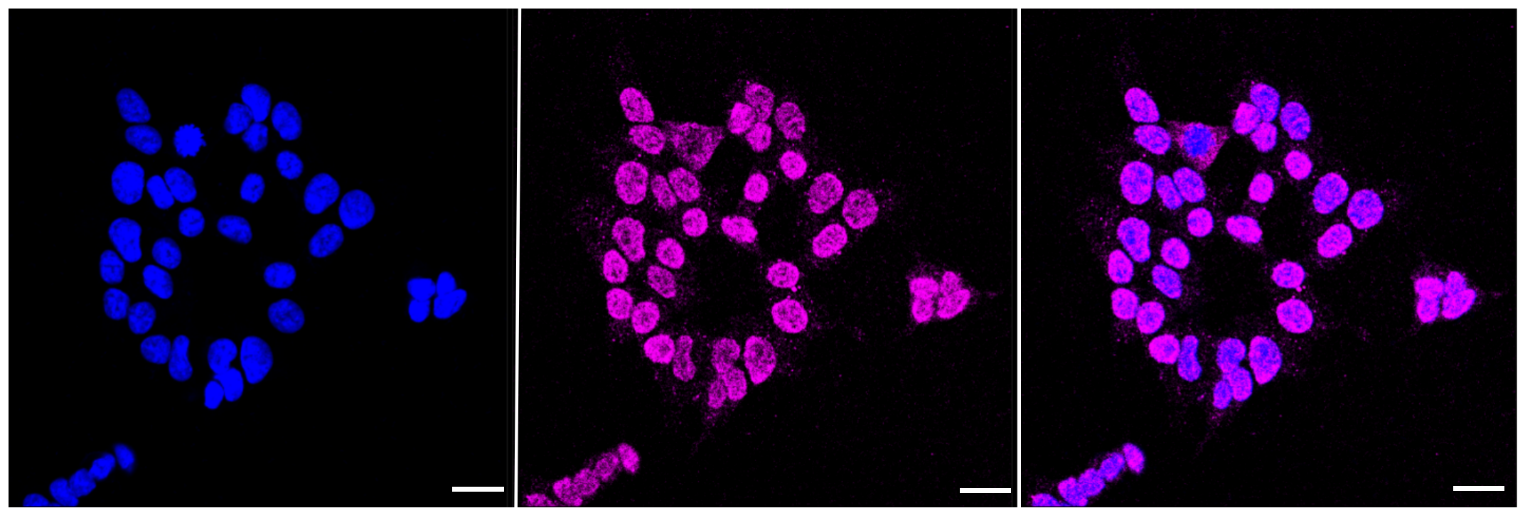
Immunocytochemical staining of HAP-1 cells with ADK antibody 1:1,000. Nuclei were stained blue with DAPI; ADK was stained magenta with Alexa Fluor® 647. Images were taken using Leica stellaris 5. Protein abundance based on laser Intensity and smart gain: Medium. Scale bar: 20 μm.
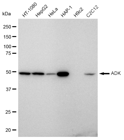
Western blotting analysis using anti-ADK antibody . Total cell lysates (30 μg) from various cell lines were loaded and separated by SDS-PAGE. The blot was incubated with anti-ADK antibody and HRP-conjugated goat anti rabbit secondary antibody respectively.
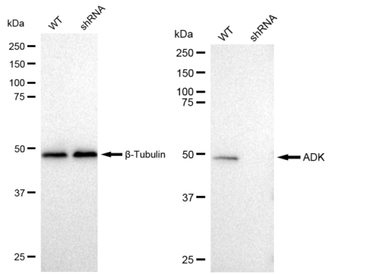
Western blotting analysis using anti-ADK antibody . ADK expression in wild type (WT) and ADK shRNA knockdown (KD) HeLa cells with 30 μg of total cell lysates. β-Tubulin serves as a loading control. The blot was incubated with anti-ADK antibody and HRP-conjugated goat anti rabbit secondary antibody respectively.

Partial purchase records(bought amounts latest0)

No one bought this product
Total 0 records, divided into1 pages First Prev Next Last

User Comment(Total0User Comment Num)

  • No comment
Total 0 records, divided into1 pages First Prev Next Last
Username: Anonymous user
E-mail:
Rank:
Content:
Verification code: captcha

Call us

+86 571 56623320

Address

Room 1-315, Kongle Changqing Building, No. 160 Guangye Road,Gongshu District, Hangzhou City, Zhejiang Province, China

Join Us with

Leave a message
* To protect against spam, please pass the CAPTCHA test below.