
Rabbit Anti-Moesin antibody
Membrane organizing extension spike protein; Moesin/anaplastic lymphoma kinase fusion protein; MSN; MSN/ALK fusion; MOES_HUMAN.
View History [Clear]
Details
Product Name Moesin Chinese Name 膜突蛋白Recombinant rabbit monoclonal anti Alias Membrane organizing extension spike protein; Moesin/anaplastic lymphoma kinase fusion protein; MSN; MSN/ALK fusion; MOES_HUMAN. Research Area Signal transduction Immunogen Species Rabbit Clonality Monoclonal Clone NO. R1F5 React Species (predicted: Human, Mouse, Rat, ) Applications WB=1:500-2000 IHC-P=1:100-500 IHC-F=1:50-1:100 (Paraffin sections need antigen repair)
not yet tested in other applications.
optimal dilutions/concentrations should be determined by the end user.Theoretical molecular weight 63kDa Cellular localization cytoplasmic Form Liquid Concentration 1mg/ml immunogen KLH conjugated synthetic peptide derived from human Moesin Lsotype IgG Purification affinity purified by Protein A Buffer Solution 0.01M TBS(pH7.4) with 1% BSA, 0.03% Proclin300 and 50% Glycerol. Storage Shipped at 4℃. Store at -20 °C for one year. Avoid repeated freeze/thaw cycles. Attention This product as supplied is intended for research use only, not for use in human, therapeutic or diagnostic applications. PubMed PubMed Product Detail Moesin (for membrane-organizing extension spike protein) is a member of the ERM family which includes ezrin and radixin. ERM proteins appear to function as cross-linkers between plasma membranes and actin-based cytoskeletons. Moesin is localized to filopodia and other membranous protrusions that are important for cell-cell recognition and signaling and for cell movement. [provided by RefSeq, Jul 2008]
Subcellular Location:
Cell membrane; Peripheral membrane protein; Cytoplasmic side. Cytoplasm, cytoskeleton. Apical cell membrane; Peripheral membrane protein; Cytoplasmic side. Cell projection, microvillus membrane; Peripheral membrane protein; Cytoplasmic side. Note=Phosphorylated form is enriched in microvilli-like structures at apical membrane. Increased cell membrane localization of both phosphorylated and non-phosphorylated forms seen after thrombin treatment.
Tissue Specificity:
In all tissues and cultured cells studied.
SWISS:
P26038
Gene ID:
4478
Database links:Entrez Gene: 4478 Human
Entrez Gene: 17698 Mouse
SwissProt: P26038 Human
SwissProt: P26041 Mouse
Product Picture 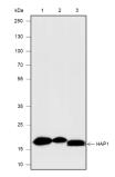 Blocking buffer: 5% NFDM/TBST
Primary Ab dilution: 1:5000
Primary Ab incubation condition: 2 hours at
room temperature
Secondary Ab: Goat Anti-Rabbit IgG H&L
(HRP)
Lysate: 1: HepG2, 2: 293T, 3: Mouse kidney
Protein loading quantity: 20 μg
Exposure time: 30 s
Predicted MW: 22 kDa
Observed MW: 18 kDa
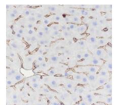 Tissue: Mouse liver
Section type: Formalin-fixed & Paraffin
-embedded section
Retrieval method: High temperature and high
pressure
Retrieval buffer: Tris/EDTA buffer, pH 9.0
Primary Ab dilution: 1:100
Primary Ab incubation condition: 1 hour at
room temperature
Secondary Ab: Anti-Rabbit and Mouse
Polymer HRP (Ready to use)
Counter stain: Hematoxylin (Blue)
Comment: Color brown is the positive signal for
SLM-60653R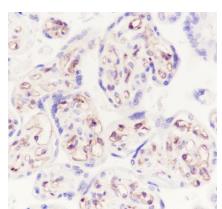 Tissue: Human placenta
Section type: Formalin-fixed & Paraffin
-embedded section
Retrieval method: High temperature and high
pressure
Retrieval buffer: Tris/EDTA buffer, pH 9.0
Primary Ab dilution: 1:50
Primary Ab incubation condition: 1 hour at
room temperature
Secondary Ab: Anti-Rabbit and Mouse
Polymer HRP (Ready to use)
Counter stain: Hematoxylin (Blue)
Comment: Color brown is the positive signal for
SLM-60653R
Cartpieces
Totalgoods,subtotals:¥Checkout
References (0)
No References
Bought notes(bought amounts latest0)
No one bought this product
User Comment(Total0User Comment Num)
- No comment


 +86 571 56623320
 +86 18668110335

