
Rabbit Anti-phospho-V-Myb+C-Myb (Ser11)antibody
Avian myeloblastosis viral (v-myb) oncogene homolog; C myb; c-Myb; MYB_HUMAN; Proto-oncogene c-Myb; Transcriptional activator Myb; v-myb avian myeloblastosis viral oncogene homolog.
View History [Clear]
Details
Product Name phospho-V-Myb+C-Myb (Ser11) Chinese Name 磷酸化转录激活因子MYBRecombinant rabbit monoclonal anti Alias Avian myeloblastosis viral (v-myb) oncogene homolog; C myb; c-Myb; MYB_HUMAN; Proto-oncogene c-Myb; Transcriptional activator Myb; v-myb avian myeloblastosis viral oncogene homolog. Product Type Phosphorylated anti Recombinant rabbit monoclonal anti Research Area Tumour immunology Chromatin and nuclear signals transcriptional regulatory factor Epigenetics Immunogen Species Rabbit Clonality Monoclonal Applications WB=1:100-500 IHC-P=1:100-500 ICC=1:50-200 IF=1:50-200 (Paraffin sections need antigen repair)
not yet tested in other applications.
optimal dilutions/concentrations should be determined by the end user.Theoretical molecular weight 84kDa Cellular localization The nucleus Form Liquid Concentration 1mg/ml immunogen KLH conjugated synthesised phosphopeptide derived from human MYB around the phosphorylation site of Ser11 Lsotype IgG Purification affinity purified by Protein A Buffer Solution 0.01M TBS(pH7.4) with 1% BSA, 0.03% Proclin300 and 50% Glycerol. Storage Shipped at 4℃. Store at -20 °C for one year. Avoid repeated freeze/thaw cycles. Attention This product as supplied is intended for research use only, not for use in human, therapeutic or diagnostic applications. PubMed PubMed Product Detail Transcriptional activator; DNA-binding protein that specifically recognize the sequence 5'-YAAC[GT]G-3'. Plays an important role in the control of proliferation and differentiation of hematopoietic progenitor cells.
Function:
Transcriptional activator; DNA-binding protein that specifically recognize the sequence 5'-YAAC[GT]G-3'. Plays an important role in the control of proliferation and differentiation of hematopoietic progenitor cells.
Subcellular Location:
Nucleus.
Post-translational modifications:
Ubiquitinated; mediated by SIAH1 and leading to its subsequent proteasomal degradation.
Phosphorylated by NLK on multiple sites, which induces proteasomal degradation.
Similarity:
Contains 3 HTH myb-type DNA-binding domains.
SWISS:
P10242
Gene ID:
4602
Database links:Entrez Gene: 4602 Human
Entrez Gene: 17863 Mouse
SwissProt: P10242 Human
SwissProt: P06876 Mouse
Product Picture 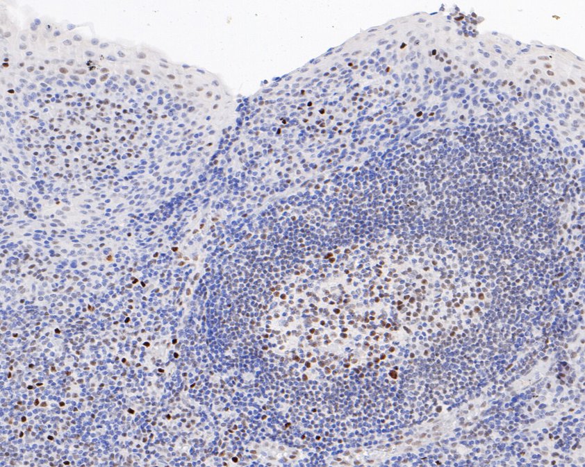 Immunohistochemical analysis of paraffin-embedded human tonsil tissue using anti-P-V-Myb+C-Myb(S11) antibody. The section was pre-treated using heat mediated antigen retrieval with sodium citrate buffer (pH 6.0) for 20 minutes. The tissues were blocked in 5% BSA for 30 minutes at room temperature, washed with ddH2O and PBS, and then probed with the primary antibody (SLM-54494R, 1/200) for 30 minutes at room temperature. The detection was performed using an HRP conjugated compact polymer system. DAB was used as the chromogen. Tissues were counterstained with hematoxylin and mounted with DPX.
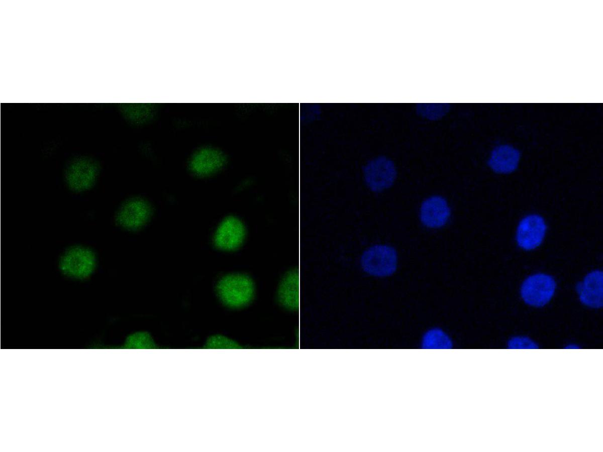 ICC staining of P-V-Myb+C-Myb(S11) in AGS cells (green). Formalin fixed cells were permeabilized with 0.1% Triton X-100 in TBS for 10 minutes at room temperature and blocked with 1% Blocker BSA for 15 minutes at room temperature. Cells were probed with the primary antibody (SLM-54494R, 1/50) for 1 hour at room temperature, washed with PBS. Alexa Fluor®488 Goat anti-Rabbit IgG was used as the secondary antibody at 1/1,000 dilution. The nuclear counter stain is DAPI (blue).
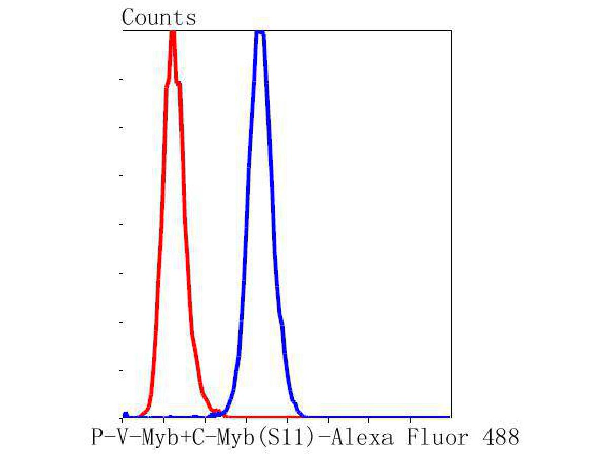 Flow cytometric analysis of P-V-Myb+C-Myb(S11) was done on MCF-7 cells. The cells were fixed, permeabilized and stained with the primary antibody (SLM-54494R, 1/50) (blue). After incubation of the primary antibody at room temperature for an hour, the cells were stained with a Alexa Fluor 488-conjugated Goat anti-Rabbit IgG Secondary antibody at 1/1000 dilution for 30 minutes.Unlabelled sample was used as a control (cells without incubation with primary antibody; red).
Cartpieces
Totalgoods,subtotals:¥Checkout
References (0)
No References
Bought notes(bought amounts latest0)
No one bought this product
User Comment(Total0User Comment Num)
- No comment


 +86 571 56623320
 +86 18668110335

