
Rabbit Anti-MARK2 antibody
ELKL motif kinase 1; ELKL motif kinase; EMK-1; EMK1; MAP/microtubule affinity regulating kinase 2; MAP/microtubule affinity-regulating kinase 2; Mark2; MARK2_HUMAN; PAR1 homolog; Serine/threonine protein kinase MARK2; Serine/threonine-protein kinase MARK2
View History [Clear]
Details
Product Name MARK2 Chinese Name 丝氨酸/苏氨酸蛋白激酶MARK2Recombinant rabbit monoclonal anti Alias ELKL motif kinase 1; ELKL motif kinase; EMK-1; EMK1; MAP/microtubule affinity regulating kinase 2; MAP/microtubule affinity-regulating kinase 2; Mark2; MARK2_HUMAN; PAR1 homolog; Serine/threonine protein kinase MARK2; Serine/threonine-protein kinase MARK2. Research Area immunology Signal transduction Kinases and Phosphatases Immunogen Species Rabbit Clonality Monoclonal Clone NO. 1F10 React Species Human, (predicted: Mouse, Rat, ) Applications WB=1:500-1000 IHC-P=1:50-200 Flow-Cyt=1:100 (Paraffin sections need antigen repair)
not yet tested in other applications.
optimal dilutions/concentrations should be determined by the end user.Theoretical molecular weight 88kDa Form Liquid Concentration 1mg/ml immunogen Recombinant human MARK2 Lsotype IgG Purification affinity purified by Protein A Buffer Solution 0.01M TBS(pH7.4) with 1% BSA, 0.03% Proclin300 and 50% Glycerol. Storage Shipped at 4℃. Store at -20 °C for one year. Avoid repeated freeze/thaw cycles. Attention This product as supplied is intended for research use only, not for use in human, therapeutic or diagnostic applications. PubMed PubMed Product Detail This gene encodes a member of the Par-1 family of serine/threonine protein kinases. The protein is an important regulator of cell polarity in epithelial and neuronal cells, and also controls the stability of microtubules through phosphorylation and inactivation of several microtubule-associating proteins. The protein localizes to cell membranes. Multiple transcript variants encoding different isoforms have been found for this gene. [provided by RefSeq, Jul 2009]. Function : Serine/threonine-protein kinase involved in cell polarity and microtubule dynamics regulation. Phosphorylates CRTC2/TORC2, DCX, HDAC7, KIF13B, MAP2, MAP4, MAPT/TAU, and RAB11FIP2. Plays a key role in cell polarity by phosphorylating the microtubule-associated proteins MAP2, MAP4 and MAPT/TAU at KXGS motifs, causing detachment from microtubules, and their disassembly. Regulates epithelial cell polarity by phosphorylating RAB11FIP2. Involved in the regulation of neuronal migration through its dual activities in regulating cellular polarity and microtubule dynamics, possibly by phosphorylating and regulating DCX. Regulates axogenesis by phosphorylating KIF13B, promoting interaction between KIF13B and 14-3-3 and inhibiting microtubule-dependent accumulation of KIF13B. Also required for neurite outgrowth and establishment of neuronal polarity. Regulates localization and activity of some histone deacetylases by mediating phosphorylation of HDAC7, promoting subsequent interaction between HDAC7 and 14-3-3 and export from the nucleus. Also acts as a positive regulator of the Wnt signaling pathway, probably by mediating phosphorylation of dishevelled proteins (DVL1, DVL2 and/or DVL3). Modulates the developmental decision to build a columnar versus a hepatic epithelial cell apparently by promoting a switch from a direct to a transcytotic mode of apical protein delivery. Essential for the asymmetric development of membrane domains of polarized epithelial cells.
Subunit:
Homodimer. Interacts with PAK7/PAK5; leading to inhibit the protein kinase activity (By similarity). Interacts (when phosphorylated at Thr-596) with YWHAZ. In case of infection, interacts with H.pylori CagA, leading to inhibit kinase activity and junctional and polarity defects.
Subcellular Location:
Cell membrane; Peripheral membrane protein. Cytoplasm. Note=Phosphorylation at Thr-596 by PRKCZ/aPKC and subsequent interaction with 14-3-3 protein YWHAZ promotes relocation from the cell membrane to the cytoplasm.
Tissue Specificity:
High levels of expression in heart, brain, skeletal muscle and pancreas, lower levels observed in lung, liver and kidney.
Post-translational modifications:
Autophosphorylated. Phosphorylated at Thr-208 by STK11/LKB1 in complex with STE20-related adapter-alpha (STRADA) pseudo kinase and CAB39. Phosphorylation at Thr-208 by TAOK1 activates the kinase activity, leading to phosphorylation and detachment of MAPT/TAU from microtubules. Phosphorylation at Ser-212 by GSK3-beta (GSK3B) inhibits the kinase activity. Phosphorylation by CaMK1 promotes activity and is required to promote neurite outgrowth. Phosphorylation at Thr-596 by PRKCZ/aPKC in polarized epithelial cells inhibits the kinase activity and promotes binding to 14-3-3 protein YWHAZ, leading to relocation from cell membrane to cytoplasm.
Similarity:
Belongs to the protein kinase superfamily. CAMK Ser/Thr protein kinase family. SNF1 subfamily.
Contains 1 KA1 (kinase-associated) domain.
Contains 1 protein kinase domain.
Contains 1 UBA domain.
SWISS:
Q7KZI7
Gene ID:
2011
Product Picture 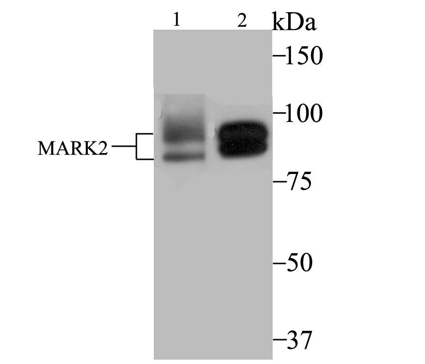 Sample:
Lane 1: MCF-7 cell lysate
Lane 2: SK-Br-3 cell lysate
Primary: Anti-MARK2 (SLM-54447R) at 1:2000 dilution
Secondary: Goat Anti-Rabbit IgG - HRP at 1:5000 dilution
Predicted band size: 88 kD
Observed band size: 77/88 kD
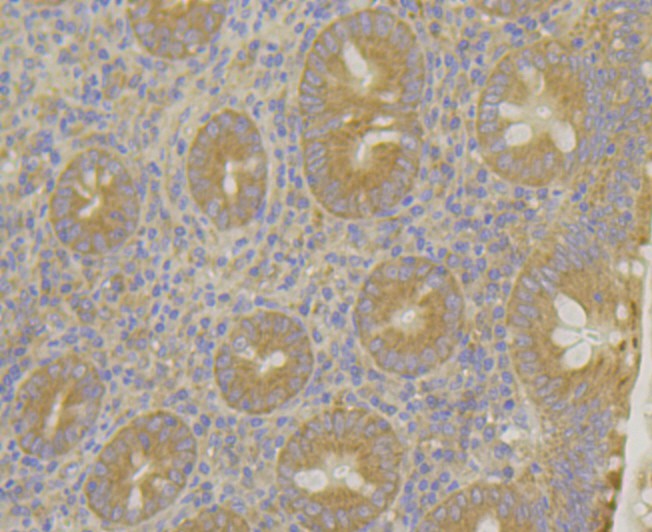 Paraformaldehyde-fixed, paraffin embedded (human appendix); Antigen retrieval by boiling in sodium citrate buffer (pH6.0) for 15min; Block endogenous peroxidase by 3% hydrogen peroxide for 20 minutes; Blocking buffer (normal goat serum) at 37°C for 30min; Antibody incubation with (MARK2) Monoclonal Antibody, Unconjugated (SLM-54447R) at 1:50 overnight at 4°C, followed by operating according to SP Kit(Rabbit) (sp-0023) instructionsand DAB staining.
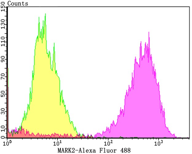 Blank control:MCF-7.
Primary Antibody (green line): Rabbit Anti-MARK2 antibody (bms-54447R)
Dilution: 1:100;
Secondary Antibody : Goat anti-rabbit IgG-AF488
Dilution: 1:1000.
Protocol
The cells were then incubated in 5%BSA to block non-specific protein-protein interactions for 30 min at room temperature .Cells stained with Primary Antibody for 30 min at room temperature. The secondary antibody used for 40 min at room temperature. Acquisition of 20,000 events was performed.
Cartpieces
Totalgoods,subtotals:¥Checkout
References (0)
No References
Bought notes(bought amounts latest0)
No one bought this product
User Comment(Total0User Comment Num)
- No comment


 +86 571 56623320
 +86 18668110335

