
Rabbit Anti-Biglycan antibody
BGN; Biglycan proteoglycan; Bone/cartilage proteoglycan I; Dermatan sulphate proteoglycan I; DSPG1; PG S1; PG-S1; PGI; PGS1_HUMAN; SLRR1A; Small leucine rich protein 1A.
View History [Clear]
Details
Product Name Biglycan Chinese Name 骨/软骨蛋白多糖1Recombinant rabbit monoclonal anti Alias BGN; Biglycan proteoglycan; Bone/cartilage proteoglycan I; Dermatan sulphate proteoglycan I; DSPG1; PG S1; PG-S1; PGI; PGS1_HUMAN; SLRR1A; Small leucine rich protein 1A. Research Area Cell biology immunology Signal transduction Stem cells Cytoskeleton Extracellular matrix Immunogen Species Rabbit Clonality Monoclonal React Species (predicted: Human, Mouse, ) Applications WB=1:500-1000 IHC-P=1:100-500 (Paraffin sections need antigen repair)
not yet tested in other applications.
optimal dilutions/concentrations should be determined by the end user.Theoretical molecular weight 42kDa Form Liquid Concentration 1mg/ml immunogen Recombinant Human Biglycan Lsotype IgG Purification affinity purified by Protein A Buffer Solution 0.01M TBS(pH7.4) with 1% BSA, 0.03% Proclin300 and 50% Glycerol. Storage Shipped at 4℃. Store at -20 °C for one year. Avoid repeated freeze/thaw cycles. Attention This product as supplied is intended for research use only, not for use in human, therapeutic or diagnostic applications. PubMed PubMed Product Detail May be involved in collagen fiber assembly.
Function:
May be involved in collagen fiber assembly (By similarity).
Subunit:
Homodimer. Forms a ternary complex with MFAP2 and ELN
Subcellular Location:
Secreted, extracellular space, extracellular.
Tissue Specificity:
Found in several connective tissues, especially in articular cartilages.
Post-translational modifications:
The two attached glycosaminoglycan chains can be either chondroitin sulfate or dermatan sulfate (By similarity).
Similarity:
Belongs to the small leucine-rich proteoglycan (SLRP) family. SLRP class I subfamily. Contains 12 LRR (leucine-rich) repeats.
SWISS:
P21810
Gene ID:
633
Database links:Entrez Gene: 633 Human
Entrez Gene: 12111 Mouse
Omim: 301870 Human
SwissProt: P21810 Human
SwissProt: P28653 Mouse
Unigene: 821 Human
Unigene: 2608 Mouse
Unigene: 783 Rat
Product Picture 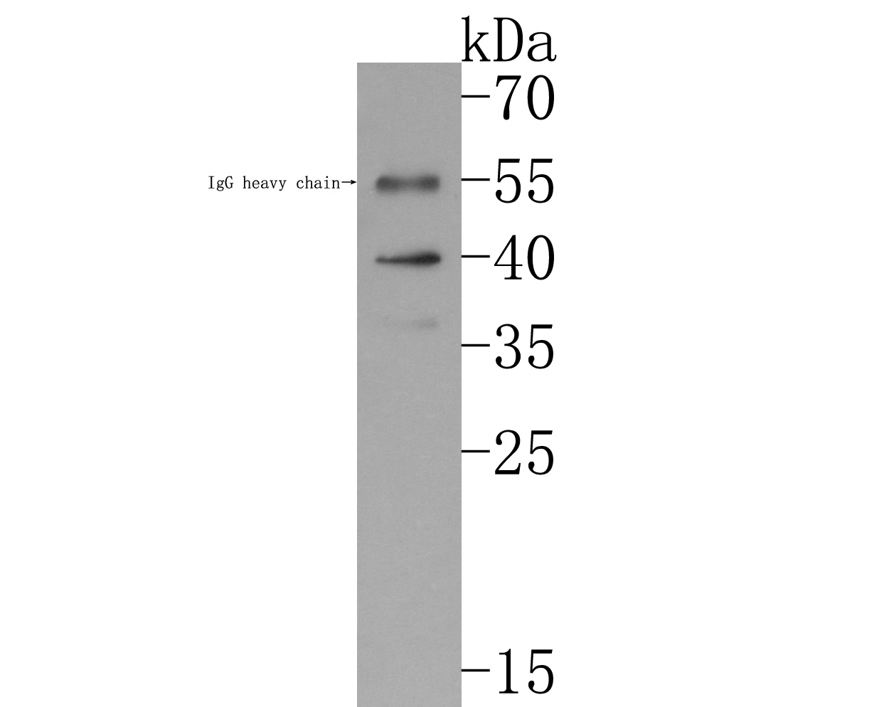 Western blot analysis of Biglycan on human skin tissue lysates. Proteins were transferred to a PVDF membrane and blocked with 5% BSA in PBS for 1 hour at room temperature. The primary antibody (SLM-54293R, 1/500) was used in 5% BSA at room temperature for 2 hours. Goat Anti-Rabbit IgG - HRP Secondary Antibody at 1:200,000 dilution was used for 1 hour at room temperature.
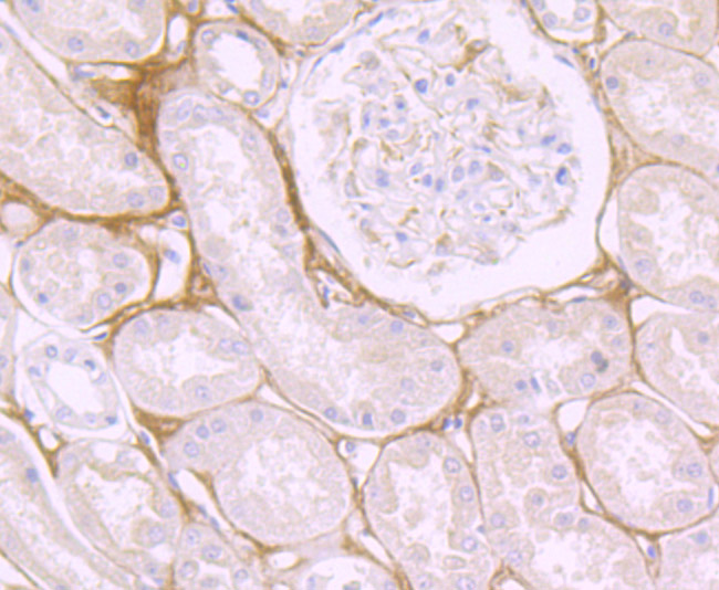 Immunohistochemical analysis of paraffin-embedded human kidney tissue using anti-Biglycan antibody. The section was pre-treated using heat mediated antigen retrieval with Tris-EDTA buffer (pH 9.0) for 20 minutes.The tissues were blocked in 1% BSA for 30 minutes at room temperature, washed with ddH2O and PBS, and then probed with the primary antibody (SLM-54293R, 1/50) for 30 minutes at room temperature. The detection was performed using an HRP conjugated compact polymer system. DAB was used as the chromogen. Tissues were counterstained with hematoxylin and mounted with DPX.
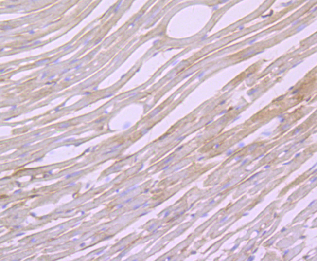 Immunohistochemical analysis of paraffin-embedded mouse heart tissue using anti-Biglycan antibody. The section was pre-treated using heat mediated antigen retrieval with Tris-EDTA buffer (pH 9.0) for 20 minutes.The tissues were blocked in 1% BSA for 30 minutes at room temperature, washed with ddH2O and PBS, and then probed with the primary antibody (SLM-54293R, 1/50) for 30 minutes at room temperature. The detection was performed using an HRP conjugated compact polymer system. DAB was used as the chromogen. Tissues were counterstained with hematoxylin and mounted with DPX.
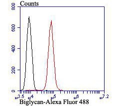 Flow cytometric analysis of Biglycan was done on HepG2 cells. The cells were fixed, permeabilized and stained with the primary antibody (SLM-54293R, 1/50) (red). After incubation of the primary antibody at room temperature for an hour, the cells were stained with a Alexa Fluor®488 conjugate-Goat anti-Rabbit IgG Secondary antibody at 1/1,000 dilution for 30 minutes.Unlabelled sample was used as a control (cells without incubation with primary antibody; black).
Cartpieces
Totalgoods,subtotals:¥Checkout
References (0)
No References
Bought notes(bought amounts latest0)
No one bought this product
User Comment(Total0User Comment Num)
- No comment


 +86 571 56623320
 +86 18668110335

