
Rabbit Anti-TYRP1 antibody
TRP; CAS2; CATB; GP75; OCA3; TRP1; TYRP; TRP-1; b-PROTEIN; tyrosinase related protein 1; TYRRP; 5,6-dihydroxyindole-2-carboxylic acid oxidase; EC:1.14.18; Catalase B; Glycoprotein 75; Melanoma antigen gp75; Tyrosinase-related protein 1; TYRP1_HUMAN;
View History [Clear]
Details
Product Name TYRP1 Chinese Name 酪氨酸酶相关蛋白1Recombinant rabbit monoclonal anti Alias TRP; CAS2; CATB; GP75; OCA3; TRP1; TYRP; TRP-1; b-PROTEIN; tyrosinase related protein 1; TYRRP; 5,6-dihydroxyindole-2-carboxylic acid oxidase; EC:1.14.18; Catalase B; Glycoprotein 75; Melanoma antigen gp75; Tyrosinase-related protein 1; TYRP1_HUMAN; Research Area Tumour Cell biology Chromatin and nuclear signals Epigenetics Immunogen Species Rabbit Clonality Monoclonal React Species (predicted: Human, ) Applications WB=1:500-2000 IP=1:10-50 IHC-P=1:100-500 ICC=1:50-100 (Paraffin sections need antigen repair)
not yet tested in other applications.
optimal dilutions/concentrations should be determined by the end user.Theoretical molecular weight 58kDa Cellular localization cytoplasmic Form Liquid Concentration 1mg/ml immunogen Recombinant human TRP1 protein Lsotype IgG Purification affinity purified by Protein A Buffer Solution 0.01M TBS(pH7.4) with 1% BSA, 0.03% Proclin300 and 50% Glycerol. Attention This product as supplied is intended for research use only, not for use in human, therapeutic or diagnostic applications. PubMed PubMed Product Detail TRP1 is a melanosomal enzyme that belongs to the tyrosinase family and plays an important role in the melanin biosynthetic pathway. Defects in this gene are the cause of rufous oculocutaneous albinism and oculocutaneous albinism type III.
SWISS:
P17643
Gene ID:
7306
Database links:Entrez Gene: 7306 Human
Entrez Gene: 22178 Mouse
Omim: 115501 Human
SwissProt: P17643 Human
SwissProt: P07147 Mouse
Unigene: 270279 Human
Unigene: 30438 Mouse
Product Picture  Western blot analysis of TRP1 on melanoma tissue lysates. Proteins were transferred to a PVDF membrane and blocked with 5% BSA in PBS for 1 hour at room temperature. The primary antibody (SLM-54216R, 1/500) was used in 5% BSA at room temperature for 2 hours. Goat Anti-Rabbit IgG - HRP Secondary Antibody (HA1001) at 1:200,000 dilution was used for 1 hour at room temperature.
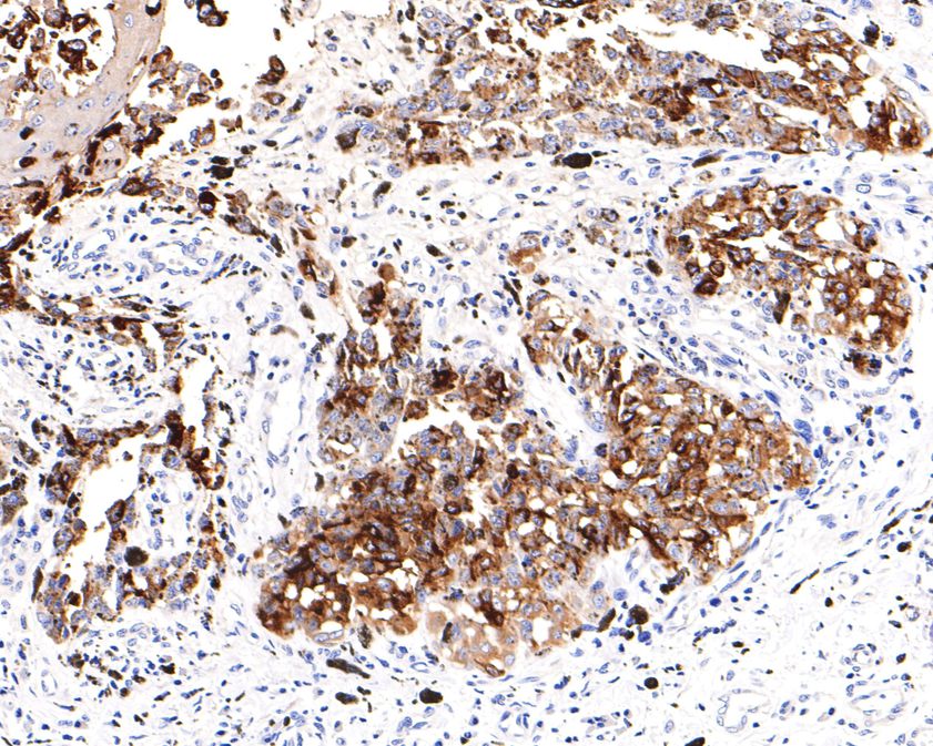 Immunohistochemical analysis of paraffin-embedded human malignant melanoma tissue using anti-TRP1 antibody. The section was pre-treated using heat mediated antigen retrieval with Tris-EDTA buffer (pH 9.0) for 20 minutes.The tissues were blocked in 1% BSA for 30 minutes at room temperature, washed with ddH2O and PBS, and then probed with the primary antibody (SLM-54216R, 1/400) for 30 minutes at room temperature. The detection was performed using an HRP conjugated compact polymer system. DAB was used as the chromogen. Tissues were counterstained with hematoxylin and mounted with DPX.
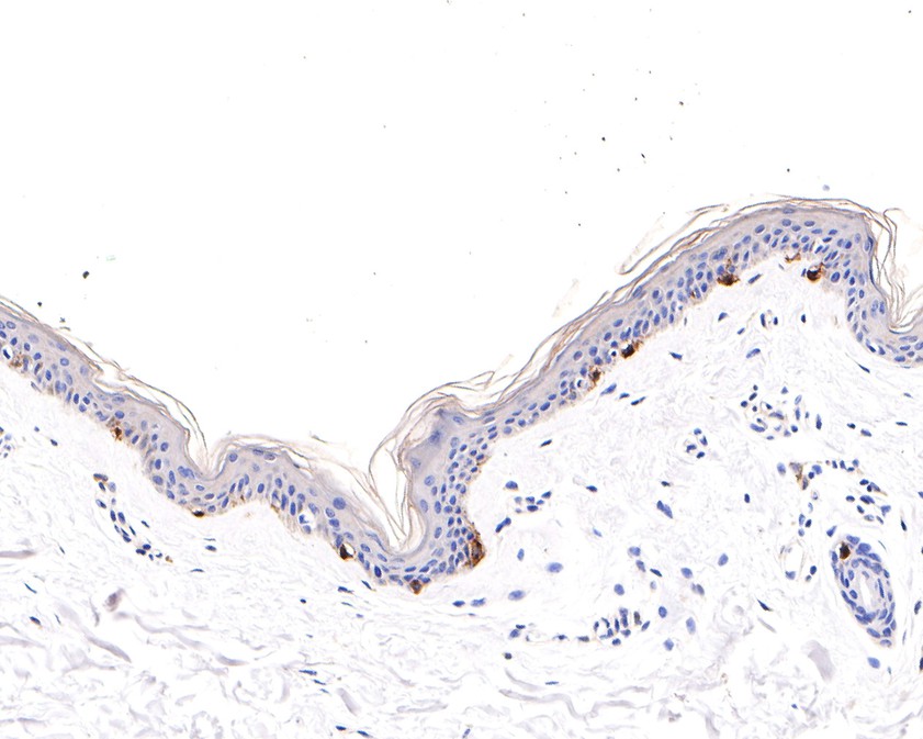 Immunohistochemical analysis of paraffin-embedded human skin tissue using anti-TRP1 antibody. The section was pre-treated using heat mediated antigen retrieval with Tris-EDTA buffer (pH 9.0) for 20 minutes.The tissues were blocked in 1% BSA for 30 minutes at room temperature, washed with ddH2O and PBS, and then probed with the primary antibody (SLM-54216R, 1/100) for 30 minutes at room temperature. The detection was performed using an HRP conjugated compact polymer system. DAB was used as the chromogen. Tissues were counterstained with hematoxylin and mounted with DPX.ICC staining of TRP1 in A431 cells (green). Formalin fixed cells were permeabilized with 0.1% Triton X-100 in TBS for 10 minutes at room temperature and blocked with 10% negative goat serum for 15 minutes at room temperature. Cells were probed with the primary antibody (SLM-54216R, 1/50) for 1 hour at room temperature, washed with PBS. Alexa Fluor®488 conjugate-Goat anti-Rabbit IgG was used as the secondary antibody at 1/1,000 dilution. The nuclear counter stain is DAPI (blue).
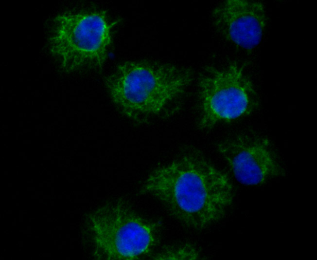 ICC staining of TRP1 in HUVEC cells (green). Formalin fixed cells were permeabilized with 0.1% Triton X-100 in TBS for 10 minutes at room temperature and blocked with 10% negative goat serum for 15 minutes at room temperature. Cells were probed with the primary antibody (SLM-54216R, 1/50) for 1 hour at room temperature, washed with PBS. Alexa Fluor®488 conjugate-Goat anti-Rabbit IgG was used as the secondary antibody at 1/1,000 dilution. The nuclear counter stain is DAPI (blue).
Cartpieces
Totalgoods,subtotals:¥Checkout
References (0)
No References
Bought notes(bought amounts latest0)
No one bought this product
User Comment(Total0User Comment Num)
- No comment


 +86 571 56623320
 +86 18668110335

