
Rabbit Anti-CD3D antibody
CD3 antigen delta subunit; CD3 delta; CD3d antigen delta polypeptide; CD3d molecule delta; CD3D_HUMAN; IMD19; OKT3 delta chain; T cell receptor T3 delta chain; T-cell surface glycoprotein CD3 delta chain; T3D.
View History [Clear]
Details
Product Name CD3D Chinese Name CD3DRecombinant rabbit monoclonal anti Alias CD3 antigen delta subunit; CD3 delta; CD3d antigen delta polypeptide; CD3d molecule delta; CD3D_HUMAN; IMD19; OKT3 delta chain; T cell receptor T3 delta chain; T-cell surface glycoprotein CD3 delta chain; T3D. Research Area Tumour immunology Stem cells t-lymphocyte b-lymphocyte Immunogen Species Rabbit Clonality Monoclonal React Species (predicted: Human, ) Applications WB=1:2000-10000 IHC-P=1:100-500 ICC=1:50-200 IF=1:50-200 (Paraffin sections need antigen repair)
not yet tested in other applications.
optimal dilutions/concentrations should be determined by the end user.Theoretical molecular weight 19kDa Cellular localization The cell membrane Form Liquid Concentration 1mg/ml immunogen KLH conjugated synthetic peptide derived from human CD3D Lsotype IgG Purification affinity purified by Protein A Buffer Solution 0.01M TBS(pH7.4) with 1% BSA, 0.03% Proclin300 and 50% Glycerol. Storage Shipped at 4℃. Store at -20 °C for one year. Avoid repeated freeze/thaw cycles. Attention This product as supplied is intended for research use only, not for use in human, therapeutic or diagnostic applications. PubMed PubMed Product Detail CD3D (CD3d Molecule) is a Protein Coding gene. Diseases associated with CD3D include Immunodeficiency 19 and T-B+ Severe Combined Immunodeficiency Due To Cd3delta/Cd3epsilon/Cd3zeta. Among its related pathways are ICos-ICosL Pathway in T-Helper Cell and CTLA4 Signaling. GO annotations related to this gene include protein heterodimerization activity and transmembrane signaling receptor activity. An important paralog of this gene is CD3G.
SWISS:
P04234
Gene ID:
915
Database links:Entrez Gene: 915 Human
Entrez Gene: 12500 Mouse
Omim: 186790 Human
SwissProt: P04234 Human
SwissProt: P04235 Mouse
Unigene: 504048 Human
Unigene: 4527 Mouse
Product Picture  Western blot analysis of CD3D on Jurkat cell lysates. Proteins were transferred to a PVDF membrane and blocked with 5% BSA in PBS for 1 hour at room temperature. The primary antibody (SLM-52744R, 1/500) was used in 5% BSA at room temperature for 2 hours. Goat Anti-Rabbit IgG - HRP Secondary Antibody at 1:200,000 dilution was used for 1 hour at room temperature.
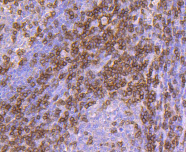 Immunohistochemical analysis of paraffin-embedded human tonsil tissue using anti-CD3D antibody. The section was pre-treated using heat mediated antigen retrieval with Tris-EDTA buffer (pH 8.0-8.4) for 20 minutes.The tissues were blocked in 5% BSA for 30 minutes at room temperature, washed with ddH2O and PBS, and then probed with the primary antibody (SLM-52744R, 1/50) for 30 minutes at room temperature. The detection was performed using an HRP conjugated compact polymer system. DAB was used as the chromogen. Tissues were counterstained with hematoxylin and mounted with DPX.
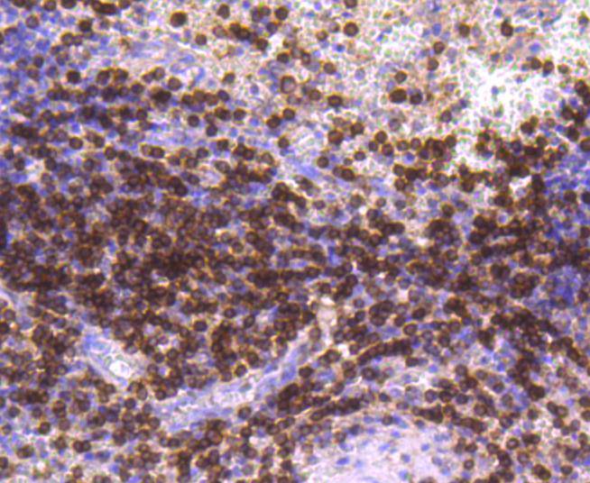 Immunohistochemical analysis of paraffin-embedded human spleen tissue using anti-CD3D antibody. The section was pre-treated using heat mediated antigen retrieval with Tris-EDTA buffer (pH 8.0-8.4) for 20 minutes.The tissues were blocked in 5% BSA for 30 minutes at room temperature, washed with ddH2O and PBS, and then probed with the primary antibody (SLM-52744R, 1/50) for 30 minutes at room temperature. The detection was performed using an HRP conjugated compact polymer system. DAB was used as the chromogen. Tissues were counterstained with hematoxylin and mounted with DPX
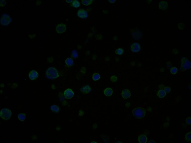 ICC staining of CD3D in Jurkat cells (green). Formalin fixed cells were permeabilized with 0.1% Triton X-100 in TBS for 10 minutes at room temperature and blocked with 1% Blocker BSA for 15 minutes at room temperature. Cells were probed with the primary antibody (SLM-52744R, 1/50) for 1 hour at room temperature, washed with PBS. Alexa Fluor®488 Goat anti-Rabbit IgG was used as the secondary antibody at 1/1,000 dilution. The nuclear counter stain is DAPI (blue).
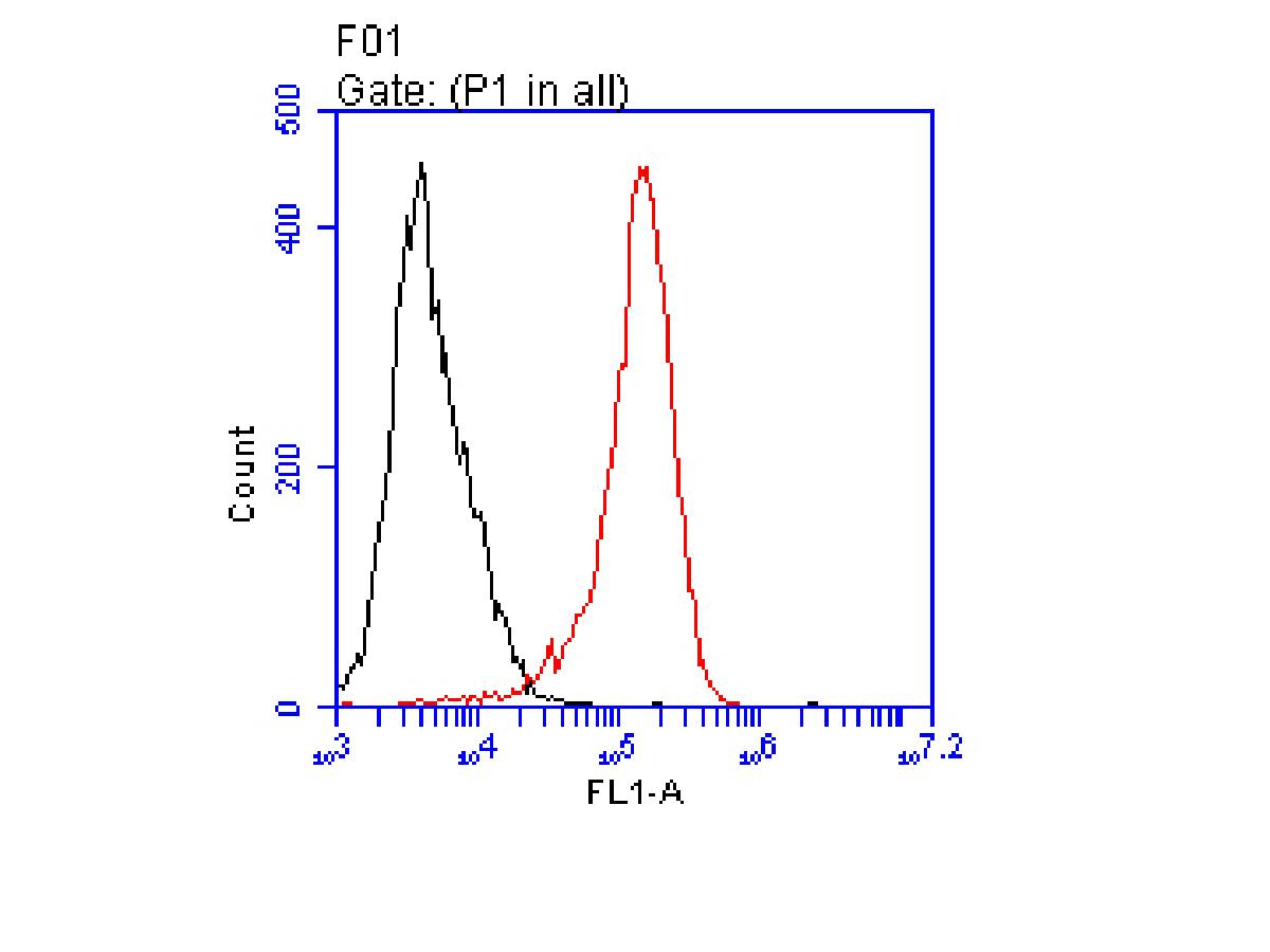 Flow cytometric analysis of CD3D was done on Jurkat cells. The cells were fixed, permeabilized and stained with the primary antibody (SLM-52744R, 1/50) (red). After incubation of the primary antibody at room temperature for an hour, the cells were stained with a Alexa Fluor 488-conjugated Goat anti-Rabbit IgG Secondary antibody at 1/1000 dilution for 30 minutes.Unlabelled sample was used as a control (cells without incubation with primary antibody; black).
Cartpieces
Totalgoods,subtotals:¥Checkout
References (0)
No References
Bought notes(bought amounts latest0)
No one bought this product
User Comment(Total0User Comment Num)
- No comment


 +86 571 56623320
 +86 18668110335

