
Rabbit Anti-ABCG2 antibody
ABC15; ABCG 2; ABCG2_HUMAN; ABCP; ATP binding cassette sub family G (WHITE) member 2; ATP binding cassette transporter G2; ATP-binding cassette sub-family G member 2; BCRP1; BMDP; Breast cancer resistance protein; CD338; CDw338; CDw338 antigen; EST157481;
View History [Clear]
Details
Product Name ABCG2 Chinese Name 三磷酸腺苷结合TransporterG超家族成员2Recombinant rabbit monoclonal anti Alias ABC15; ABCG 2; ABCG2_HUMAN; ABCP; ATP binding cassette sub family G (WHITE) member 2; ATP binding cassette transporter G2; ATP-binding cassette sub-family G member 2; BCRP1; BMDP; Breast cancer resistance protein; CD338; CDw338; CDw338 antigen; EST157481; GOUT1; MGC102821; Mitoxantrone resistance associated protein; Mitoxantrone resistance-associated protein; MRX; Multi drug resistance efflux transport ATP binding cassette sub family G (WHITE) member 2; MXR; MXR1; Placenta specific ATP binding cassette transporter; Placenta specific MDR protein; Placenta-specific ATP-binding cassette transporter; UAQTL1. Research Area immunology Neurobiology Stem cells Transporter Cell Surface Molecule Immunogen Species Rabbit Clonality Monoclonal React Species (predicted: Human, Mouse, ) Applications WB=1:500-2000 IHC-P=1:100-500 ICC=1:50-200 IF=1:50-200 (Paraffin sections need antigen repair)
not yet tested in other applications.
optimal dilutions/concentrations should be determined by the end user.Theoretical molecular weight 72kDa Cellular localization The nucleus The cell membrane Form Liquid Concentration 1mg/ml immunogen KLH conjugated synthetic peptide derived from human ABCG2 Lsotype IgG Purification affinity purified by Protein A Buffer Solution 0.01M TBS(pH7.4) with 1% BSA, 0.03% Proclin300 and 50% Glycerol. Storage Shipped at 4℃. Store at -20 °C for one year. Avoid repeated freeze/thaw cycles. Attention This product as supplied is intended for research use only, not for use in human, therapeutic or diagnostic applications. PubMed PubMed Product Detail The membrane-associated protein encoded by this gene is included in the superfamily of ATP-binding cassette (ABC) transporters. ABC proteins transport various molecules across extra- and intra-cellular membranes. ABC genes are divided into seven distinct subfamilies (ABC1, MDR/TAP, MRP, ALD, OABP, GCN20, White). This protein is a member of the White subfamily. Alternatively referred to as a breast cancer resistance protein, this protein functions as a xenobiotic transporter which may play a major role in multi-drug resistance. It likely serves as a cellular defense mechanism in response to mitoxantrone and anthracycline exposure. Significant expression of this protein has been observed in the placenta, which may suggest a potential role for this molecule in placenta tissue. Multiple transcript variants encoding different isoforms have been found for this gene. [provided by RefSeq, Apr 2012]
Function:
Xenobiotic transporter that may play an important role in the exclusion of xenobiotics from the brain. May be involved in brain-to-blood efflux. Appears to play a major role in the multidrug resistance phenotype of several cancer cell lines. When overexpressed, the transfected cells become resistant to mitoxantrone, daunorubicin and doxorubicin, display diminished intracellular accumulation of daunorubicin, and manifest an ATP-dependent increase in the efflux of rhodamine 123.
Subunit:
Monomer or homodimer; disulfide-linked.
Subcellular Location:
Cell membrane; Multi-pass membrane protein.
Tissue Specificity:
Highly expressed in placenta. Low expression in small intestine, liver and colon.
Similarity:
Belongs to the ABC transporter superfamily. ABCG family. Eye pigment precursor importer (TC 3.A.1.204) subfamily.
Contains 1 ABC transmembrane type-2 domain.
Contains 1 ABC transporter domain.
SWISS:
Q9UNQ0
Gene ID:
9429
Database links:Entrez Gene: 9429 Human
Omim: 603756 Human
SwissProt: Q9UNQ0 Human
Unigene: 480218 Human
Product Picture 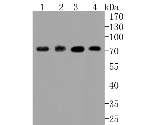 Western blot analysis of ABCG2 on different lysates. Proteins were transferred to a PVDF membrane and blocked with 5% BSA in PBS for 1 hour at room temperature. The primary antibody (SLM-52821R, 1/500) was used in 5% BSA at room temperature for 2 hours. Goat Anti-Rabbit IgG - HRP Secondary Antibody (HA1001) at 1:5,000 dilution was used for 1 hour at room temperature.
Positive control:
Lane 1: HepG2 cell lysate
Lane 2: human placenta tissue lysate
Lane 3: Hela cell lysate
Lane 4: 293T cell lysate
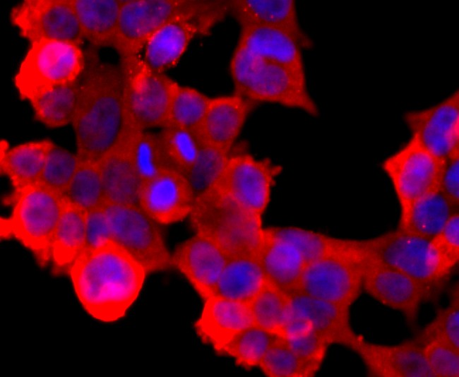 ICC staining of ABCG2 in 293T cells (red). Formalin fixed cells were permeabilized with 0.1% Triton X-100 in TBS for 10 minutes at room temperature and blocked with 1% Blocker BSA for 15 minutes at room temperature. Cells were probed with the primary antibody (SLM-52821R, 1/50) for 1 hour at room temperature, washed with PBS. Alexa Fluor®594 Goat anti-Rabbit IgG was used as the secondary antibody at 1/1,000 dilution. The nuclear counter stain is DAPI (blue).
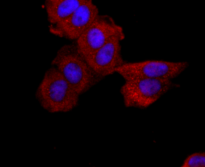 ICC staining of ABCG2 in MCF-7 cells (red). Formalin fixed cells were permeabilized with 0.1% Triton X-100 in TBS for 10 minutes at room temperature and blocked with 1% Blocker BSA for 15 minutes at room temperature. Cells were probed with the primary antibody (SLM-52821R, 1/50) for 1 hour at room temperature, washed with PBS. Alexa Fluor®594 Goat anti-Rabbit IgG was used as the secondary antibody at 1/1,000 dilution. The nuclear counter stain is DAPI (blue).
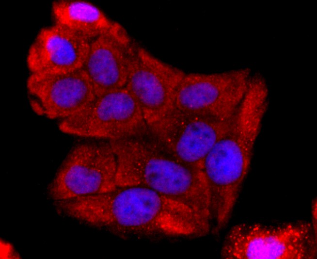 ICC staining of ABCG2 in Hela cells (red). Formalin fixed cells were permeabilized with 0.1% Triton X-100 in TBS for 10 minutes at room temperature and blocked with 1% Blocker BSA for 15 minutes at room temperature. Cells were probed with the primary antibody (SLM-52821R, 1/50) for 1 hour at room temperature, washed with PBS. Alexa Fluor®594 Goat anti-Rabbit IgG was used as the secondary antibody at 1/1,000 dilution. The nuclear counter stain is DAPI (blue).
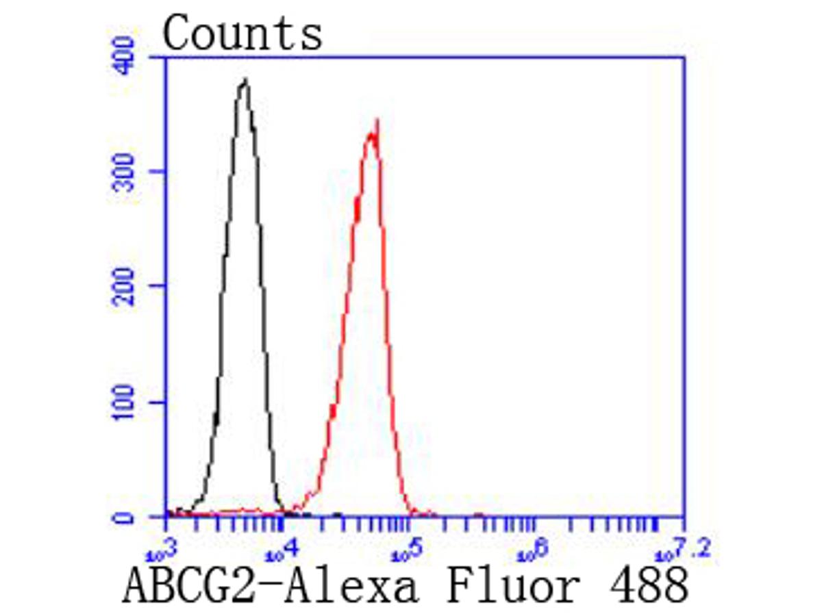 Flow cytometric analysis of ABCG2 was done on 293T cells. The cells were fixed, permeabilized and stained with the primary antibody (SLM-52821R, 1/50) (red). After incubation of the primary antibody at room temperature for an hour, the cells were stained with a Alexa Fluor 488-conjugated Goat anti-Rabbit IgG Secondary antibody at 1/1000 dilution for 30 minutes.Unlabelled sample was used as a control (cells without incubation with primary antibody; black).
Cartpieces
Totalgoods,subtotals:¥Checkout
References (0)
No References
Bought notes(bought amounts latest0)
No one bought this product
User Comment(Total0User Comment Num)
- No comment


 +86 571 56623320
 +86 18668110335

