
Rabbit Anti-PAX8 antibody
Paired Box Gene 8; paired box homeotic gene 8; Paired box protein Pax 8; Paired Domain Gene 8; PAX 8;PAX-8; PAX8_HUMAN.
View History [Clear]
Details
Product Name PAX8 Chinese Name 配对盒基因8Recombinant rabbit monoclonal anti Alias Paired Box Gene 8; paired box homeotic gene 8; Paired box protein Pax 8; Paired Domain Gene 8; PAX 8;PAX-8; PAX8_HUMAN. Research Area Developmental biology Neurobiology transcriptional regulatory factor Epigenetics Immunogen Species Rabbit Clonality Monoclonal React Species (predicted: Human, ) Applications WB=1:500-2000 IHC-P=1:100-500 ICC=1:50-200 (Paraffin sections need antigen repair)
not yet tested in other applications.
optimal dilutions/concentrations should be determined by the end user.Theoretical molecular weight 48kDa Cellular localization The nucleus Form Liquid Concentration 1mg/ml immunogen Recombinant human PAX8 protein : 101-450/450 Lsotype IgG Purification affinity purified by Protein A Buffer Solution 0.01M TBS(pH7.4) with 1% BSA, 0.03% Proclin300 and 50% Glycerol. Storage Shipped at 4℃. Store at -20 °C for one year. Avoid repeated freeze/thaw cycles. Attention This product as supplied is intended for research use only, not for use in human, therapeutic or diagnostic applications. PubMed PubMed Product Detail This protein is a member of the paired box (PAX) family of transcription factors, typically containing a paired box domain, an octapeptide, and a paired-type homeodomain. This family plays critical roles during fetal development and cancer growth. The specific function of the PAX8 is unknown but it may involve kidney cell differentiation, thyroid development, or thyroid dysgenesis. Alternative splicing in the gene by inclusion or exclusion of exons 7 and/or 8 has produced several known products but the biological significance of the variants is unknown. Several other splice variants have been proposed but the full nature of these products has not been described. Pax8 is also a marker of otic progenitor cells.
SWISS:
Q06710
Gene ID:
7849
Database links:Entrez Gene: 7849 Human
Entrez Gene: 18510 Mouse
Omim: 167415 Human
SwissProt: Q06710 Human
SwissProt: Q00288 Mouse
Unigene: 469728 Human
Unigene: 2533 Mouse
Unigene: 10392 Rat
Product Picture 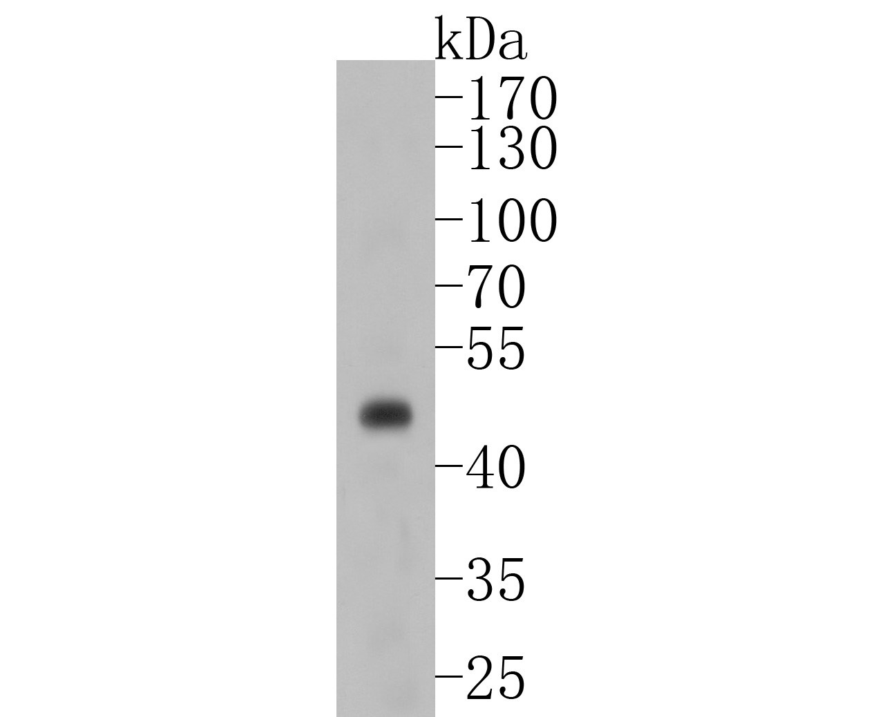 Western blot analysis of PAX8 on SKOV-3 cell lysates. Proteins were transferred to a PVDF membrane and blocked with 5% BSA in PBS for 1 hour at room temperature. The primary antibody (SLM-52737R, 1/500) was used in 5% BSA at room temperature for 2 hours. Goat Anti-Rabbit IgG - HRP Secondary Antibody (HA1001) at 1:200,000 dilution was used for 1 hour at room temperature.
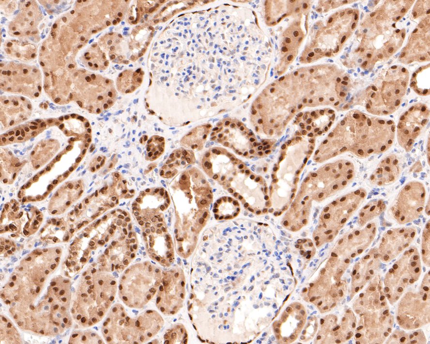 Immunohistochemical analysis of paraffin-embedded human kidney tissue with Rabbit anti-PAX8 antibody (SLM-52737R) at 1/500 dilution. The section was pre-treated using heat mediated antigen retrieval with sodium citrate buffer (pH 6.0) for 2 minutes. The tissues were blocked in 1% BSA for 20 minutes at room temperature, washed with ddH2O and PBS, and then probed with the primary antibody (SLM-52737R) at 1/500 dilution for 1 hour at room temperature. The detection was performed using an HRP conjugated compact polymer system. DAB was used as the chromogen. Tissues were counterstained with hematoxylin and mounted with DPX.
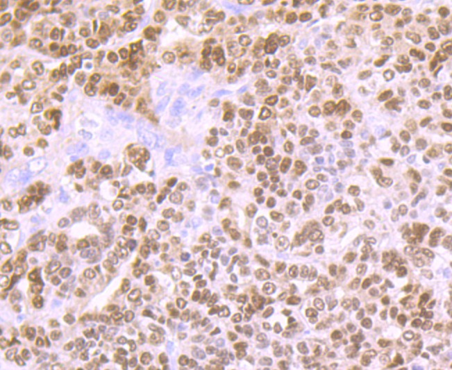 Immunohistochemical analysis of paraffin-embedded human ovarian carcinoma tissue using anti-PAX8 antibody. The section was pre-treated using heat mediated antigen retrieval with Tris-EDTA buffer (pH 9.0) for 20 minutes.The tissues were blocked in 5% BSA for 30 minutes at room temperature, washed with ddH2O and PBS, and then probed with the primary antibody (SLM-52737R, 1/50) for 30 minutes at room temperature. The detection was performed using an HRP conjugated compact polymer system. DAB was used as the chromogen. Tissues were counterstained with hematoxylin and mounted with DPX.
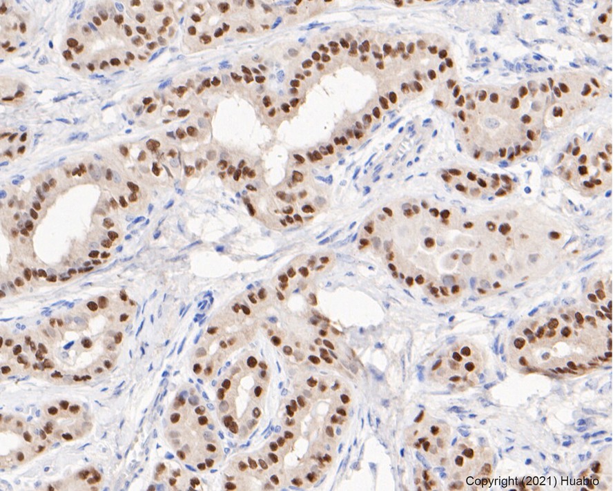 Immunohistochemical analysis of paraffin-embedded human thyroid cancer tissue using anti-PAX8 antibody. The section was pre-treated using heat mediated antigen retrieval with sodium citrate buffer (pH 6.0) for 20 minutes. The tissues were blocked in 5% BSA for 30 minutes at room temperature, washed with ddH2O and PBS, and then probed with the primary antibody (SLM-52737R, 1/50) for 30 minutes at room temperature. The detection was performed using an HRP conjugated compact polymer system. DAB was used as the chromogen. Tissues were counterstained with hematoxylin and mounted with DPX.
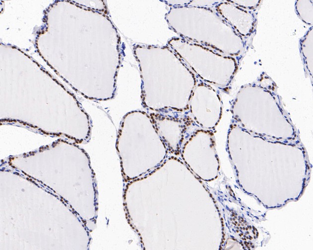 Immunohistochemical analysis of paraffin-embedded human thyroid tissue using anti-PAX8 antibody. The section was pre-treated using heat mediated antigen retrieval with sodium citrate buffer (pH 6.0) for 20 minutes. The tissues were blocked in 5% BSA for 30 minutes at room temperature, washed with ddH2O and PBS, and then probed with the primary antibody (SLM-52737R, 1/200) for 30 minutes at room temperature. The detection was performed using an HRP conjugated compact polymer system. DAB was used as the chromogen. Tissues were counterstained with hematoxylin and mounted with DPX.
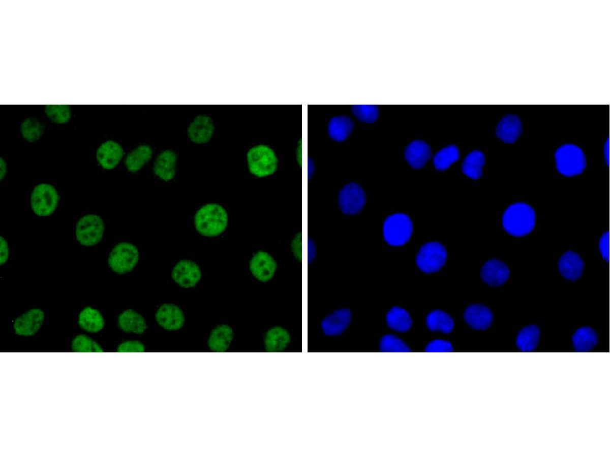 ICC staining of PAX8 in SKOV-3 cells (green). Formalin fixed cells were permeabilized with 0.1% Triton X-100 in TBS for 10 minutes at room temperature and blocked with 10% negative goat serum for 15 minutes at room temperature. Cells were probed with the primary antibody (SLM-52737R, 1/50) for 1 hour at room temperature, washed with PBS. Alexa Fluor®488 conjugate-Goat anti-Rabbit IgG was used as the secondary antibody at 1/1,000 dilution. The nuclear counter stain is DAPI (blue).
Cartpieces
Totalgoods,subtotals:¥Checkout
References (0)
No References
Bought notes(bought amounts latest0)
No one bought this product
User Comment(Total0User Comment Num)
- No comment


 +86 571 56623320
 +86 18668110335

