
Rabbit Anti-Furin antibody
FURIN_HUMAN; Furin; EC:3.4.21.75; FURIN; FUR; PACE; PCSK3; Dibasic-processing enzyme; Paired basic amino acid residue-cleaving enzyme (PACE);
View History [Clear]
Details
Product Name Furin Chinese Name 弗林蛋白酶Recombinant rabbit monoclonal anti Alias FURIN_HUMAN; Furin; EC:3.4.21.75; FURIN; FUR; PACE; PCSK3; Dibasic-processing enzyme; Paired basic amino acid residue-cleaving enzyme (PACE); Research Area Cell biology Signal transduction Ubiquitin Immunogen Species Rabbit Clonality Monoclonal React Species (predicted: Human, Mouse, Rat, ) Applications WB=1:300-500 IHC-P=1:100-500 (Paraffin sections need antigen repair)
not yet tested in other applications.
optimal dilutions/concentrations should be determined by the end user.Theoretical molecular weight 74kDa Cellular localization cytoplasmic The cell membrane Extracellular matrix Form Liquid Concentration 1mg/ml immunogen KLH conjugated synthetic peptide derived from human Furin Lsotype IgG Purification affinity purified by Protein A Buffer Solution 0.01M TBS(pH7.4) with 1% BSA, 0.03% Proclin300 and 50% Glycerol. Storage Shipped at 4℃. Store at -20 °C for one year. Avoid repeated freeze/thaw cycles. Attention This product as supplied is intended for research use only, not for use in human, therapeutic or diagnostic applications. PubMed PubMed Product Detail Furin is a calcium-dependent serine endoprotease that belongs to the subtilisin-like proprotein convertase family. The members of this family process latent precursor proteins into their biologically active products. Furin cleaves at paired basic amino acid processing sites within proparathyroid hormone, transforming growth factor β 1 precursor, proalbumin, pro-β-secretase, membrane type-1 matrix metalloproteinase, β subunit of pro-nerve growth factor and von Willebrand factor. Furin can directly cleave proMMP-2 within the ttrans-Golgi network leading to an inactive form of matrix metalloproteinase-2 (MMP-2). Furin is synthesized as an inactive zymogen that may minimize the occurrence of premature enzymatic activity that would lead to alternative protein activation or degradation. The inhibitory mechanism is based on the presence of an inactivating prosegment at the NH2 terminal of the Furin. After initial autocatalytic cleavage, the prosegment remains tightly associated until it reaches the trans-Golgi network where the dissociation of the prosegment and activation of furin occurs.
Function:
Furin is likely to represent the ubiquitous endoprotease activity within constitutive secretory pathways and capable of cleavage at the RX(K/R)R consensus motif.
Subunit:
Interacts with FLNA (By similarity). Binds to PACS1 which mediates TGN localization and connection to clathrin adapters.
Subcellular Location:
Golgi apparatus > trans-Golgi network membrane. Cell membrane. Shuttles between the trans-Golgi network and the cell surface. Propeptide cleavage is a prerequisite for exit of furin molecules out of the endoplasmic reticulum (ER). A second cleavage within the propeptide occurs in the trans Golgi network (TGN), followed by the release of the propeptide and the activation of furin.
Tissue Specificity:
Seems to be expressed ubiquitously.
Post-translational modifications:
The inhibition peptide, which plays the role of an intramolecular chaperone, is autocatalytically removed in the endoplasmic reticulum (ER) and remains non-covalently bound to furin as a potent autoinhibitor. Following transport to the trans Golgi, a second cleavage within the inhibition propeptide results in propeptide dissociation and furin activation.
Similarity:
Belongs to the peptidase S8 family. Furin subfamily.
Contains 1 homo B/P domain.
SWISS:
P09958
Gene ID:
5045
Database links:Entrez Gene: 5045 Human
Entrez Gene: 18550 Mouse
SwissProt: P09958 Human
SwissProt: P23188 Mouse
在真核生物细胞中,许多具有生物活性的多肽和蛋白是在其分泌过程中由前体蛋白经内切蛋白酶切割后激活形成的.弗林蛋白酶(Furin)就是这个内切蛋白酶家族重要成员之一,它可以识别剪切多种蛋白质,如生长医子、血清蛋白、基质金属蛋白酶、受体、病毒囊膜蛋白和细菌外毒素等.近年来Furin得到了迅速而广泛的研究,本文简介了它的表达与加工运输、生物学功能、与病毒侵染的关系,以及它的抑制剂.Product Picture 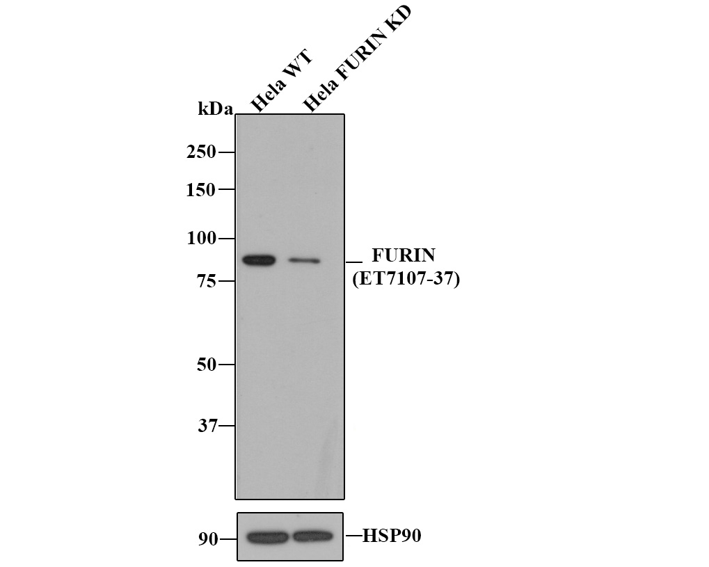 All lanes: Western blot analysis of FURIN with anti-Furin antibody (SLM-54283R) at 1:500 dilution. Lane 1: Wild-type Hela whole cell lysate. Lane 2: FURIN knockdown Hela whole cell lysate.
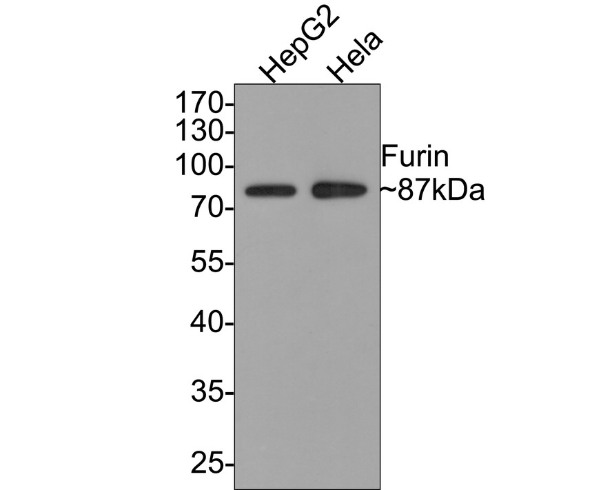 Western blot analysis of Furin on different lysates with Rabbit anti-Furin antibody (SLM-54283R) at 1/500 dilution. Lane 1: HepG2 cell lysate Lane 2: Hela cell lysate Lysates/proteins at 10 µg/Lane. Predicted band size: 87 kDa Observed band size: 87 kDa Exposure time: 1 minute; 10% SDS-PAGE gel.
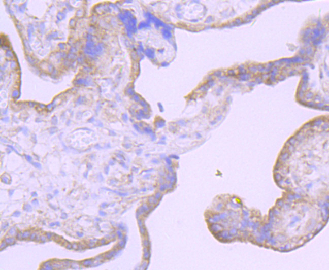 Immunohistochemical analysis of paraffin-embedded human placenta tissue using anti-Furin antibody. The section was pre-treated using heat mediated antigen retrieval with Tris-EDTA buffer (pH 8.0-8.4) for 20 minutes.The tissues were blocked in 5% BSA for 30 minutes at room temperature, washed with ddH2O and PBS, and then probed with the primary antibody (ET7107-37, 1/50) for 30 minutes at room temperature. The detection was performed using an HRP conjugated compact polymer system. DAB was used as the chromogen. Tissues were counterstained with hematoxylin and mounted with DPX.
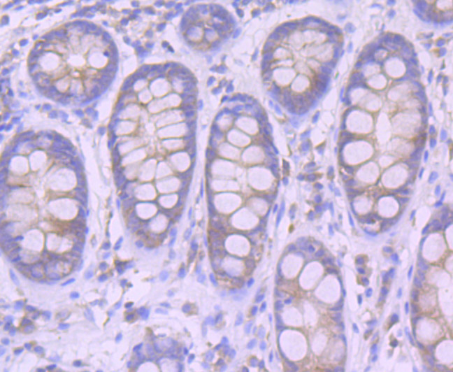 Immunohistochemical analysis of paraffin-embedded human colon tissue using anti-Furin antibody. The section was pre-treated using heat mediated antigen retrieval with Tris-EDTA buffer (pH 8.0-8.4) for 20 minutes.The tissues were blocked in 5% BSA for 30 minutes at room temperature, washed with ddH2O and PBS, and then probed with the primary antibody (SLM-54283R, 1/50) for 30 minutes at room temperature. The detection was performed using an HRP conjugated compact polymer system. DAB was used as the chromogen. Tissues were counterstained with hematoxylin and mounted with DPX.
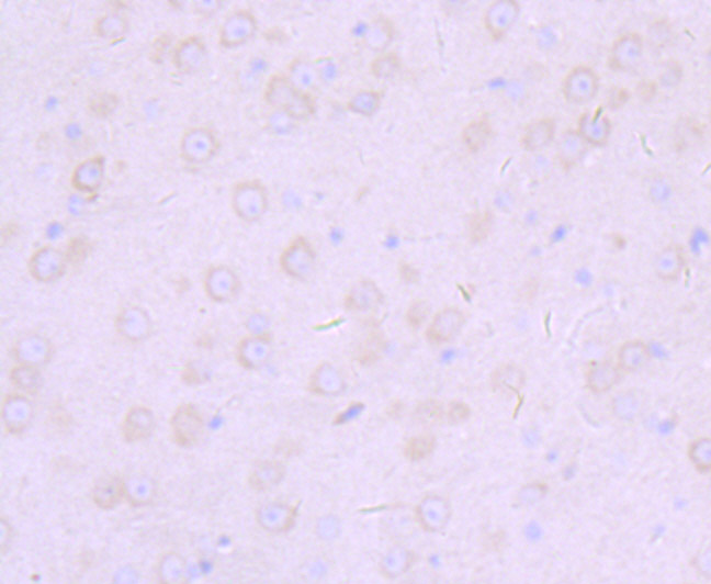 Immunohistochemical analysis of paraffin-embedded mouse brain tissue using anti-Furin antibody. The section was pre-treated using heat mediated antigen retrieval with Tris-EDTA buffer (pH 8.0-8.4) for 20 minutes.The tissues were blocked in 5% BSA for 30 minutes at room temperature, washed with ddH2O and PBS, and then probed with the primary antibody (SLM-54283R, 1/50) for 30 minutes at room temperature. The detection was performed using an HRP conjugated compact polymer system. DAB was used as the chromogen. Tissues were counterstained with hematoxylin and mounted with DPX.
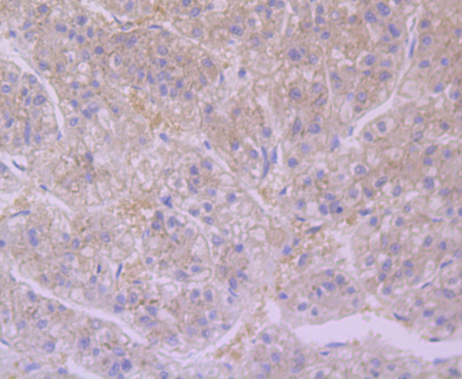 Immunohistochemical analysis of paraffin-embedded human liver tissue using anti-Furin antibody. The section was pre-treated using heat mediated antigen retrieval with Tris-EDTA buffer (pH 8.0-8.4) for 20 minutes.The tissues were blocked in 5% BSA for 30 minutes at room temperature, washed with ddH2O and PBS, and then probed with the primary antibody (SLM-54283R, 1/50) for 30 minutes at room temperature. The detection was performed using an HRP conjugated compact polymer system. DAB was used as the chromogen. Tissues were counterstained with hematoxylin and mounted with DPX.
Cartpieces
Totalgoods,subtotals:¥Checkout
References (0)
No References
Bought notes(bought amounts latest0)
No one bought this product
User Comment(Total0User Comment Num)
- No comment


 +86 571 56623320
 +86 18668110335

