
Rabbit Anti-STAT5a antibody
signal transducers and activators of transduction 5; MGF; Signal Transducer and Activator of Transcription 5A; STAT 5; STAT 5A; STAT 5B; STAT5A; STAT5B; Transcription factor STAT5A; STA5B_HUMAN; STA5A_HUMAN.
View History [Clear]
Details
Product Name STAT5a Chinese Name Signal transduction和转录激活因子5Recombinant rabbit monoclonal anti Alias signal transducers and activators of transduction 5; MGF; Signal Transducer and Activator of Transcription 5A; STAT 5; STAT 5A; STAT 5B; STAT5A; STAT5B; Transcription factor STAT5A; STA5B_HUMAN; STA5A_HUMAN. Research Area immunology Signal transduction transcriptional regulatory factor Immunogen Species Rabbit Clonality Monoclonal Clone NO. 6E5 React Species (predicted: Human, Mouse, Rat, ) Applications WB=1:1000-2000 IHC-P=1:100-500 IHC-F=1:50-200 ICC=1:50-200 IF=1:50-200 (Paraffin sections need antigen repair)
not yet tested in other applications.
optimal dilutions/concentrations should be determined by the end user.Theoretical molecular weight 90kDa Cellular localization The nucleus cytoplasmic Form Liquid Concentration 1mg/ml immunogen Recombinant human STAT5a protein, around C-terminal 100aa Lsotype IgG Purification affinity purified by Protein A Buffer Solution 0.01M TBS(pH7.4) with 1% BSA, 0.03% Proclin300 and 50% Glycerol. Storage Shipped at 4℃. Store at -20 °C for one year. Avoid repeated freeze/thaw cycles. Attention This product as supplied is intended for research use only, not for use in human, therapeutic or diagnostic applications. PubMed PubMed Product Detail The protein encoded by this gene is a member of the STAT family of transcription factors. In response to cytokines and growth factors, STAT family members are phosphorylated by the receptor associated kinases, and then form homo- or heterodimers that translocate to the cell nucleus where they act as transcription activators. This protein is activated by, and mediates the responses of many cell ligands, such as IL2, IL3, IL7 GM-CSF, erythropoietin, thrombopoietin, and different growth hormones. Activation of this protein in myeloma and lymphoma associated with a TEL/JAK2 gene fusion is independent of cell stimulus and has been shown to be essential for the tumorigenesis. The mouse counterpart of this gene is found to induce the expression of BCL2L1/BCL-X(L), which suggests the antiapoptotic function of this gene in cells. [provided by RefSeq, Jul 2008]
Function:
Carries out a dual function: signal transduction and activation of transcription. Mediates cellular responses to the cytokine KITLG/SCF and other growth factors. Mediates cellular responses to ERBB4. May mediate cellular responses to activated FGFR1, FGFR2, FGFR3 and FGFR4. Binds to the GAS element and activates PRL-induced transcription. Regulates the expression of milk proteins during lactation.
Subunit:
Forms a homodimer or a heterodimer with a related family member. Binds NR3C1. Interacts with NCOA1 and SOCS7. Interacts with ERBB4.
Subcellular Location:
Cytoplasm. Nucleus. Note=Translocated into the nucleus in response to phosphorylation.
Post-translational modifications:
Tyrosine phosphorylated in response to KITLG/SCF, IL2, IL3, IL7, IL15, CSF2/GMCSF, GH1, PRL, EPO and THPO. Activated KIT promotes phosphorylation on tyrosine residues and subsequent translocation to the nucleus. Tyrosine phosphorylated in response to constitutively activated FGFR1, FGFR2, FGFR3 and FGFR4. Tyrosine phosphorylation is required for DNA-binding activity and dimerization. Serine phosphorylation is also required for maximal transcriptional activity (By similarity). Tyrosine phosphorylated in response to signaling via activated FLT3; wild-type FLT3 results in much weaker phosphorylation than constitutively activated mutant FLT3. Alternatively, can be phosphorylated by JAK2 at Tyr-694.
Similarity:
Belongs to the transcription factor STAT family.
Contains 1 SH2 domain.
SWISS:
P42229
Gene ID:
6776
Database links:Entrez Gene: 6776 Human
Entrez Gene: 6777 Human
Entrez Gene: 20850 Mouse
Entrez Gene: 20851 Mouse
Omim: 601511 Human
Omim: 604260 Human
SwissProt: P42229 Human
SwissProt: P51692 Human
SwissProt: P42230 Mouse
SwissProt: P42232 Mouse
Unigene: 437058 Human
Unigene: 277403 Mouse
Unigene: 154399 Rat
Product Picture 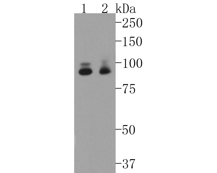 Western blot analysis of STAT5a on different lysates. Proteins were transferred to a PVDF membrane and blocked with 5% BSA in PBS for 1 hour at room temperature. The primary antibody (SLM-52237R, 1/500) was used in 5% BSA at room temperature for 2 hours. Goat Anti-Rabbit IgG - HRP Secondary Antibody (HA1001) at 1:5,000 dilution was used for 1 hour at room temperature.
Positive control:
Lane 1: Hela cell lysate
Lane 2: K562 cell lysate
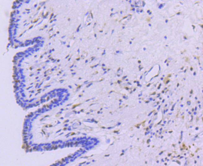 Immunohistochemical analysis of paraffin-embedded human breast carcinoma tissue using anti-STAT5a antibody. The section was pre-treated using heat mediated antigen retrieval with Tris-EDTA buffer (pH 8.0-8.4) for 20 minutes.The tissues were blocked in 5% BSA for 30 minutes at room temperature, washed with ddH2O and PBS, and then probed with the primary antibody (SLM-52237R, 1/50) for 30 minutes at room temperature. The detection was performed using an HRP conjugated compact polymer system. DAB was used as the chromogen. Tissues were counterstained with hematoxylin and mounted with DPX.
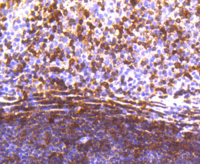 Immunohistochemical analysis of paraffin-embedded human tonsil tissue using anti-STAT5a antibody. The section was pre-treated using heat mediated antigen retrieval with Tris-EDTA buffer (pH 8.0-8.4) for 20 minutes.The tissues were blocked in 5% BSA for 30 minutes at room temperature, washed with ddH2O and PBS, and then probed with the primary antibody (SLM-52237R, 1/50) for 30 minutes at room temperature. The detection was performed using an HRP conjugated compact polymer system. DAB was used as the chromogen. Tissues were counterstained with hematoxylin and mounted with DPX.
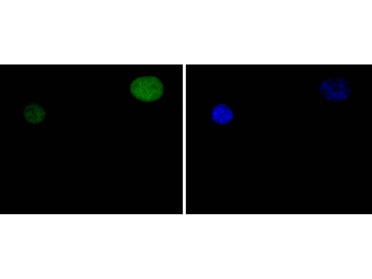 ICC staining of STAT5a in C2C12 cells (green). Formalin fixed cells were permeabilized with 0.1% Triton X-100 in TBS for 10 minutes at room temperature and blocked with 1% Blocker BSA for 15 minutes at room temperature. Cells were probed with the primary antibody (SLM-52237R, 1/50) for 1 hour at room temperature, washed with PBS. Alexa Fluor®488 Goat anti-Rabbit IgG was used as the secondary antibody at 1/1,000 dilution. The nuclear counter stain is DAPI (blue).
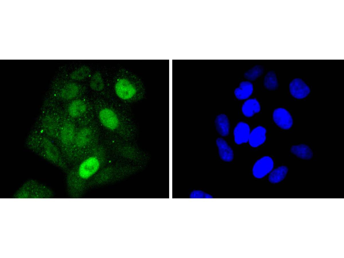 ICC staining of STAT5a in Hela cells (green). Formalin fixed cells were permeabilized with 0.1% Triton X-100 in TBS for 10 minutes at room temperature and blocked with 1% Blocker BSA for 15 minutes at room temperature. Cells were probed with the primary antibody (SLM-52237R, 1/50) for 1 hour at room temperature, washed with PBS. Alexa Fluor®488 Goat anti-Rabbit IgG was used as the secondary antibody at 1/1,000 dilution. The nuclear counter stain is DAPI (blue).
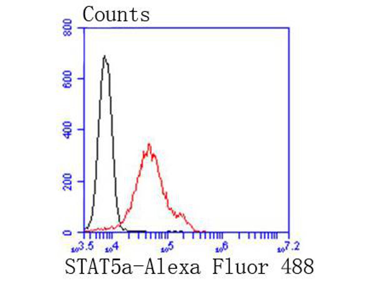 Flow cytometric analysis of STAT5a was done on Jurkat cells. The cells were fixed, permeabilized and stained with the primary antibody (SLM-52237R, 1/50) (red). After incubation of the primary antibody at room temperature for an hour, the cells were stained with a Alexa Fluor 488-conjugated Goat anti-Rabbit IgG Secondary antibody at 1/1000 dilution for 30 minutes.Unlabelled sample was used as a control (cells without incubation with primary antibody; black).
Cartpieces
Totalgoods,subtotals:¥Checkout
References (0)
No References
Bought notes(bought amounts latest0)
No one bought this product
User Comment(Total0User Comment Num)
- No comment


 +86 571 56623320
 +86 18668110335

