
Rabbit Anti-ITGB3 antibody
Integrin beta-3; Integrin beta chain, β3 precursor; Integrin Beta 3; CD 61; CD61; CD61 antigen; GP3A; GPIIIa; HPA 1; HPA 4; Integrin beta 3 (platelet glycoprotein IIIa antigen CD61); Integrin beta chain beta 3; ITG B3; ITGB 3; NAIT; Platelet fibrinogen re
View History [Clear]
Details
Product Name ITGB3 Chinese Name 整合素β3/CD61Recombinant rabbit monoclonal anti Alias Integrin beta-3; Integrin beta chain, β3 precursor; Integrin Beta 3; CD 61; CD61; CD61 antigen; GP3A; GPIIIa; HPA 1; HPA 4; Integrin beta 3 (platelet glycoprotein IIIa antigen CD61); Integrin beta chain beta 3; ITG B3; ITGB 3; NAIT; Platelet fibrinogen receptor beta subunit; Platelet glycoprotein IIIa; platelet glycoprotein IIIa precursor; Platelet membrane glycoprotein IIIa; PTP; ITB3_HUMAN. Integrin β3; Integrin-β3; Integrin β 3; Integrinβ3; Research Area Cell biology immunology Signal transduction Stem cells Cell adhesion molecule Immunogen Species Rabbit Clonality Monoclonal Clone NO. 2B1 React Species (predicted: Human, Mouse, Rat, ) Applications WB=1:500-1000 IHC-P=1:100-500 ICC=1:50-200 IF=1:50-200 (Paraffin sections need antigen repair)
not yet tested in other applications.
optimal dilutions/concentrations should be determined by the end user.Theoretical molecular weight 84kDa Cellular localization The cell membrane Form Liquid Concentration 1mg/ml immunogen KLH conjugated synthetic peptide derived from human ITGB3 : 729-788/788 Lsotype IgG Purification affinity purified by Protein A Buffer Solution 0.01M TBS(pH7.4) with 1% BSA, 0.03% Proclin300 and 50% Glycerol. Storage Shipped at 4℃. Store at -20 °C for one year. Avoid repeated freeze/thaw cycles. Attention This product as supplied is intended for research use only, not for use in human, therapeutic or diagnostic applications. PubMed PubMed Product Detail The ITGB3 protein product is the integrin beta chain beta 3. Integrins are integral cell-surface proteins composed of an alpha chain and a beta chain. A given chain may combine with multiple partners resulting in different integrins. Integrin beta 3 is found along with the alpha IIb chain in platelets. Integrins are known to participate in cell adhesion as well as cell-surface mediated signalling. [provided by RefSeq, Jul 2008]
SWISS:
P05106
Gene ID:
3690
Database links:Entrez Gene: 3690 Human
Entrez Gene: 16416 Mouse
Omim: 173470 Human
SwissProt: P05106 Human
SwissProt: O54890 Mouse
Unigene: 218040 Human
Unigene: 87150 Mouse
Product Picture 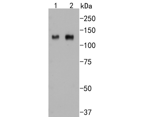 Western blot analysis of Integrin beta 3 on different lysates. Proteins were transferred to a PVDF membrane and blocked with 5% BSA in PBS for 1 hour at room temperature. The primary antibody (SLM-52374R, 1/500) was used in 5% BSA at room temperature for 2 hours. Goat Anti-Rabbit IgG - HRP Secondary Antibody (HA1001) at 1:5,000 dilution was used for 1 hour at room temperature.
Positive control:
Lane 1: HUVEC cell lysate
Lane 2: Mouse marrow tissue lysate
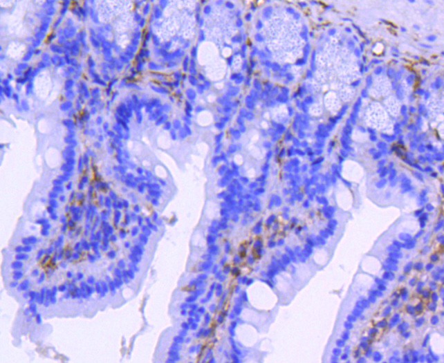 Immunohistochemical analysis of paraffin-embedded mouse colon tissue using anti-Integrin beta 3 antibody. The section was pre-treated using heat mediated antigen retrieval with Tris-EDTA buffer (pH 8.0-8.4) for 20 minutes.The tissues were blocked in 5% BSA for 30 minutes at room temperature, washed with ddH2O and PBS, and then probed with the primary antibody (SLM-52374R, 1/50) for 30 minutes at room temperature. The detection was performed using an HRP conjugated compact polymer system. DAB was used as the chromogen. Tissues were counterstained with hematoxylin and mounted with DPX.
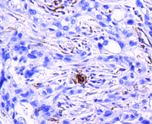 Immunohistochemical analysis of paraffin-embedded human breast carcinoma tissue using anti-Integrin beta 3 antibody. The section was pre-treated using heat mediated antigen retrieval with Tris-EDTA buffer (pH 8.0-8.4) for 20 minutes.The tissues were blocked in 5% BSA for 30 minutes at room temperature, washed with ddH2O and PBS, and then probed with the primary antibody (SLM-52374R, 1/50) for 30 minutes at room temperature. The detection was performed using an HRP conjugated compact polymer system. DAB was used as the chromogen. Tissues were counterstained with hematoxylin and mounted with DPX.
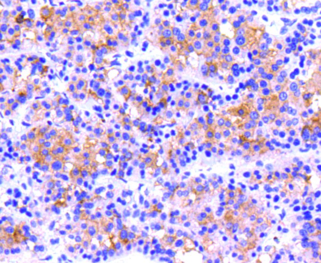 Immunohistochemical analysis of paraffin-embedded human liver carcinoma tissue using anti-Integrin beta 3 antibody. The section was pre-treated using heat mediated antigen retrieval with Tris-EDTA buffer (pH 8.0-8.4) for 20 minutes.The tissues were blocked in 5% BSA for 30 minutes at room temperature, washed with ddH2O and PBS, and then probed with the primary antibody (SLM-52374R, 1/50) for 30 minutes at room temperature. The detection was performed using an HRP conjugated compact polymer system. DAB was used as the chromogen. Tissues were counterstained with hematoxylin and mounted with DPX.
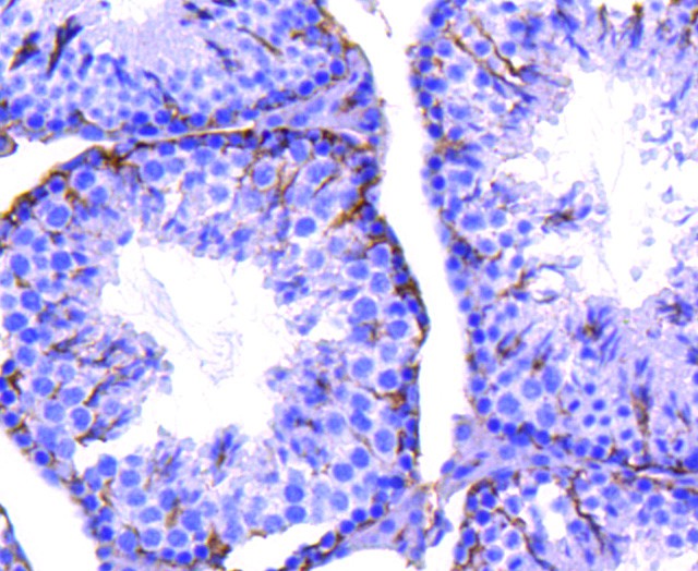 Immunohistochemical analysis of paraffin-embedded mouse testis tissue using anti-Integrin beta 3 antibody. The section was pre-treated using heat mediated antigen retrieval with Tris-EDTA buffer (pH 8.0-8.4) for 20 minutes.The tissues were blocked in 5% BSA for 30 minutes at room temperature, washed with ddH2O and PBS, and then probed with the primary antibody (SLM-52374R, 1/50) for 30 minutes at room temperature. The detection was performed using an HRP conjugated compact polymer system. DAB was used as the chromogen. Tissues were counterstained with hematoxylin and mounted with DPX.
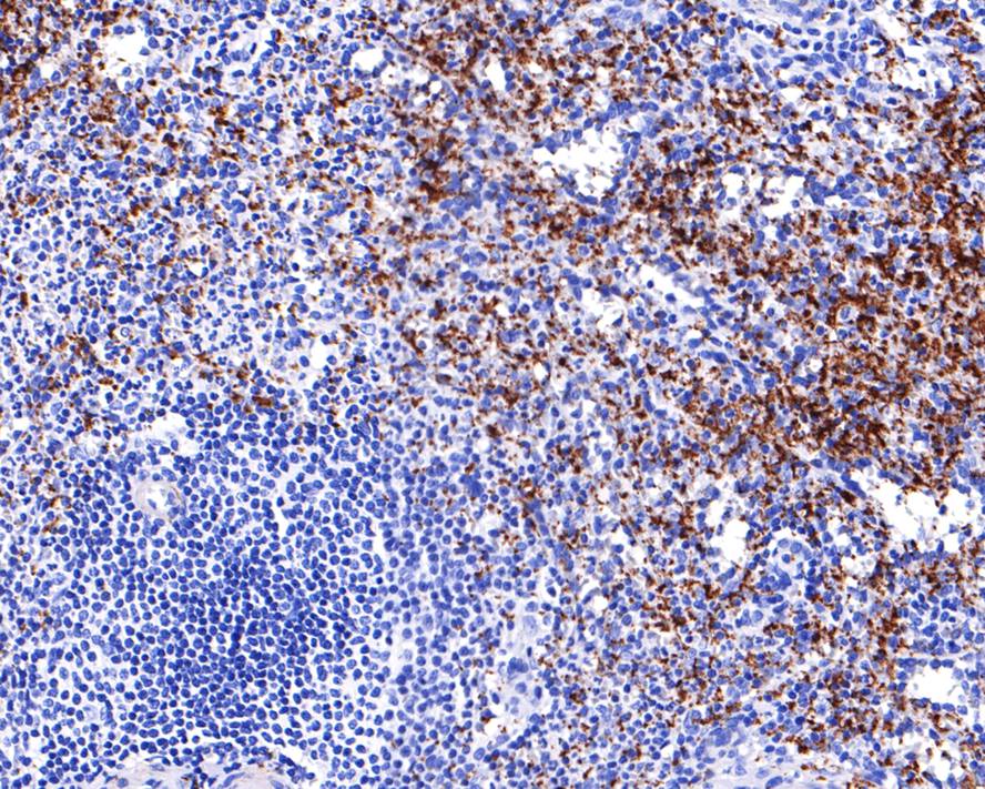 Immunohistochemical analysis of paraffin-embedded human spleen tissue with Rabbit anti-Integrin beta 3 antibody (SLM-52374R) at 1/400 dilution. The section was pre-treated using heat mediated antigen retrieval with Tris-EDTA buffer (pH 9.0) for 20 minutes. The tissues were blocked in 1% BSA for 20 minutes at room temperature, washed with ddH2O and PBS, and then probed with the primary antibody (SLM-52374R) at 1/400 dilution for 1 hour at room temperature. The detection was performed using an HRP conjugated compact polymer system. DAB was used as the chromogen. Tissues were counterstained with hematoxylin and mounted with DPX.
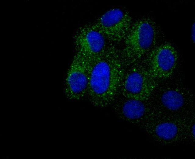 ICC staining of Integrin beta 3 in HepG2 cells (green). Formalin fixed cells were permeabilized with 0.1% Triton X-100 in TBS for 10 minutes at room temperature and blocked with 1% Blocker BSA for 15 minutes at room temperature. Cells were probed with the primary antibody (SLM-52374R, 1/50) for 1 hour at room temperature, washed with PBS. Alexa Fluor®488 Goat anti-Rabbit IgG was used as the secondary antibody at 1/1,000 dilution. The nuclear counter stain is DAPI (blue).
Cartpieces
Totalgoods,subtotals:¥Checkout
References (0)
No References
Bought notes(bought amounts latest0)
No one bought this product
User Comment(Total0User Comment Num)
- No comment


 +86 571 56623320
 +86 18668110335

